. 2015 May 20;137(19):6232-44.
doi: 10.1021/ja5132648. Epub 2015 May 5.

A generalizable platform for interrogating target- and signal-specific consequences of electrophilic modifications in redox-dependent cell signaling

Affiliations

A generalizable platform for interrogating target- and signal-specific consequences of electrophilic modifications in redox-dependent cell signaling

Hong-Yu Lin et al. J Am Chem Soc. .

Abstract

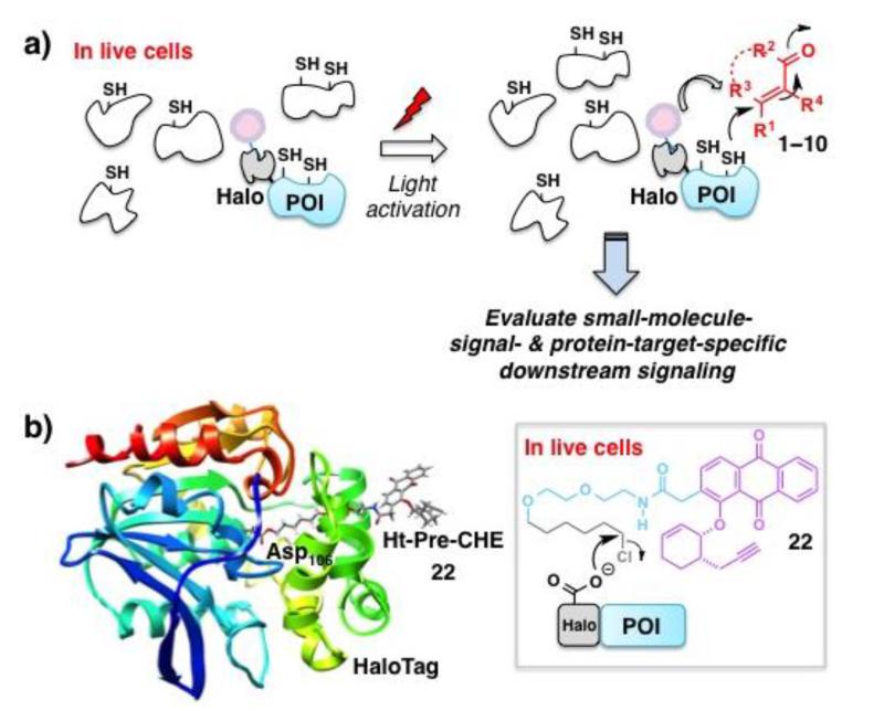
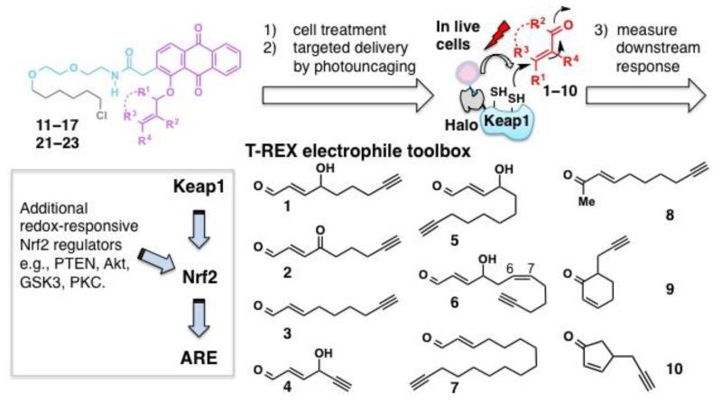
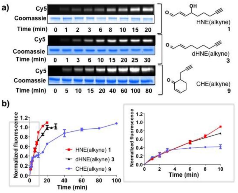
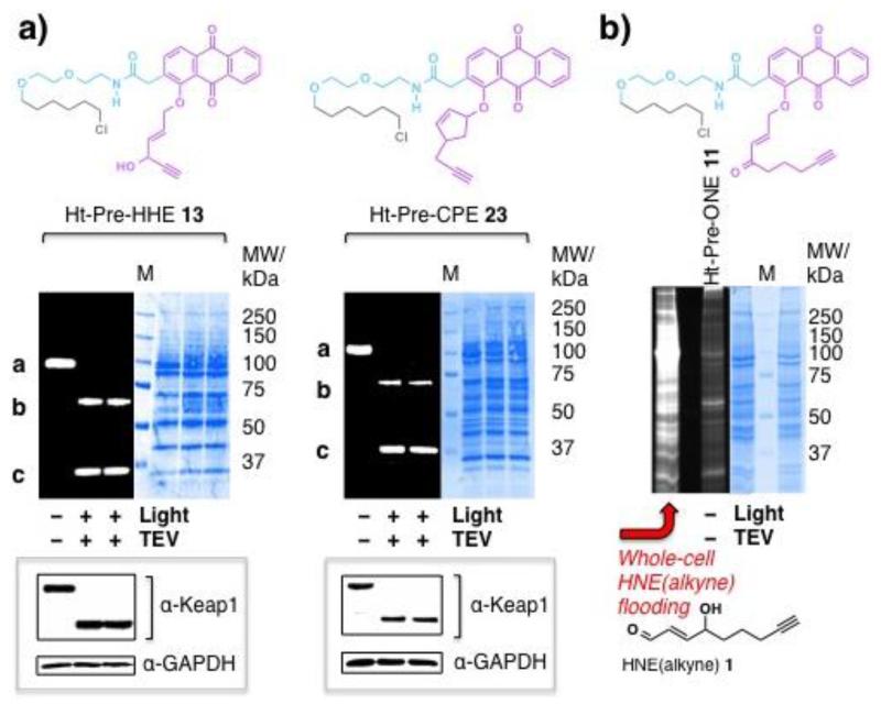
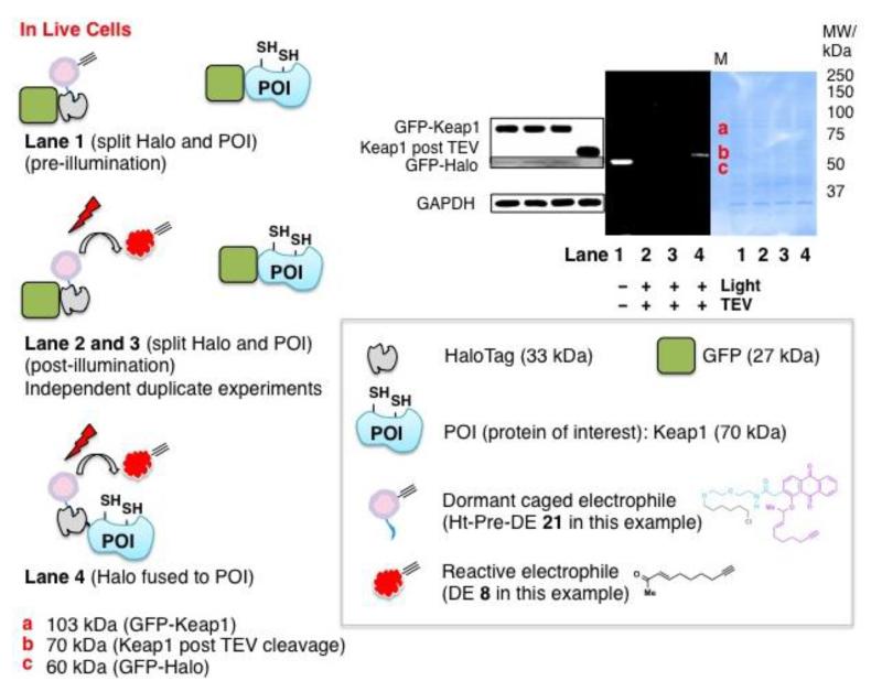
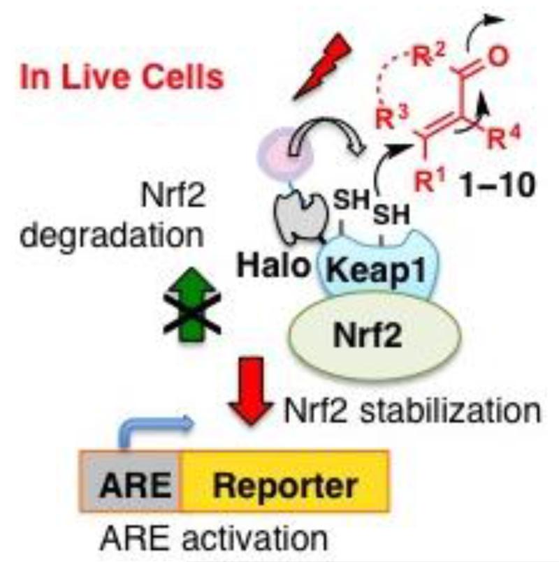
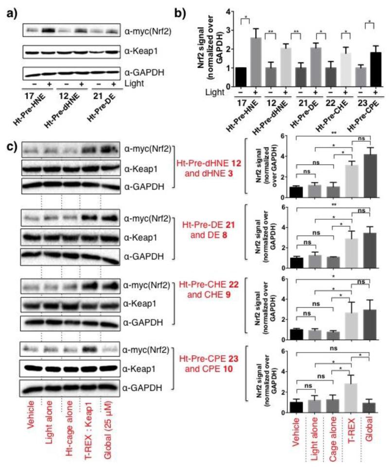
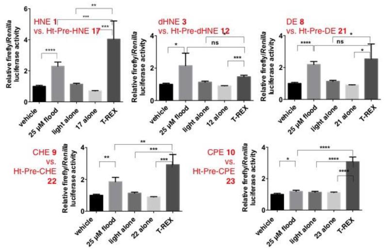
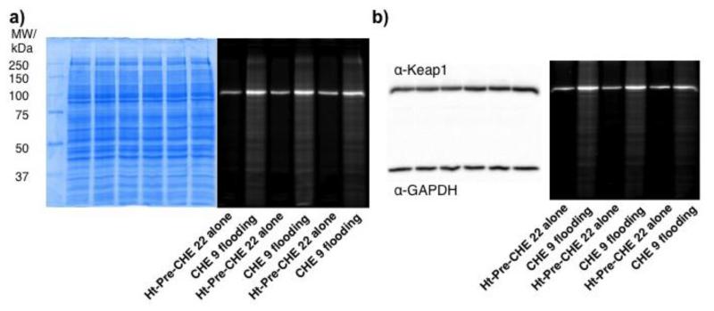
Despite the known propensity of small-molecule electrophiles to react with numerous cysteine-active proteins, biological actions of individual signal inducers have emerged to be chemotype-specific. To pinpoint and quantify the impacts of modifying one target out of the whole proteome, we develop a target-protein-personalized "electrophile toolbox" with which specific intracellular targets can be selectively modified at a precise time by specific reactive signals. This general methodology, T-REX (targetable reactive electrophiles and oxidants), is established by (1) constructing a platform that can deliver a range of electronic and sterically different bioactive lipid-derived signaling electrophiles to specific proteins in cells; (2) probing the kinetics of targeted delivery concept, which revealed that targeting efficiency in cells is largely driven by initial on-rate of alkylation; and (3) evaluating the consequences of protein-target- and small-molecule-signal-specific modifications on the strength of downstream signaling. These data show that T-REX allows quantitative interrogations into the extent to which the Nrf2 transcription factor-dependent antioxidant response element (ARE) signaling is activated by selective electrophilic modifications on Keap1 protein, one of several redox-sensitive regulators of the Nrf2-ARE axis. The results document Keap1 as a promiscuous electrophile-responsive sensor able to respond with similar efficiencies to discrete electrophilic signals, promoting comparable strength of Nrf2-ARE induction. T-REX is also able to elicit cell activation in cases in which whole-cell electrophile flooding fails to stimulate ARE induction prior to causing cytotoxicity. The platform presents a previously unavailable opportunity to elucidate the functional consequences of small-molecule-signal- and protein-target-specific electrophilic modifications in an otherwise unaffected cellular background.


Figures

Figure 1
Figure 1
(a) The generalizable T-REX (targetable reactive electrophiles and oxidants) approach selectively delivers a reactive signaling electrophile of choice to specific redox-sensor protein of interest (POI) at a precise time in live cells. Photocaged precursor (pink sphere) is covalently linked to HaloTag fused to POI. Low-energy light activation (365 nm, 0.6 mW/cm2) liberates the desired reactive signal in stoichiometric amount to the microenvironment of the POI enabling targeted modification. The spatiotemproally precise modification upstream in turn enables quantitative evaluation of single- and target-specific response downstream. (b) Energy minimized model of Halo (PDB: 1BN6) with covalently bound caged precursor Ht-Pre-CHE 22 showed the caged motif is solvent exposed. Inset exemplifies covalent conjugation of 22 to the active site Asp residue on Halo.
Figure 2
Figure 2
T-REX electrophile toolbox enables assessment of downstream signaling strength triggered by target-specific delivery of specific bioactive LDEs (110) to specific proteins in cells (e.g., Keap1) at a precise time. Inset: The simplified model for Nrf2–ARE pathway. The downstream phenotypic responses—Nrf2 stabilization and ARE upregulation—are observed from whole-cell LDE flooding. Electrophilic modifications of various upstream redox-sensitive targets, including Keap1, PTEN, Akt, GSK3, PKC, etc., have been implicated to play roles in modulating Nrf2–ARE signaling. Using T-REX, this study directly probes the Nrf2–ARE signaling strength selectively elicited by LDE-signal-specific and Keap1-protein-specific electrophilic modifications in an otherwise unmodified proteome.
Figure 3
Figure 3
Synthetic strategies for the construction of T-REX electrophile toolbox. (a) The Williamson ether synthesis approach. (b) The Mitsunobu approach. See also Scheme S2–S10. All compounds are racemic where applicable.
Figure 4
Figure 4
Light-activatable release efficiencies of HNE 1 and HHE 4 from (a) 17 and (b) 13 covalently bound to HaloTag in solution, respectively. See also Figure S1 and Table S1.
Figure 5
Figure 5
(a) Representative in-gel fluorescence data for the time-dependent alkylation of recombinant human His6-Keap1 with 1, 3, and 9. (b) Quantitation of the data in (a) (n = 2) as progress curve plots. Inset shows the first 10 min. The “normalized fluorescence” was derived by first calculating the ratio of Cy5 : Coomassie signals for each time point, and normalizing the resultant values to the respective mean of the last three time points for each LDE.
Figure 6
Figure 6
(a) Representative data for T-REX-assisted Keap1-specific electrophilic adduction (a) vs. whole-cell electrophile flooding (b). (a) Live HEK-293 cells stably expressing Halo-Keap1 were treated with 25 μM 13 and 23, and the excess washed out. After 20-min light exposure, cells were lysed, and the lysate was treated with TEV-protease (separating Halo and Keap1), followed by Cy5 click assay reagents. Band a, Halo–Keap1 (104 kDa), b, Keap1 (70 kDa, post TEV cleavage), c, Halo (33 kDa). Dark gel is the fluorescence gel of the Coomassie-stained PVDF membrane on the right. Independent duplicates are shown for light-activated TEV-treated samples. “M” indicates the ladder lane. Insets show western blot. See also Figure S2, Table 1, and Eq. 1. (b) Whole-cell HNE 1 (25 μM) treatment target non-specifically. Ht-Pre-ONE 11 reacts before uncaging.
Figure 7
Figure 7
The mechanistic basis of T-REX platform supports proximity-induced reactivity concept within the “target–signal encounter complex”. In-cell T-REX experiment in which Halo and Keap1 were not fused failed to deliver electrophile DE 8 to Keap1 using Ht-Pre-DE 21 and subsequent illumination (Lane 2 and 3, independent duplicates). By contrast, successful targeted delivery was achieved when Halo and Keap1 were fused (Lane 4 and also see Figre S2b). Schematic representations for each Lane are shown on the left. DE adduction of Keap1 (post TEV-protease cleavage) was observed (band b on gel) as expected in Lane 4. Western blot data and coomassie-stained PVDF were also shown. Bands a, b, and c correspond to GFP-Keap1 (103 kDa), Keap1 (70 kDa, post TEV-cleavage), and GFP-Halo (60 kDa), respectively. “M” indicates the ladder lane. Inset shows reference to the schematics. Rabbit polyclonal anti-GFP primary antibody (sc-8334) was used to probe GFP-Halo protein. All forms of proteins containing Keap1 were probed by mouse monoclonal anti-Keap1 primary antibody (Ab119403). See also Figure S3 and S4 for analogous data sets with non-GFP-fulsed Keap1.
Figure 8
Figure 8
Two independent readouts for quantitating the downstream signaling strength selectively elicited by target- and signal-specific electrophilic modification of a specific upstream target in cells. T-REX-assisted temporally controlled Keap1-specific modifications resulted in blockage of Nrf2 degradation. Accumulating Nrf2 (assessed by western blot) consequently enabled ARE activation (assessed by ARE-luciferase assay).
Figure 9
Figure 9
The upregulation of Nrf2 protein expression levels elicited by T-REX-assisted Keap1-specific electrophilic modification in HEK-293 cells expressing Halo-Keap1 and Nrf2. See also Figure S5. (a) Representative western blot data from T-REX conditions in comparison with Ht-caged precursor treatment alone (i.e., no light). (b) Quantitation of data in (a, c) and in Figure S5a–b. (c) Representative western blot data (left) and corresponding quantitation (right) from T-REX conditions in comparison with whole-cell electrophile (25 μM) soaking. Error bars designate S.D. over 3 independent biological replicates. Nrf2 levels were assessed after 18 h incubation period post 20 min light exposure. Vehicle, DMSO.
Figure 10
Figure 10
Reporter gene assay data on ARE upregulation in HEK-293 cells stably expressing Halo-Keap1 and transiently expressing Nrf2 alongside ARE-inducible firefly luciferase and constitutive Renilla luciferase. For each LDE, the magnitude of ARE induction is compared between T-REX conditions and whole-cell electrophile (25 μM) flooding. Vehicle, DMSO. See also Figure S6–S7 and S13 for titration of ARE response and cell viability analyses. Error bars designate S.D. over 3 independent biological replicates. The ARE response was analyzed after 18 h incubation period post 20-min light exposure.
Figure 11
Figure 11
Representative data from in-gel fluorescence analysis evaluating the extent of Keap1 modifications under whole-cell LDE flooding. Signal from samples designated as “Ht-Pre-CHE 22 alone“ equates to 1 equiv of CHE-alkyne 9 stoichiometrically labeled to Halo-Keap1. See discussion in Text. The data in each case originate from three independent biological replicates. (a) “Ht-Pre-CHE 22 alone“ designates live HEK-293 cells transiently expressing Halo-Keap1 treated with Ht-Pre-CHE 22 alone without exposure to light (see SI for details), followed by cell lysis and Click coupling to Cy5 azide. “CHE 9 flooding“ designates whole-cell CHE 9 treatment (25 μM, 20 min), followed by cell lysis and Click coupling with Cy5 azide. The corresponding Coomassie stained gel is shown on the left. (b) Remaining lysate samples from experiment in (a) were subjected to Click coupling for in-gel fluorescence analysis followed by western blot using antibodies to Keap1 and GAPDH (loading control). See also Figure S2d.

References

    1. Jacob C, Winyard PG, editors. Redox Signaling and Regulation in Biology and Medicine. WILEY-VCH; Weinheim: 2009.
    2. Rudolph TK, Freeman BA. Sci. Signal. 2009;2:re7. - PMC - PubMed
    3. Jacobs AT, Marnett LJ. Acc. Chem. Res. 2010;43:673–683. - PMC - PubMed
    1. Walsh CT. Posttranslational Modification of Proteins: Expanding Nature’s Inventory. Roberts & Co.; 2005.
    2. Tarrant MK, Cole PA. Annu. Rev. Biochem. 2009;78:797–825. - PMC - PubMed
    1. Schopfer FJ, Cipollina C, Freeman BA. Chem. Rev. 2011;111:5997–6021. - PMC - PubMed
    2. Fritz KZ, Petersen DR. Free. Radic. Biol. Med. 2013;59:85–91. - PMC - PubMed
    1. Wang C, Weerapana E, Blewett MM, Cravatt BF. Nat. Methods. 2014;11:79–85. - PMC - PubMed
    2. Condreanu SG, Zhang B, Sobecki SM, Billheimer DD, Liebler DC. Mol. Cell. Proteomics. 2009;8:670–680. - PMC - PubMed
    3. Yang J, Tallman KA, Porter NA, Liebler DC. Anal. Chem. 2015;87:2535–2541. - PMC - PubMed
    1. Ruggieri S, Tortorella C, Gasperini C. Ther. Clin. Risk Manag. 2014;10:229–239. - PMC - PubMed
    2. Phillips JT, Fox RJ. Semin. Neurol. 2013;33:56–65. - PubMed
    3. Liby KT, Sporn MB, Crunkhorn S. Pharmacol. Rev. Nat. Rev. Drug Discov. 2012;2012;6411:972–1003. - PMC - PubMed
    4. Liby KT, Yore MM, Sporn MB. Nat. Rev. Cancer. 2007;7:357–369. - PubMed

Publication types

MeSH terms

Substances

LinkOut - more resources