. 2015 Jul 17;10(7):1718-28.
doi: 10.1021/acschembio.5b00222. Epub 2015 May 1.

Covalent Inhibition of Ubc13 Affects Ubiquitin Signaling and Reveals Active Site Elements Important for Targeting

Affiliations

Covalent Inhibition of Ubc13 Affects Ubiquitin Signaling and Reveals Active Site Elements Important for Targeting

Curtis D Hodge et al. ACS Chem Biol. .

Abstract

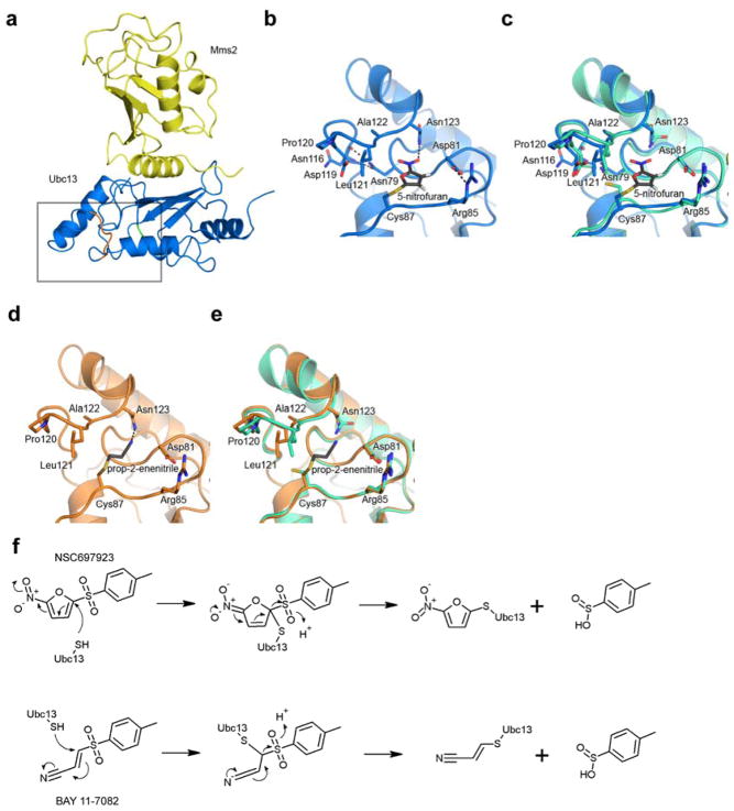
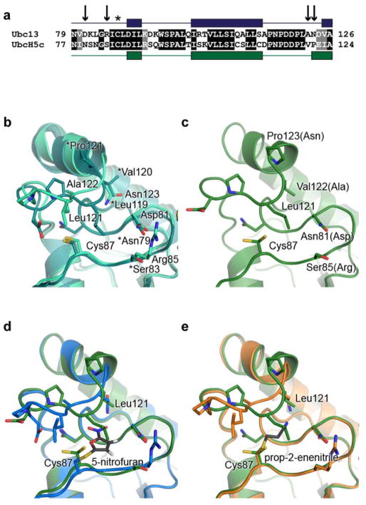
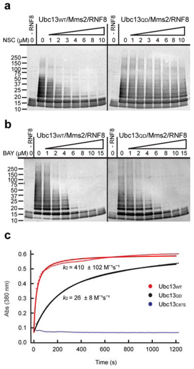
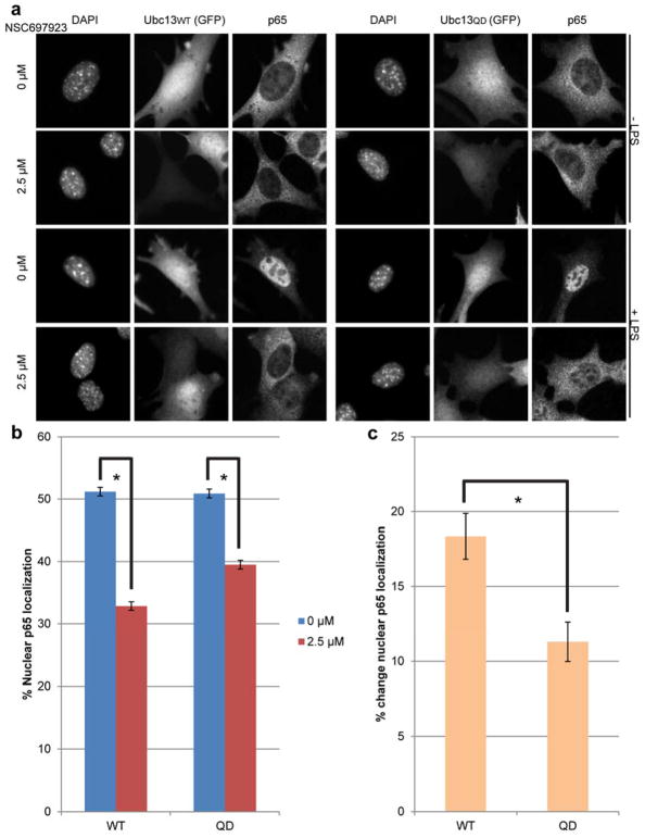
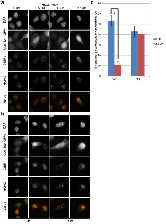
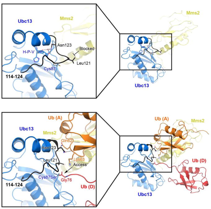
Ubc13 is an E2 ubiquitin conjugating enzyme that functions in nuclear DNA damage signaling and cytoplasmic NF-κB signaling. Here, we present the structures of complexes of Ubc13 with two inhibitors, NSC697923 and BAY 11-7082, which inhibit DNA damage and NF-κB signaling in human cells. NSC697923 and BAY 11-7082 both inhibit Ubc13 by covalent adduct formation through a Michael addition at the Ubc13 active site cysteine. The resulting adducts of both compounds exploit a binding groove unique to Ubc13. We developed a Ubc13 mutant which resists NSC697923 inhibition and, using this mutant, we show that the inhibition of cellular DNA damage and NF-κB signaling by NSC697923 is largely due to specific Ubc13 inhibition. We propose that unique structural features near the Ubc13 active site could provide a basis for the rational development and design of specific Ubc13 inhibitors.


Conflict of interest statement

Notes: The authors declare no competing financial interest.

Figures

Figure 1
Figure 1
NSC697923 and BAY 11-7082 covalently modify the Ubc13 active site. (a) Overview of Ubc13 (blue)/Mms2 (yellow) bound by the 5-nitrofuran moiety of NSC697923. Active site is boxed with the 114–124 loop in orange and the Cys87 region in green (Protein Data Bank accession 4ONM). (b) The active site view of Ubc13 bound by the 5-nitrofuran moiety of NSC697923. (c) Overlay of wild type Ubc13, PDB 1J7D (green-cyan), and 5-nitrofuran-bound Ubc13. (d) Active site view of Ubc13 bound by the prop-2-enenitrile moiety of BAY 11-7082 (PDB 4ONN). (e) Overlay of wild type Ubc13 and prop-2-enenitrile bound Ubc13. In panels (b) – (e), the view is rotated 90° from the orientation in (a). (f) Mechanisms of covalent attachment by NSC697923 and BAY 11-7082.
Figure 2
Figure 2
Design and structure of a NSC697923 resistant Ubc13 mutant. (a) Amino acid sequence alignment of important active site residues in Ubc13 and UbcH5c, with secondary structural characteristics shown above. A line signifies a loop region and a box denotes an α helix. Arrows indicate mutations made to Ubc13 to mimic UbcH5c. The asterisk is above the active site cysteine. (b) Overlay of UbcH5c, PDB 1X23 (deep-teal), and Ubc13 (light green) shows their different active site loop conformations. Asterisks denote UbcH5c residues. (c) Active site view of the mutant Ubc13QD (green) with the UbcH5c-type loop conformation (PDB 4ONL). Brackets denote wildtype residues. (d) Overlay of Ubc13 5-nitrofuran adduct (blue) and the resistant Ubc13QD (green). (e) Overlay of Ubc13 prop-2-enenitrile adduct (orange) and Ubc13QD.
Figure 3
Figure 3
Ubc13QD is resistant to NSC697923 but not BAY 11-7082. (a and b) In vitro ubiquitination assays in which purified Ubc13/Mms2 (Ubc13WT or Ubc13QD) was incubated with ubiquitin, ATP, E1 enzyme, RNF8 and the indicated concentrations of inhibitor. Results were visualized by Western blotting with an antiubiquitin antibody. (a) Results for NSC697923. (b) Results for BAY 11-7082. (c) Representative graph of an in vitro inhibition assay monitored by absorbance at 380 nm. Reactions containing either Ubc13WT, Ubc13QD, or Ubc13C87S were mixed with the NSC697923 inhibitor, and the resulting absorbance monitored. The experiment was done in triplicate and the average second-order rate constants (k2) and standard errors are reported. Dotted lines indicate experimental data, curves indicate the fit to a second-order rate model.
Figure 4
Figure 4
Inhibition of Ubc13 is required for significant disruption of cellular NF-κB signaling by NSC697923. (a) Representative images of Ubc13WT (left) or Ubc13QD (right) reconstituted mouse embryonic fibroblast cells before and after lipopolysaccharide (LPS) stimulation, with and without NSC697923 treatment (2.5 μM). (b) Quantitation of p65 translocation represented as a percent of intensity localized to the nuclei and (c) the difference in p65 translocation between NSC697923-untreated and treated cells (P-value = 0.02). Unstimulated cells have approximately 30% background nuclear p65 translocation. Data from three independent experiments were pooled with at least 200 cells per condition, and standard error of image averages is included. The tonal range of whole images was rescaled from 0 to 255 in Photoshop to increase the overall contrast for display.
Figure 5
Figure 5
Normalized inhibition of Ubc13-dependent, NF-κB-driven cytokine release by NSC697923. Unstimulated (−LPS) or stimulated (+LPS) Ubc13WT or Ubc13QD MEF cells were treated with either DMSO, or increasing concentrations of NSC697923 from 0.5 μM to 4 μM and cytokine levels in the culture medium were quantified. The background unstimulated (−LPS) level was subtracted from each treatment (DMSO to 4 μM NSC697923) and the stimulated (+LPS) DMSO treated level was normalized to 100% for optimal direct comparison of the two cell lines. The assay was done in triplicate, and the standard error of the mean for each treatment is included.
Figure 6
Figure 6
Inhibition of Ubc13 is required for disruption of cellular DNA damage signaling by NSC697923. (a) Representative images of Ubc13WT or (b) Ubc13QD reconstituted mouse embryonic fibroblast cells plus/minus 3 Gy of ionizing radiation, with or without NSC697923 treatment (2.5 μM). (c) Quantitation of 53BP1 localization represented as a percentage of total cells positive (≥3 foci) for γH2AX/53BP1 colocalization. Data from three independent experiments were pooled with at least 300 cells per condition, and standard error of image averages is included. The tonal range of whole images was rescaled from 0 to 255 in Photoshop to increase the overall contrast for display.
Figure 7
Figure 7
Conformational changes in Ubc13 loop 114–124 upon ubiquitin binding. The top panel is hUbc13/hMms2 (PDB 1J7D) and the bottom panel is yUbc13~hUb/yMms2/hUb (2GMI). Ubc13 is blue, Mms2 is yellow, donor ubiquitin is red, and acceptor ubiquitin is orange for both panels and the 114–124 loop is in black. The position of Leu121 in the unbound structure (top panel) is expected to block the approach of Lys63 of the acceptor ubiquitin toward the active site cysteine

References

    1. Haas AL, Siepmann TJ. Pathways of ubiquitin conjugation. FASEB J. 1997;11:1257–1268. - PubMed
    1. Hershko A, Ciechanover A. The ubiquitin system. Annu Rev Biochem. 1998;67:425–479. - PubMed
    1. Pickart CM, Eddins MJ. Ubiquitin: structures, functions, mechanisms. Biochim Biophys Acta. 2004;1695:55–72. - PubMed
    1. Dye BT, Schulman BA. Structural mechanisms underlying posttranslational modification by ubiquitin-like proteins. Annu Rev Biophys Biomol Struct. 2007;36:131–150. - PubMed
    1. Michelle C, Vourc'h P, Mignon L, Andres CR. What was the set of ubiquitin and ubiquitin-like conjugating enzymes in the eukaryote common ancestor? J Mol Evol. 2009;68:616–628. - PMC - PubMed

Publication types

MeSH terms