. 2015 Nov;22(11):1812-23.
doi: 10.1038/cdd.2015.34. Epub 2015 Apr 23.

Chaperone-like protein p32 regulates ULK1 stability and autophagy

Affiliations

Chaperone-like protein p32 regulates ULK1 stability and autophagy

H Jiao et al. Cell Death Differ. 2015 Nov.

Abstract

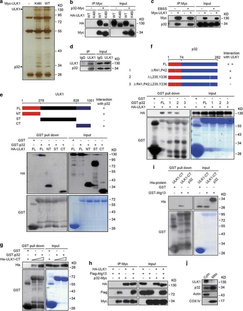
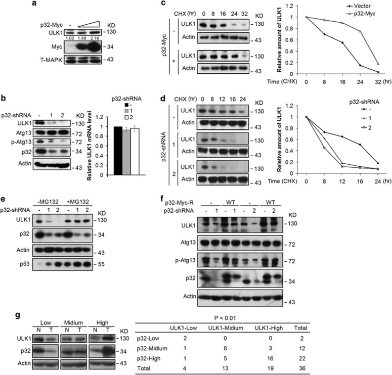
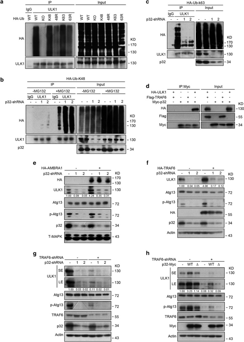
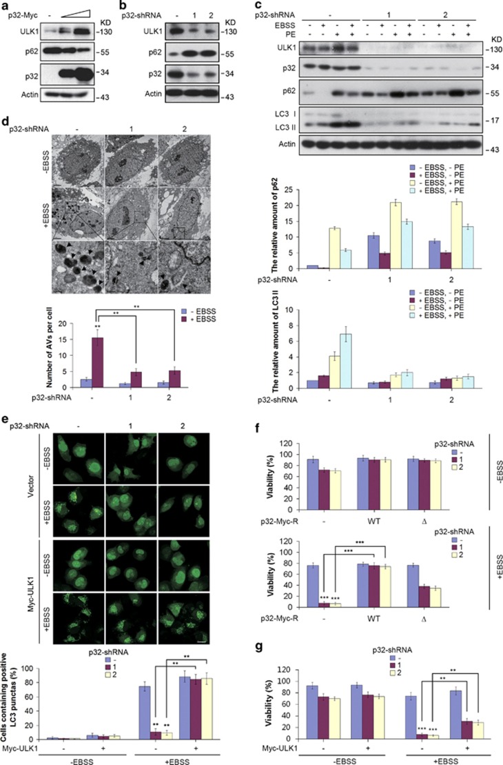
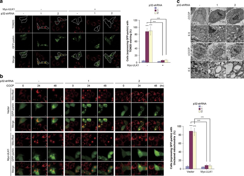
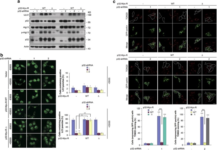
Mitophagy mediates clearance of dysfunctional mitochondria, and represents one type of mitochondrial quality control, which is essential for optimal mitochondrial bioenergetics. p32, a chaperone-like protein, is crucial for maintaining mitochondrial membrane potential and oxidative phosphorylation. However, the relationship between p32 and mitochondrial homeostasis has not been addressed. Here, we identified p32 as a key regulator of ULK1 stability by forming complex with ULK1. p32 depletion potentiated K48-linked but impaired K63-linked polyubiquitination of ULK1, leading to proteasome-mediated degradation of ULK1. As a result, silencing p32 profoundly impaired starvation-induced autophagic flux and the clearance of damaged mitochondria caused by mitochondrial uncoupler. Importantly, restoring ULK1 expression in p32-depleted cells rescued autophagy and mitophagy defects. Our findings highlight a cytoprotective role of p32 under starvation conditions by regulating ULK1 stability, and uncover a crucial role of the p32-ULK1-autophagy axis in coordinating stress response, cell survival and mitochondrial homeostasis.


Figures

Figure 1
Figure 1
p32 interacts with ULK1. (a) HEK293T cells were transiently transfected with the indicated expression constructs. The anti-Myc immunoprecipitates were resolved by SDS-PAGE, and the proteins were visualized by silver staining, and indicated bands were analyzed by mass spectrometry. (b) Western blotting analysis of input and anti-Myc IP derived from HEK293T cells that were transiently transfected with WT or mutant ULK1 (K46I) and p32-Myc. (c) Hela cells expressing Myc-ULK1 were grown either in normal or in EBSS medium for 6 h. Cell lysates were immunoprecipitated with anti-Myc antibody followed by immunoblotting with anti-p32 antibody. (d) The interaction of endogenous ULK1 and p32 was detected in Hela cells. Normal rabbit IgG was used as a negative control for the immunoprecipitation procedure. (e) Purified GST-p32 was incubated with cell lysates derived from HEK293T cells transfected with the indicated HA-ULK1 constructs. Proteins retained on Sepharose were then blotted with the indicated antibodies. (f) Extracts from HEK293T cells transfected with HA-ULK1 were incubated with recombinant full-length (FL) GST-p32 or GST-p32 mutants coupled to GSH-Sepharose. Proteins retained on Sepharose were then blotted with the indicated antibodies. (g) Purified recombinant His-ULK1-CT was incubated with GST-p32. Proteins retained on Sepharose were then blotted with the indicated antibodies. (h) HEK293T cells were transfected with vectors encoding HA-ULK1, Flag-Atg13 and p32-Myc, as indicated. Cell lysates were immunoprecipitated with anti-Myc antibody. Western blotting was performed using the indicated antibodies. (i) Purified His-ULK1-CT or His-p32 was incubated with purified GST-Atg13. Proteins retained on Sepharose were then blotted with the indicated antibodies. (j) Hela cells were fractionated and analyzed by immunoblotting using antibodies against the indicated proteins. Cyto, cytosol; Mito, mitochondria
Figure 2
Figure 2
p32 regulates ULK1 stability and kinase activity. (a) Hela cells were infected with lentiviral vectors expressing p32-Myc. Cell extracts were then prepared and analyzed by western blotting with the indicated antibodies. (b) Protein extracts from Hela cells infected with the indicated lentiviral shRNA constructs were analyzed by western blotting. Total RNA was isolated, and qRT-PCR was performed. (c) Hela cells were infected with the indicated lentiviral vector encoding p32-Myc. Cells were then treated with CHX (25 μg/ml) for the indicated time course. Cell lysates were harvested and analyzed by western blotting. Semiquantification with Actin as a loading control and relative ULK1 levels at time 0 set as 1. (d) Hela cells expressing p32-shRNAs were treated with CHX for the indicated time course. The levels of ULK1 were determined by immunoblotting the total cell lysates and quantification with Actin as a loading control. Results plotted are the amounts of ULK1 at each time point relative to the level at time 0. (e) Hela cells infected with lentivirus encoding the indicated shRNAs were treated with DMSO or MG132 (20 μM) for 6 h. Cell extracts were analyzed by western blotting using the indicated antibodies. p53 immunoblotting was shown as a positive control for inhibited proteasomal degradation by MG132. (f) Hela cells stably expressing shRNA-resistant forms of p32 were infected with the indicated lentiviral shRNA constructs. Cells were then collected and lysates were subjected to immunoblotting with the indicated antibodies. (g) Immunoblotting analysis of p32 and ULK1 levels from tumor tissues (T) and their matched surrounding normal mucosal tissues (N). Actin was used as a loading control. A total of 36 samples of human colorectal cancer were classified into nine groups based on the protein levels of p32 and ULK1 in tumor tissues compared with surrounding normal tissues. The correlation between p32 and ULK1 expression was analyzed by Fisher's exact test
Figure 3
Figure 3
p32 interferes with the polyubiquitination of ULK1. (a) HeLa cells were transfected with HA-tagged WT or mutant ubiquitin constructs. Protein extracts were immunoprecipitated using anti-ULK1 antibody or rabbit IgG as a negative control (IgG). Immunoprecipitates were analyzed by western blotting using anti-ULK1 and anti-HA antibodies. (b) Hela cells expressing HA-Ubiquitin-k48 (HA-Ub-k48) were infected with the indicated p32-shRNAs. Cells were then treated with DMSO or MG132 (20 μM) for 6 h before being collected. Protein extracts were immunoprecipitated using anti-ULK1 antibody. The immunoprecipitates were subjected to western blotting with the indicated antibodies. (c) HeLa cells expressing HA-Ubiquitin-k63 (HA-Ub-K63) were infected with p32 shRNA lentiviral vector. Endogenous ULK1 protein was immunoprecipitated and immunoblotted with the indicated antibodies. Of note, ULK1 levels in the immunoprecipitation experiment do not change in this case in the p32 shRNA lane, owing to saturation of the anti-ULK1 antibody. (d) The indicated expression constructs were transiently transfected into HEK293T cells. Protein extracts were immunoprecipitated using anti-Myc antibody. The immunoprecipitates were subjected to western blotting with the indicated antibodies. (e) Hela cells stably expressing HA-AMBRA1 were infected with p32 shRNA lentiviral vector. Cell lysates were collected and analyzed by western blotting with the indicated antibodies. (f) Hela cells stably expressing HA-TRAF6 were infected with p32 shRNA lentiviral vector as in e. Cell lysates were collected and analyzed by western blotting with the indicated antibodies. (g) Hela cells infected with lentivirus bearing TRAF6-shRNA and p32-shRNA were collected. Protein extracts were subjected to western blotting with the indicated antibodies
Figure 4
Figure 4
p32 regulates starvation-induced autophagy. (a) Protein extracts from Hela cells infected with p32-Myc lentiviral construct were analyzed by western blotting with the indicated antibodies. (b) Hela cells infected with the indicated lentiviral p32 shRNAs were collected, and cell lysates were subjected to western blotting with the indicated antibodies. (c) Hela cells stably expressing the indicated shRNAs were placed in normal or in EBSS starvation medium in the presence or absence of lysosomal protease inhibitors PE for 6 h. Lysates were immunoblotted as indicated (upper panel). p62 and LC3-II levels were quantified by densitometry and plotted as mean±S.D. of three independent experiments (middle and lower panel). (d) p32 knockdown Hela cells were untreated or treated with EBSS medium for 6 h. Cells were then fixed and analyzed by transmission electron microscopy (TEM). The bottom panels show enlarged views of the boxed areas. Quantification of the autophagic vacuoles (AVs) was shown as mean±S.D. of 15 random areas. **P<0.01. (e) H1299 cells stably expressing GFP-LC3 were infected with p32-shRNA and stably reconstituted with lentiviral Myc-ULK1 vector. Cells were then placed in EBSS medium for 6 h. GFP-LC3 positive autophagosomes were determined by confocal microscopy. The scale bar represents 20 μM (upper panel). Histogram represents the percentage of cells containing positive LC3 punctas. The error bars represent the mean±S.D. of five random areas (lower panel). **P<0.01. (f) Hela cells stably expressing the indicated shRNA-resistant forms of p32 were infected with p32-shRNA lentiviruses. Histogram represents the percentage of viable cells scored by Annexin V staining and FACS analysis after being cultured in EBSS medium for 6 h. The error bars represent the mean±S.D. of three independent experiments. ***P<0.001. (g) Hela cells were infected with p32-shRNA and stably reconstituted with lentiviral Myc-ULK1 vector. Histogram represents the percentage of viable cells scored by Annexin V staining and FACS analysis after being cultured in EBSS medium for 6 h. The error bars represent the mean±S.D. of three independent experiments. **P<0.01, ***P<0.001
Figure 5
Figure 5
p32 depletion leads to mitophagy defects. (a) Hela cells stably expressing Myc-ULK1, GFP-Parkin and p32-shRNA were treated with CCCP (10 μM) for 48 h. Cells were then immunostained with anti-TOM20 antibody (left) and scored for the percentage of cells expressing GFP-parkin with TOM20 staining, which was shown as mean±S.D. of five random areas (right). The scale bar represents 10 μM. ***P<0.001. (b) Hela cells stably expressing Mito-Red were infected with the indicated lentiviral vectors. Cells were then transiently transfected with GFP-Parkin followed by CCCP treatment (10 μM) for the indicated time course. The process of removing damaged mitochondria in living cells was monitored by live-cell imaging (left panel). Histogram represents the percentage of cells expressing GFP-parkin with uncleared mitochondria after 48 h of CCCP treatment (right panel). The error bars represent the mean±S.D. of five random areas. ***P<0.001. (c) Hela cells stably expressing Myc-parkin were infected with lentivirus encoding control shRNA or p32-shRNA. Cells were then placed in medium containing DMSO or CCCP (10 μM) for 24 h, followed by TEM analysis
Figure 6
Figure 6
p32 regulates autophagy and mitophagy via interacting with ULK1. (a) Hela cells stably reconstituted with lentivirus bearing the indicated p32 cDNA were infected with p32-shRNA lentiviruses. Lysates were immunoblotted with the indicated antibodies. (b) H1299-GFP-LC3 cells stably expressing the indicated shRNA-resistant forms of p32 were infected with p32-shRNA lentiviruses. Cells were then treated with EBSS medium for 6 h. GFP-LC3 positive autophagosomes were examined by confocal microscopy. The scale bar represents 20 μM (left panel). Histogram represents the percentage of cells containing positive LC3 punctas (right panel). The error bars represent the mean±S.D. of five random areas. **P<0.01. (c) Hela cells stably expressing GFP-Parkin and the indicated shRNA-resistant forms of p32 were infected with p32-shRNA lentiviruses. Cells then were treated with CCCP (10 μM) for 48 h. Cells were immunostained with anti-TOM20 antibody (upper) and scored for the percentage of cells expressing GFP-parkin with TOM20 staining, which was shown as mean±S.D. of five random areas (lower). The scale bar represents 10 μM. ***P<0.001

References

    1. 1Fogal V, Richardson AD, Karmali PP, Scheffler IE, Smith JW, Ruoslahti E. Mitochondrial p32 protein is a critical regulator of tumor metabolism via maintenance of oxidative phosphorylation. Mol Cell Biol 2010; 30: 1303–1318. - PMC - PubMed
    1. 2Matthews DA, Russell WC. Adenovirus core protein V interacts with p32—a protein which is associated with both the mitochondria and the nucleus. J Gen Virol 1998; 79: 1677–1685. - PubMed
    1. 3Jiang J, Zhang Y, Krainer AR, Xu RM. Crystal structure of human p32, a doughnut-shaped acidic mitochondrial matrix protein. Proc Natl Acad Sci USA 1999; 96: 3572–3577. - PMC - PubMed
    1. 4Soltys BJ, Kang D, Gupta RS. Localization of P32 protein (gC1q-R) in mitochondria and at specific extramitochondrial locations in normal tissues. Histochem Cell Biol 2000; 114: 245–255. - PubMed
    1. 5van Leeuwen HC, O'Hare P. Retargeting of the mitochondrial protein p32/gC1Qr to a cytoplasmic compartment and the cell surface. J Cell Sci 2001; 114: 2115–2123. - PubMed

Publication types

MeSH terms

Substances