Review
. 2015 Apr 23;161(3):450-457.
doi: 10.1016/j.cell.2015.03.049.

Single-Particle Cryo-EM at Crystallographic Resolution

Affiliations
Review

Single-Particle Cryo-EM at Crystallographic Resolution

Yifan Cheng. Cell. .

Abstract

Until only a few years ago, single-particle electron cryo-microscopy (cryo-EM) was usually not the first choice for many structural biologists due to its limited resolution in the range of nanometer to subnanometer. Now, this method rivals X-ray crystallography in terms of resolution and can be used to determine atomic structures of macromolecules that are either refractory to crystallization or difficult to crystallize in specific functional states. In this review, I discuss the recent breakthroughs in both hardware and software that transformed cryo-microscopy, enabling understanding of complex biomolecules and their functions at atomic level.


Figures

Figure 1
Figure 1. Single-particle cryo-EM
A: Purified biological molecules are embedded in a thin layer of vitreous ice, in which they ideally adopt random orientations. The orientations are specified by the in-plane position parameters, x and y, and three Euler angles α, β and γ, which are refined iteratively to high accuracies. The defocus values of the images are currently often determined separately. B: Typical image of frozen-hydrated archaeal 20S proteasomes. C: 3D reconstruction of the 20S proteasome at 3.3Å resolution. D: Side-chain densities of the map shown in B are comparable with those seen in maps determined by X-ray crystallography at a similar resolution.
Figure 2
Figure 2. Influence of CTF on image contrast and resolution
A and B: Image of human transferrin receptor – transferrin complex recorded using a scintillator camera. The microscope was equipped with a FEG and operated at 200kV. Particles in image recorded with a defocus of 1.2μm (A) are almost invisible, but shown with strong contrast in the image recorded with a defocus of 3.0μm (B). C: Simulations of CTF at 1.2μm (red) and 3.0μm (blue) defocuses, with an acceleration voltage of 200kV and angular spread of 0.07mrad. Note that 3um defocus generates sufficient contrast for particles with a molecular weight of ~300kDa, CTF envelop drops to close to zero at 3 ~ 4Å resolution.
Figure 3
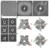
Figure 3. Direct electron detection camera enabled major breakthroughs in single particle cryo-EM
A. An image of frozen hydrated T. acidophilum 20S proteasome recorded using K2 Summit camera with a 300kV microscope and a defocus of ~0.9 μm. B. Fourier transform of a typical imperfect image of frozen hydrated 20S proteasome, showing a predominant resolution cutoff caused by beam-induced motion. C. Fourier transform of the same image after motion correction. Thon ring is restored to resolution of ~3Å. Panels A – C are reproduced from (Li et al., 2013). D. 2D class averages of TRPV1 ion channel calculated from images recorded with a scintillator camera (left) and K2 Summit camera (right) (Liao et al., 2013). E. Two different views of TRPV1 3D reconstruction determined from a dataset collected with a scintillator camera. F. Same views of the TRPV1 3D reconstruction determined from a dataset collected with a K2 Summit camera.

References

    1. Agard DA, Cheng Y, Glaeser RM, Subramaniam S. Single-particle cryo-electron microscopy (cryo-EM): progress, challenges, and perspectives for future improvment. Advances in Imaging and Electron Physics. 2014:185.
    1. Allegretti M, Mills DJ, McMullan G, Kuhlbrandt W, Vonck J. Atomic model of the F420-reducing [NiFe] hydrogenase by electron cryo-microscopy using a direct electron detector. eLife. 2014;3:e01963. - PMC - PubMed
    1. Amunts A, Brown A, Bai XC, Llacer JL, Hussain T, Emsley P, Long F, Murshudov G, Scheres SH, Ramakrishnan V. Structure of the yeast mitochondrial large ribosomal subunit. Science. 2014;343:1485–1489. - PMC - PubMed
    1. Bai XC, Fernandez IS, McMullan G, Scheres SH. Ribosome structures to near-atomic resolution from thirty thousand cryo-EM particles. eLife. 2013;2:e00461. - PMC - PubMed
    1. Baker LA, Rubinstein JL. Radiation damage in electron cryomicroscopy. Methods in enzymology. 2010;481:371–388. - PubMed

Publication types

MeSH terms