. 2015 Apr 23;161(3):459-469.
doi: 10.1016/j.cell.2015.03.051.

Selective elimination of mitochondrial mutations in the germline by genome editing

Affiliations

Selective elimination of mitochondrial mutations in the germline by genome editing

Pradeep Reddy et al. Cell. .

Abstract

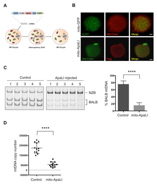
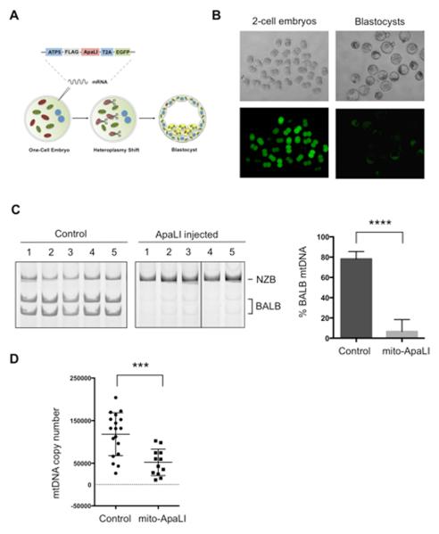
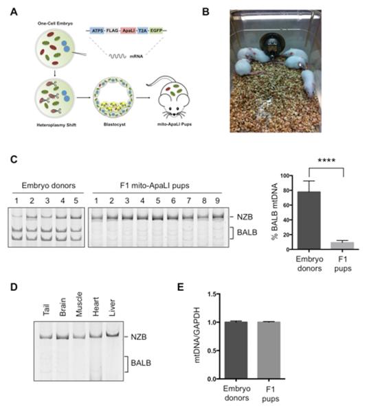
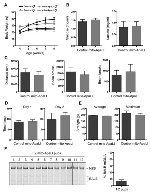
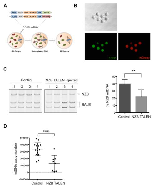
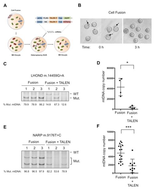
Mitochondrial diseases include a group of maternally inherited genetic disorders caused by mutations in mtDNA. In most of these patients, mutated mtDNA coexists with wild-type mtDNA, a situation known as mtDNA heteroplasmy. Here, we report on a strategy toward preventing germline transmission of mitochondrial diseases by inducing mtDNA heteroplasmy shift through the selective elimination of mutated mtDNA. As a proof of concept, we took advantage of NZB/BALB heteroplasmic mice, which contain two mtDNA haplotypes, BALB and NZB, and selectively prevented their germline transmission using either mitochondria-targeted restriction endonucleases or TALENs. In addition, we successfully reduced human mutated mtDNA levels responsible for Leber's hereditary optic neuropathy (LHOND), and neurogenic muscle weakness, ataxia, and retinitis pigmentosa (NARP), in mammalian oocytes using mitochondria-targeted TALEN (mito-TALENs). Our approaches represent a potential therapeutic avenue for preventing the transgenerational transmission of human mitochondrial diseases caused by mutations in mtDNA. PAPERCLIP.


Figures

Figure 1
Figure 1. Heteroplasmy shift in NZB/BALB MII oocytes using mito-ApaLI
(A) Injection of mito-ApaLI mRNA in oocytes for induction of heteroplasmy shift. (B) Mitochondrial co-localization of mito-GFP and mito-ApaLI with Mitotracker in injected oocytes by immunofluorescence. Scale bars, 10μm. (C) RFLP analysis and quantification of mtDNA heteroplasmy in control and mito-ApaLI injected MII oocytes after 48 h (Control n=16; mito-ApaLI n=12). Representative gel. (D) Quantification of mtDNA copy number by qPCR in control and mito-ApaLI injected oocytes MII after 48 h (Control n=12; mito-ApaLI n=12). Error bars represent ± SEM. ****p<0.0001. See also Figure S1.
Figure 2
Figure 2. Heteroplasmy shift in NZB/BALB embryos using mito-ApaLI
(A) Injection of mito-ApaLI mRNA in one-cell embryos for induction of heteroplasmy shift. (B) In vitro development of mito-ApaLI injected embryos to blastocyst stage. Time-lapse images of EGFP reporter expression at different developmental stages. (C) RFLP analysis and quantification of mtDNA heteroplasmy in control and mito-ApaLI injected embryos (Control n=10; mito-ApaLI n=8). Representative gel. (D) Quantification of mtDNA copy number by qPCR in control and mito-ApaLI injected embryos (Control n=18; mito-ApaLI n=12). Error bars represent ± SEM. ***p<0.001. ****p<0.0001. See also Figure S2.
Figure 3
Figure 3. Generation of live animals after induction of heteroplasmy shift in NZB/BALB embryos using mito-ApaLI
(A) Outline for the generation of live animals after injection of mito-ApaLI mRNA in one-cell embryos. (B) Representative photograph of F1 mito-ApaLI mice. (C) RFLP analysis and quantification of mtDNA heteroplasmy in tail tip biopsies of embryo donors and generated F1 mito-ApaLI pups. (Donor n=10; mito-ApaLI n=9). (D) RFLP analysis and quantification of mtDNA heteroplasmy in tail, brain, muscle, heart and liver of F1 mito-ApaLI mice. (E) Quantification of mtDNA copy number by qPCR in F1 mito-ApaLI pups (Donor n=10; F1 mito-ApaLI n=9). Error bars represent ± SEM. ****p<0.0001. See also Figure S3.
Figure 4
Figure 4. Characterization of F1 mito-ApaLI mice
(A) Body weight of mito-ApaLI males (Control n=5 and mito-ApaLI n=3) and mito-ApaLI females (Control n=5 and mito-ApaLI n=6) at different time points. ns. (B) Biochemical analysis of glucose and lactate in blood of control (n=10) and mito-ApaLI (n=9) mice. ns. (C) Open field test measuring baseline levels of locomotor activity in freely moving mice quantifying distance traveled, ambulatory counts and vertical counts. (D) Rotarod test evaluating locomotor coordination based on the latency at which a fall occurs on a gradually accelerating spinning rod. (E) Grip strength test measuring average and maximum grip force in the forelimbs. (F) RFLP analysis and quantification of mtDNA heteroplasmy in tail tip biopsies of F2 mito-ApaLI pups. (F2 mito-ApaLI n=12). Error bars represent ± SEM. See also Figure S4 and Table S1.
Figure 5
Figure 5. Heteroplasmy shift in NZB/BALB MII oocytes using NZB mito-TALEN
(A) Injection of NZB mito-TALEN mRNA in oocytes for induction of heteroplasmy shift. (B) Expression of fluorescent reporters of NZB TALEN monomer in MII oocytes. (C) RFLP analysis and quantification of mtDNA heteroplasmy in control and NZB TALEN injected oocytes after 48 h (Control n=9; NZB TALEN n=7). Representative gel. (D) Quantification of mtDNA copy number by qPCR in control and NZB TALEN injected oocytes after 48 h (Control n=16; NZB TALEN n=8). Error bars represent ± SEM. **p<0.01. ***p<0.001. See also Figure S5.
Figure 6
Figure 6. Specific elimination of human LHOND m.14459G>A and NARP m.9176T>C mutations in mammalian oocytes using mito-TALENs
(A) Fusion of human cells harboring LHOND m.14459G>A and NARP m.9176T>C mutations with mouse MII oocytes followed by the injection of mito-TALENs for induction of heteroplasmy shift. (B) Representative images of MII oocytes before and after cell fusion. (C) RFLP analysis and quantification of LHOND heteroplasmy in individual MII oocytes with and without LHOND TALEN injection after 48 h (Fusion n=3; Fusion + TALEN n=3). (D) Quantification of human mtDNA copy number by qPCR in individual MII oocytes with and without LHOND TALEN injection after 48 h (Fusion n=4; Fusion + TALEN n=4). (E) RFLP analysis and quantification of NARP heteroplasmy in individual MII oocytes with and without NARP TALEN injection after 48 h (Fusion n=7; Fusion + TALEN n=3). (F) Quantification of human mtDNA copy number by qPCR in individual MII oocytes with and without NARP TALEN injection after 48 h (Fusion n=17; Fusion + TALEN n=9). Error bars represent ± SEM. *p<0.05. ***p<0.001. See also Figure S6.

Comment in

References

    1. Alexeyev MF, Venediktova N, Pastukh V, Shokolenko I, Bonilla G, Wilson GL. Selective elimination of mutant mitochondrial genomes as therapeutic strategy for the treatment of NARP and MILS syndromes. Gene Ther. 2008;15:516–523. - PMC - PubMed
    1. Alexeyev M, Shokolenko I, Wilson G, LeDoux S. The Maintenance of Mitochondrial DNA Integrity--Critical Analysis and Update. Cold Spring Harb Perspect Biol. 2013;5:a012641–a012641. - PMC - PubMed
    1. Anderson S, Bankier AT, Barrell BG, Debruijn M, Coulson AR, Drouin J, Eperon IC, Nierlich DP, Roe BA, Sanger F, et al. Sequence and Organization of the Human Mitochondrial Genome. Nature. 1981;290:457–465. - PubMed
    1. Bacman SR, Williams SL, Duan D, Moraes CT. Manipulation of mtDNA heteroplasmy in all striated muscles of newborn mice by AAV9-mediated delivery of a mitochondria-targeted restriction endonuclease. Gene Ther. 2012;19:1101–1106. - PMC - PubMed
    1. Bacman SR, Williams SL, Garcia S, Moraes CT. Organ-specific shifts in mtDNA heteroplasmy following systemic delivery of a mitochondria-targeted restriction endonuclease. Gene Ther. 2010;17:713–720. - PMC - PubMed

Publication types