. 2015 Apr 24:5:9633.
doi: 10.1038/srep09633.

Loss of PTPRM associates with the pathogenic development of colorectal adenoma-carcinoma sequence

Affiliations

Loss of PTPRM associates with the pathogenic development of colorectal adenoma-carcinoma sequence

Putty-Reddy Sudhir et al. Sci Rep. .

Abstract

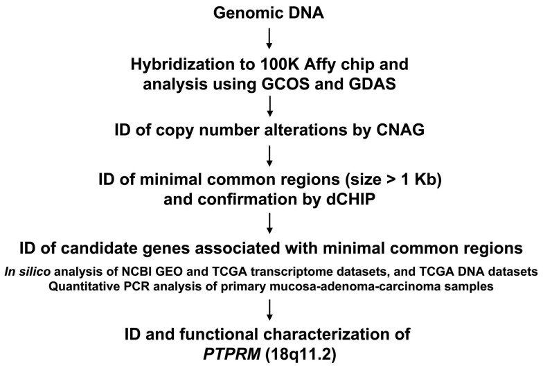
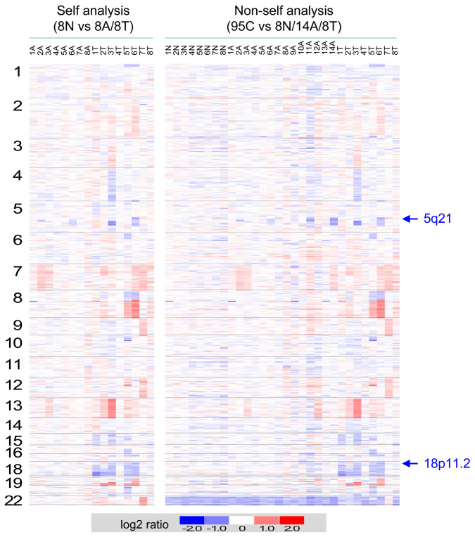
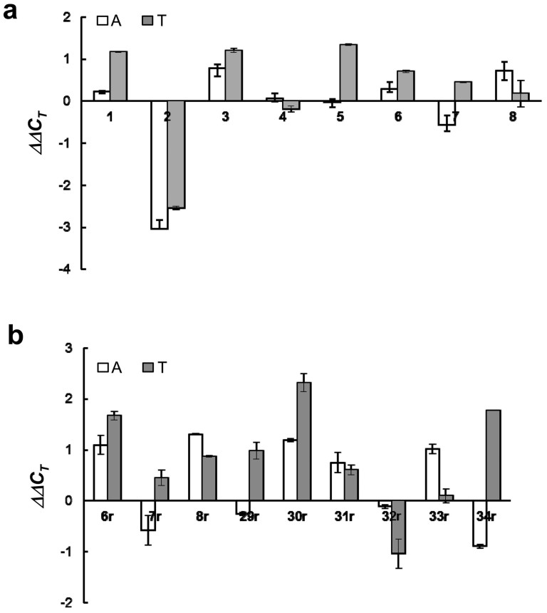
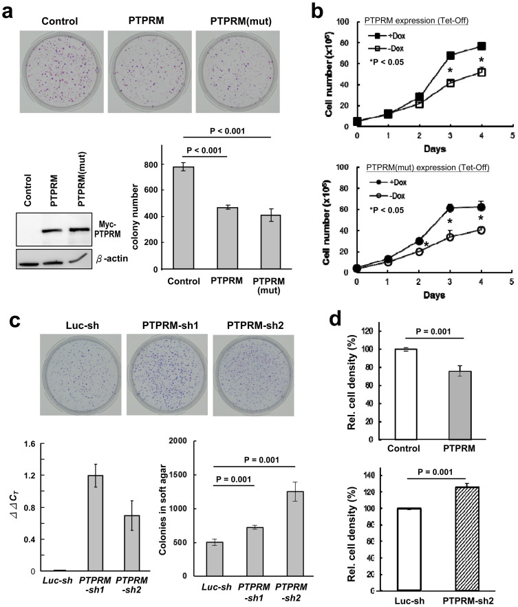
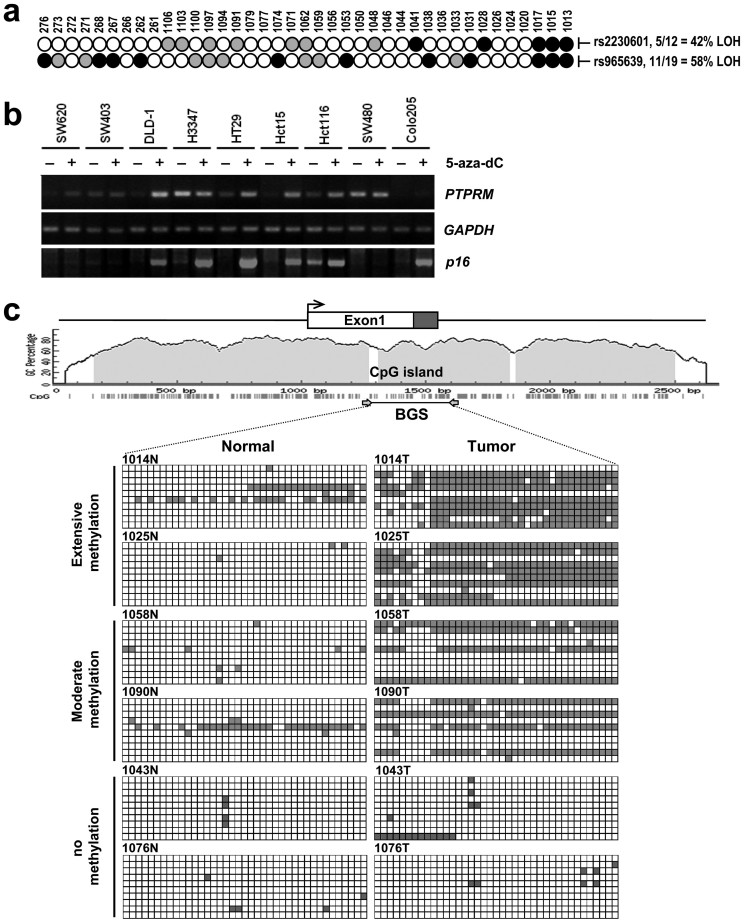
Identification and functional analysis of genes from genetically altered chromosomal regions would suggest new molecular targets for cancer diagnosis and treatment. Here we performed a genome-wide analysis of chromosomal copy number alterations (CNAs) in matching sets of colon mucosa-adenoma-carcinoma samples using high-throughput oligonucleotide microarray analysis. In silico analysis of NCBI GEO and TCGA datasets allowed us to uncover the significantly altered genes (p ≤ 0.001) associated with the identified CNAs. We performed quantitative PCR analysis of the genomic and complementary DNA derived from primary mucosa, adenoma, and carcinoma samples, and confirmed the recurrent loss and down-regulation of PTPRM in colon adenomas and carcinomas. Functional characterization demonstrated that PTPRM negatively regulates cell growth and colony formation, whereas loss of PTPRM promotes oncogenic cell growth. We further showed that, in accordance to Knudson's two-hit hypothesis, inactivation of PTPRM in colon cancer was mainly attributed to loss of heterozygosity and promoter hypermethylation. Taken together, this study demonstrates a putative tumor suppressive role for PTPRM and that genetic and epigenetic alterations of PTPRM may contribute to early step of colorectal tumorigenesis.


Conflict of interest statement

The authors declare no competing financial interests.

Figures

Figure 1
Figure 1. Strategy for identification of CNAs and associated genes in colon adenoma-carcinoma sequence.
Figure 2
Figure 2. Genome-wide DNA copy-number alterations associated with colon adenoma-carcinoma sequence.
Heatmaps of Log2 ratio DNA copy number alterations of the adenoma (A) and carcinoma (T) samples in relation to the non-tumorous mucosa (N) by pairwise analyses and to normal control (C) by non-pairwise analyses are shown. Chromosomes 1-22 are depicted from top to bottom, and individual samples are shown from left to right. The relative DNA copy number alterations are shown in blue as deletion and red as amplification.
Figure 3
Figure 3. Expression level and copy number alteration of PTPRM in primary colon non-tumorous mucosa-adenoma-carcinoma samples.
(a) Semi-quantitative PCR analysis of PTPRM DNA in 8 trio sets of colon mucosa, adenoma, and carcinoma tissues. Relative levels of genomic DNA and transcripts in adenomatous polyps (A) and carcinomas (T) are shown as ΔΔCT in relation to the levels in matching non-tumor mucosa tissue after normalization to the controls, GAPDH (for genomic DNA) and DDX5 (for cDNA). The identification numbers of patients are shown at the bottom of the panel.
Figure 4
Figure 4. Ectopic expression of PTPRM suppresses cell growth and colony formation, whereas knockdown of PTPRM promotes anchorage-independent cell growth.
(a) Ectopic expression of PTPRM suppresses colony formation. HCT116 cells were transfected with vectors containing the full-length cDNA of PTPRM in right (PTPRM) and reverse (Control) orientations as well as the PTP domain mutant (PTPRM(mut)), and cultured in neomycin-containing medium. Colonies were counted after two weeks and presented in bar graph. Western blot shows the expression of PTPRM. (b) PTPRM suppresses cell growth independent of its phosphatase activity. Tet-Off HeLa cell clones stably harboring PTPRM cDNA (Top panel, the wild-type PTPRM; Bottom panel, PTPRM mutant) were selected, and cultured in medium containing G418, puromycin and doxycycline. For cell growth assay, cells were incubated with fresh medium without doxycycline for 48 hrs to induce the expression of PTPRM, and cell numbers were counted using CCK-8 reagent at indicated time. (c) Knockdown of PTPRM promotes anchorage-independent growth in soft agar. Soft agar colony formation assay was performed in colon cancer SW620 cell clones stably harboring lentivirus-encoded control shRNA or shRNA targeting PTPRM as indicated. Two individual shRNAs targeting at PTPRM were employed. Expression of PTPRM transcripts in the cell clones harboring the individual shRNAs was measured by semi-quantitative RT-PCR analysis. ΔΔCT was derived by comparing the CT of the PTPRM-KD clones to that of the luciferase-KD clone after normalization to GAPDH. Data shown are means ± SD of three independent experiments. *P < 0.05 by Student's t test. (d) Saturation density was measured in the Tet-Off HeLa cell clone expressing PTPRM after removal of doxycycline (Top) and SW620/Luc-KD and PTPRM-KD clones (Bottom). Cells were cultured, with fresh medium changed every two days till reaching confluency. Cells were changed into fresh medium and cell numbers were counted by CCK-8 assay after culturing for two more days.
Figure 5
Figure 5. Genetic and epigenetic alterations of PTPRM in colorectal cancer.
(a) Assessment of LOH of PTPRM locus on chromosome 18p11.2 in CRC. Two PRPRM gene-specific SNP markers, rs2230601 and rs965639, were assessed for LOH (closed circle), retention of heterozygosity (gray colored circle), or non-informative (open circles) in 39 primary CRC samples. The frequency of LOH for each marker in the informative cases is shown at right. (b) RNA was prepared from nine CRC cell lines treated with (+) and without (-) 5-aza-dC and subjected to RT-PCR analyses of the expression of PTPRM, p16 and GAPDH transcripts. (c) Bisulfite sequencing analysis of methylation of PTPRM promoter in primary colorectal carcinoma and corresponding mucosa samples. The genomic structure of PTPRM and the relative position of CpG island are depicted. Exon 1 is shown as the open box, with coding region shown in the shaded area. An enlarged representation of the CpG island is sketched as a solid line with relative positions of the CpG sites depicted as short vertical lines. Bisulfite-modified genomic DNA was amplified by PCR using primer set indicated, PCR products were subcloned, and ten isolates were sequenced for each sample. The CpG sites examined are shown in open (for unmethylated) and filled (for methylated) squares. Tumors displaying methylated CpG sites in at least 5 clones were designated as extensively methylated, 2-5 clones as moderately methylated, and scattered methylation as unmethylated. Representative tumors from each category are shown.

References

    1. Fearon E. R. & Vogelstein B. A genetic model for colorectal tumorigenesis. Cell 61, 759–767 (1990). - PubMed
    1. Kinzler K. W. & Vogelstein B. Lessons from hereditary colorectal cancer. Cell 87, 159–170 (1996). - PubMed
    1. Leslie A., Carey F. A., Pratt N. R. & Steele R. J. The colorectal adenoma-carcinoma sequence. Br J Surg 89, 845–860 (2002). - PubMed
    1. Vogelstein B. et al. Genetic alterations during colorectal-tumor development. N Engl J Med 319, 525–532 (1988). - PubMed
    1. Habermann J. K. et al. Stage-specific alterations of the genome, transcriptome, and proteome during colorectal carcinogenesis. Genes Chromosomes Cancer 46, 10–26 (2007). - PubMed

Publication types

MeSH terms

Substances