. 2015 May;6(5):351-62.
doi: 10.1007/s13238-015-0163-3. Epub 2015 Apr 25.

Insight into the Ebola virus nucleocapsid assembly mechanism: crystal structure of Ebola virus nucleoprotein core domain at 1.8 Å resolution

Affiliations

Insight into the Ebola virus nucleocapsid assembly mechanism: crystal structure of Ebola virus nucleoprotein core domain at 1.8 Å resolution

Shishang Dong et al. Protein Cell. 2015 May.

Abstract

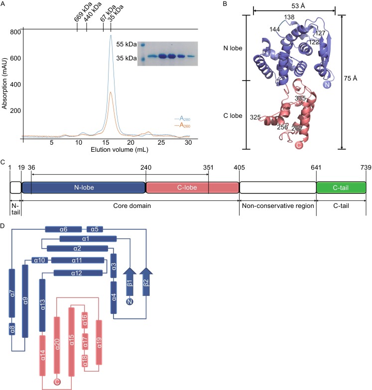
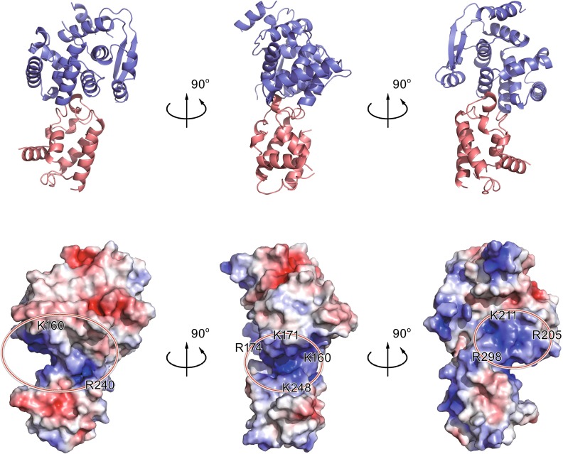
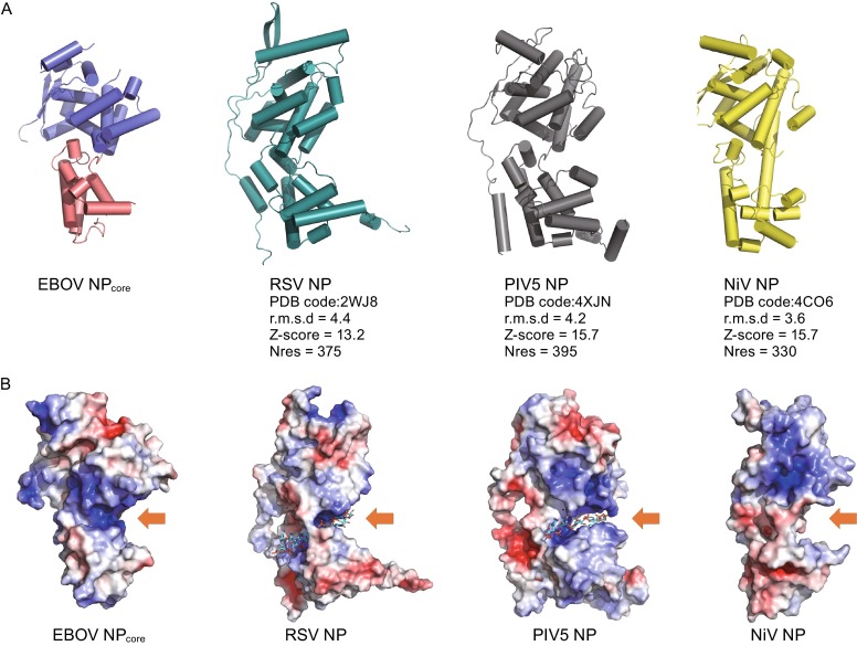
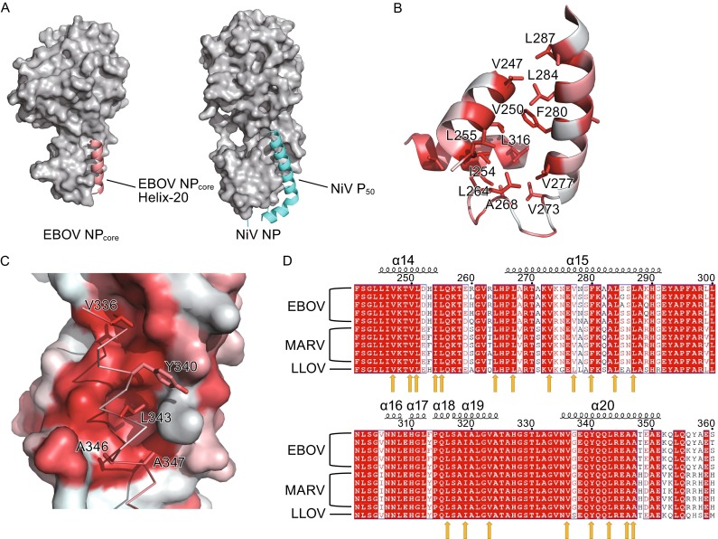
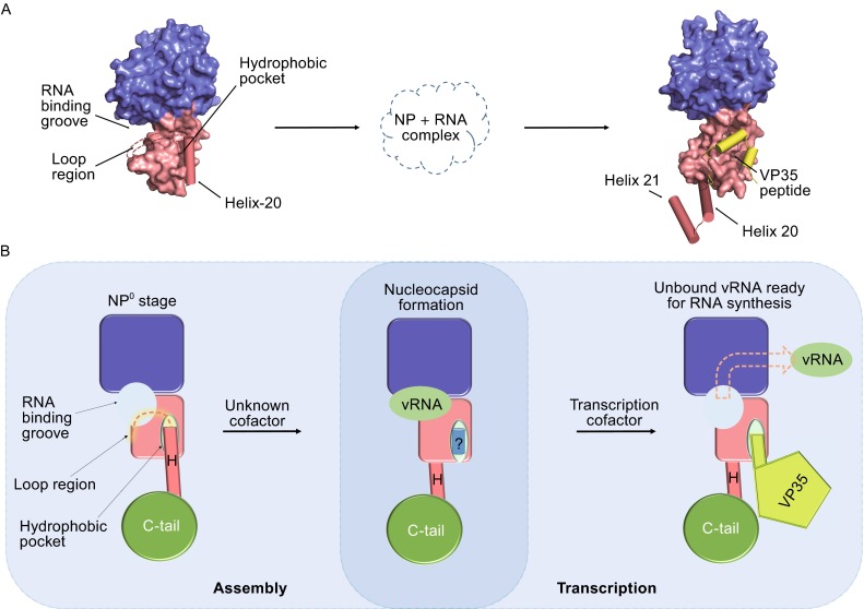
Ebola virus (EBOV) is a key member of Filoviridae family and causes severe human infectious diseases with high morbidity and mortality. As a typical negative-sense single-stranded RNA (-ssRNA) viruses, EBOV possess a nucleocapsid protein (NP) to facilitate genomic RNA encapsidation to form viral ribonucleoprotein complex (RNP) together with genome RNA and polymerase, which plays the most essential role in virus proliferation cycle. However, the mechanism of EBOV RNP formation remains unclear. In this work, we solved the high resolution structure of core domain of EBOV NP. The polypeptide of EBOV NP core domain (NP(core)) possesses an N-lobe and C-lobe to clamp a RNA binding groove, presenting similarities with the structures of the other reported viral NPs encoded by the members from Mononegavirales order. Most strikingly, a hydrophobic pocket at the surface of the C-lobe is occupied by an α-helix of EBOV NP(core) itself, which is highly conserved among filoviridae family. Combined with other biochemical and biophysical evidences, our results provides great potential for understanding the mechanism underlying EBOV RNP formation via the mobility of EBOV NP element and enables the development of antiviral therapies targeting EBOV RNP formation.


Figures

Figure 1
Figure 1
Purification and structure of EBOV NP core. (A) Size exclusion chromatography (SEC) of EBOV NPcore. The sample containing EBOV NPcore was injected into a Superdex-200 column. The molecular weights of standard protein markers are shown on the top. Blue and red lines denote A280 and A260, respectively. SDS-PAGE analysis of the peak fractions is shown inset. (B) Overall structure of EBOV NPcore. The polypeptide of EBOV NPcore is shown as colored cartoon. The N-lobe and C-lobe are colored as blue and red, respectively. Missing residues are linked by dotted lines. (C) Schematic diagram of the domain organization in the primary sequence of EBOV NP. N-lobe, C-lobe, and C-tail are colored as slate blue, salmon red, and green, respectively. N-tail and non-conservative region are colored as blank. (D) Topology diagram of EBOV NPcore helices are presented as rectangulars and strands are shown as arrows. The color scheme is the same as that in (B) and (C)
Figure 2
Figure 2
Potential RNA binding region of EBOV NP core. (A) Cartoon representation of EBOV NPcore. The N-lobe and C-lobe are colored as slate blue and salmon red, respectively. (B) Electrostatic surface potential of EBOV NPcore. The electrostatic surface potential of EBOV NPcore was calculated using APBS tools, with limits ±5 kbT/ec. Positive residues are highlighted by red circle on EBOV NPcore, suggesting the presence of several positively charged grooves that may be involved in RNA binding
Figure 3
Figure 3
Structural comparison of NPs from Mononegavirales viruses. (A) Structural comparison of EBOV NPcore with RSV, PIV5, and NiV NP. NPs are displayed as cartoon. RSV (PDB code: 2WJ8), PIV5 (PDB code: 4XJN), and NiV (PDB code: 4CO6) NPs are colored cyan, gray, and yellow, respectively. Alignment information is listed under each molecule. All molecules are aligned to the structure of EBOV NPcore and shown in the same orientation. (B) Electrostatic potential comparison of EBOV NPcore with RSV, PIV5, and NiV NP. Positively charged pockets for RNA binding are indicated with orange arrows. RNA molecules in RSV and PIV5 NPs are shown as colored sticks
Figure 4
Figure 4
Helix-20 in EBOV NP core indicates an essential hydrophobic pocket. (A) Comparison of EBOV NPcore and NiV NP. Both structures are shown as surface and colored gray. EBOV NPcore Helix-20 and NiV P50 are shown as cartoon, colored salmon red and cyan, respectively. (B) Formation of the hydrophobic pocket. Main chain is shown in cartoon and hydrophobic residues are shown in stick, colored in red by hydrophobicity. (C) Interaction of Helix-20 with hydrophobic pocket. Hydrophobic pocket are shown in surface, Helix-20 are displayed in ribbon and stick, colored in red by hydrophobicity. (D) Primary sequence alignment of members of the filoviridae family. Highly conserved residue in Helix-14, Helix-15, Helix-19, and Helix-20 are indicated by yellow arrow
Figure 5
Figure 5
Structural mobility of EBOV NP core within virus proliferation. (A) Structural comparison of EBOV NPcore and complex with VP35 peptide. The molecule of EBOV NPcore alone and in complex with VP35 peptide are shown in the left and right panels. The N-lobe and C-lobe are colored as blue and red, respectively. Helices 20–21 are shown as cylinder, and the loop region (A326–V334) are linked by dotted lines. Small peptide derived from VP35 is shown as yellow cylinders. (B) A proposed model of conformation change during nucleocapsid assembly and transcription process. RNA binding groove and hydrophobic pocket are indicated by arrow

References

    1. Adams PD, et al. PHENIX: building new software for automated crystallographic structure determination. Acta Crystallogr D Biol Crystallogr. 2002;58(Pt 11):1948–1954. doi: 10.1107/S0907444902016657. - DOI - PubMed
    1. Albertini AA, et al. Crystal structure of the rabies virus nucleoprotein-RNA complex. Science. 2006;313(5785):360–363. doi: 10.1126/science.1125280. - DOI - PubMed
    1. Ariza A, et al. Nucleocapsid protein structures from orthobunyaviruses reveal insight into ribonucleoprotein architecture and RNA polymerization. Nucleic Acids Res. 2013;41(11):5912–5926. doi: 10.1093/nar/gkt268. - DOI - PMC - PubMed
    1. Arranz R, et al. The structure of native influenza virion ribonucleoproteins. Science. 2013;338(6114):1634–1637. doi: 10.1126/science.1228172. - DOI - PubMed
    1. Bharat TA, et al. Cryo-electron tomography of Marburg virus particles and their morphogenesis within infected cells. PLoS Biol. 2011;9(11):e1001196. doi: 10.1371/journal.pbio.1001196. - DOI - PMC - PubMed

Publication types