. 2015 Jun 12;290(24):14826-40.
doi: 10.1074/jbc.M114.630640. Epub 2015 Apr 24.

Genome Protection by the 9-1-1 Complex Subunit HUS1 Requires Clamp Formation, DNA Contacts, and ATR Signaling-independent Effector Functions

Affiliations

Genome Protection by the 9-1-1 Complex Subunit HUS1 Requires Clamp Formation, DNA Contacts, and ATR Signaling-independent Effector Functions

Pei Xin Lim et al. J Biol Chem. .

Abstract

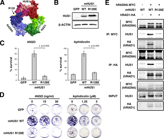
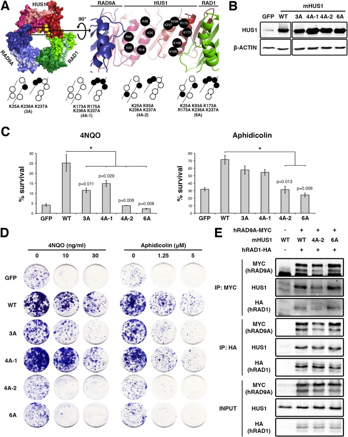
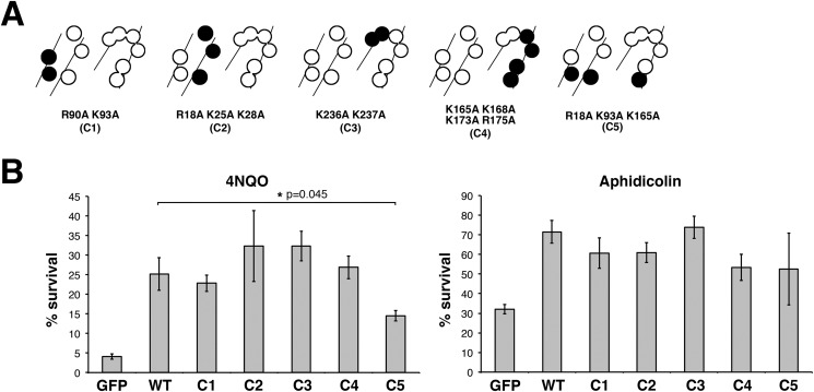
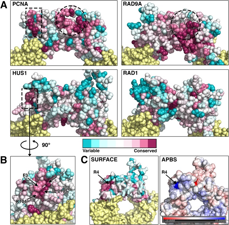
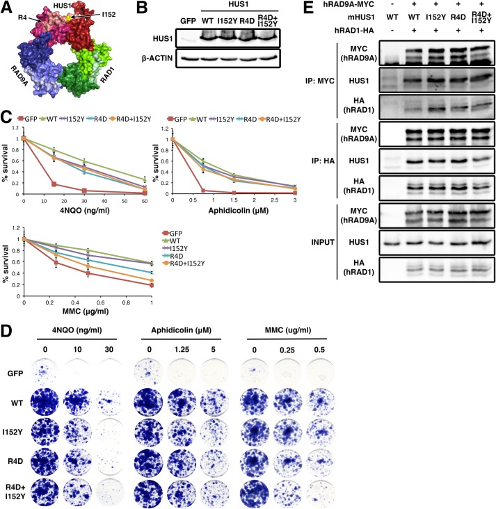
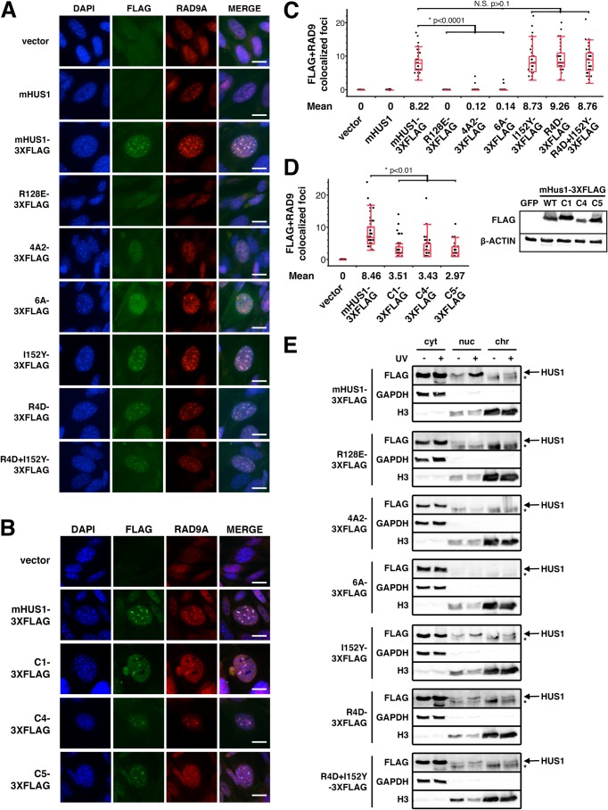
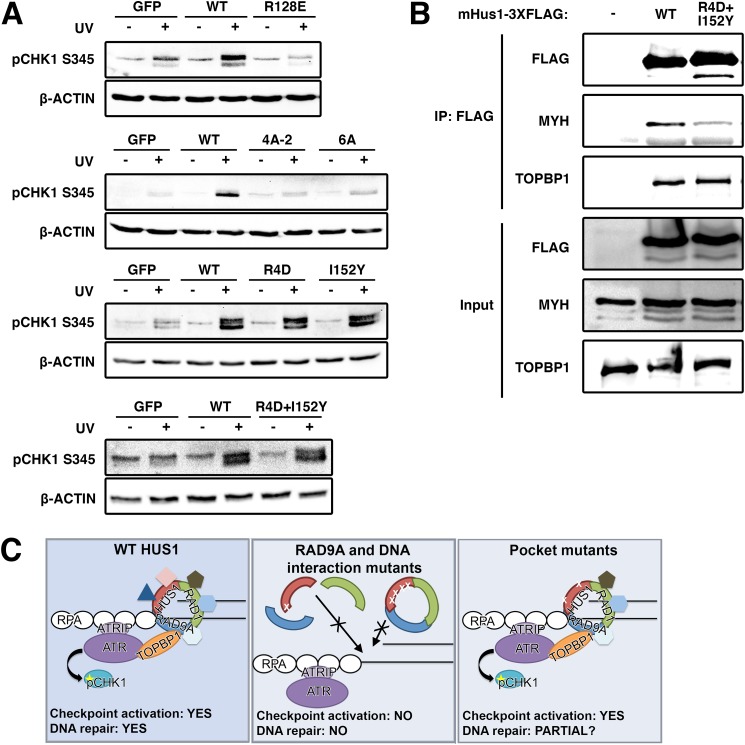
The RAD9A-HUS1-RAD1 (9-1-1) complex is a heterotrimeric clamp that promotes checkpoint signaling and repair at DNA damage sites. In this study, we elucidated HUS1 functional residues that drive clamp assembly, DNA interactions, and downstream effector functions. First, we mapped a HUS1-RAD9A interface residue that was critical for 9-1-1 assembly and DNA loading. Next, we identified multiple positively charged residues in the inner ring of HUS1 that were crucial for genotoxin-induced 9-1-1 chromatin localization and ATR signaling. Finally, we found two hydrophobic pockets on the HUS1 outer surface that were important for cell survival after DNA damage. Interestingly, these pockets were not required for 9-1-1 chromatin localization or ATR-mediated CHK1 activation but were necessary for interactions between HUS1 and its binding partner MYH, suggesting that they serve as interaction domains for the recruitment and coordination of downstream effectors at damage sites. Together, these results indicate that, once properly loaded onto damaged DNA, the 9-1-1 complex executes multiple, separable functions that promote genome maintenance.

Keywords: DNA damage, DNA damage response, checkpoint control, proliferating cell nuclear antigen (PCNA), genomic instability, HUS1, 9-1-1 complex.


Figures

FIGURE 1.
FIGURE 1.
mHUS1 residue Arg-128 is crucial for 9-1-1 clamp formation. A, Arg-128 (arrow) is located at the HUS1-RAD9A interface (Protein Data Bank code 3GGR). B, immunoblotting using antibodies specific for HUS1 or β-actin was performed to compare the stability of WT and R128E mHUS1 proteins in HEK293T cells. C and D, short term viability and clonogenic survival were measured for Hus1-null MEFs stably expressing mHUS1 R128E after 4NQO or aphidicolin treatments. MEFs expressing GFP or WT mHUS1 served as negative and positive controls, respectively. Each experiment in C was repeated five times with two independently generated cell lines. E, interaction of mHUS1 R128E with hRAD9 and hRAD1 was assessed by co-IP. Error bars, S.D.
FIGURE 2.
FIGURE 2.
Multiple positively charged residues on the HUS1 inner ring are synergistically important for genotoxic stress responses. A, mHUS1 has 11 arginines and lysines (black circles) distributed on four α-helices in the inner ring surface (Protein Data Bank code 3GGR). Alanine substitutions of these residues (3A-6A) were made. B, protein expression was measured as in Fig. 1B. C and D, genotoxin sensitivity was measured as in Fig. 1, C and D. Each experiment in C was repeated three times with three independently generated cell lines. E, interaction of mHUS1 mutant proteins with hRAD9 and hRAD1 was assessed by co-IP. Error bars, S.D.
FIGURE 3.
FIGURE 3.
Certain configurations of positively charged residues in the inner ring of HUS1 are dispensable for genotoxic stress responses. A, alanine substitutions of positively charged residues were made in various combinations (C1–C5), as indicated by the filled circles. B, short term viability of Hus1−/− p21−/− MEFs stably expressing mHUS1 inner ring mutants C1–C5 after 4NQO or aphidicolin treatments was measured. Each experiment was repeated four times using three independently generated cell lines. No significant differences in survival between wild-type mHUS1 and mutants C1–C5 were identified, except for C5 with 4NQO treatment. Error bars, S.D.
FIGURE 4.
FIGURE 4.
The identification of a novel conserved hydrophobic pocket on the outer surface of HUS1. A and B, the evolutionary conservation values of each amino acid position in the protein structures of PCNA, RAD9A, HUS1, and RAD1 were calculated using the Consurf bioinformatics server (see “Experimental Procedures”). Multiple sequence alignments of 44 organisms that encompass a wide range of taxa were used (see Table 2). Residues pseudocolored in cyan have diverged and are variable, whereas those in magenta are conserved. Dotted lines outline conserved regions that potentially mediate protein-protein associations. The circled regions correspond to the PIP box-binding hydrophobic pocket of PCNA and the analogous conserved region of RAD9A. A second conserved region for PCNA and HUS1 is outlined with a rectangle and in the case of HUS1 corresponds to a novel pocket on the side of globular domain 1 composed of 3 conserved hydrophobic residues. C, HUS1 atomic surface and surface electrostatic potential models reveal a positively charged groove at the base of the novel pocket. The charge is contributed by an arginine at position 4.
FIGURE 5.
FIGURE 5.
Two hydrophobic pockets on the outer surface of HUS1 are required for genotoxic stress responses. A, Arg-4 and Ile-152 (arrows) are located on the outer ring surface (Protein Data Bank code 3GGR). B, protein expression was measured as in Fig. 1B. C and D, genotoxin sensitivity was measured as in Fig. 1, C and D, as well as with MMC treatment. Each experiment in C was repeated two times with two independently generated cell lines. E, interaction of the pocket mutants with hRAD9 and hRAD1 was assessed by co-IP. Error bars, S.D.
FIGURE 6.
FIGURE 6.
DNA damage-induced HUS1 localization is defective in HUS1 clamp-forming and DNA-interacting mutants but not in HUS1 pocket mutants. A and B, MMC-treated Hus1-null MEFs stably expressing the indicated constructs were stained with DAPI (blue) and α-FLAG (green) and α-RAD9A (red) antibodies. Scale bar, 10 μm. C and D, quantifications of colocalized FLAG and RAD9A foci are presented in quartile box and dot plots. NS, not significant. D, immunoblotting using antibodies specific for FLAG or β-actin was performed to compare the stability of 3XFLAG-tagged WT and mutant mHUS1 proteins (C1, C4, and C5) in HEK293T cells. E, cells were UV-treated; fractionated into cytoplasmic (cyt), nuclear (nuc), and chromatin (chr) fractions; and immunoblotted. GAPDH and histone 3 served as fractionation controls. Arrows, HUS1 band; asterisks, nonspecific band.
FIGURE 7.
FIGURE 7.
9-1-1-dependent checkpoint signaling requires clamp formation and DNA associations but not HUS1 outer surface pocket function, which is necessary for effector interactions. A, DNA damage-induced CHK1 phosphorylation is hampered in HUS1 clamp formation and DNA interaction mutants but is intact for HUS1 outer surface pocket mutants. Lysates from cells treated with 0 or 100 J/m2 UV were immunoblotted using antibodies specific for phospho-CHK1 or β-actin. B, HUS1 pocket mutant R4D,I152Y is impaired for interaction with base excision repair protein MYH. Lysates prepared from HEK293T cells overexpressing 3XFLAG-tagged WT or R4D,I152Y mHUS1 proteins were immunoprecipitated with antibody specific for FLAG and immunoblotted using antibodies specific for MYH and TOPBP1. C, model for HUS1-mediated function in DNA damage response. WT HUS1 forms 9-1-1 clamps, localizes to DNA damage sites, and mediates ATR checkpoint signaling and DNA repair functions. When the RAD9A-interacting residue is dysfunctional, HUS1 cannot form 9-1-1 clamp, causing loss of all downstream functions. HUS1 mutants defective for DNA interactions are still able to form 9-1-1 clamps but cannot localize to DNA damage sites, similarly causing loss of all downstream functions. Only HUS1 pocket mutants are able to form 9-1-1 clamps, localize to DNA lesions, and activate ATR for checkpoint signaling. However, checkpoint-independent functions of HUS1 are perturbed in the pocket mutants, probably causing increased genotoxin hypersensitivity due to impaired DNA repair.

References

    1. Jackson S. P., Bartek J. (2009) The DNA-damage response in human biology and disease. Nature 461, 1071–1078 - PMC - PubMed
    1. Cimprich K. A., Cortez D. (2008) ATR: an essential regulator of genome integrity. Nat. Rev. Mol. Cell Biol. 9, 616–627 - PMC - PubMed
    1. Eichinger C. S., Jentsch S. (2011) 9-1-1: PCNA's specialized cousin. Trends Biochem. Sci. 36, 563–568 - PubMed
    1. Hopkins K. M., Auerbach W., Wang X. Y., Hande M. P., Hang H., Wolgemuth D. J., Joyner A. L., Lieberman H. B. (2004) Deletion of mouse rad9 causes abnormal cellular responses to DNA damage, genomic instability, and embryonic lethality. Mol. Cell Biol. 24, 7235–7248 - PMC - PubMed
    1. Han L., Hu Z., Liu Y., Wang X., Hopkins K. M., Lieberman H. B., Hang H. (2010) Mouse Rad1 deletion enhances susceptibility for skin tumor development. Mol. Cancer 9, 67. - PMC - PubMed

Publication types