. 2015 Jun 12;290(24):14915-26.
doi: 10.1074/jbc.M115.651380. Epub 2015 Apr 24.

Redox Control of Protein Arginine Methyltransferase 1 (PRMT1) Activity

Affiliations

Redox Control of Protein Arginine Methyltransferase 1 (PRMT1) Activity

Yalemi Morales et al. J Biol Chem. .

Abstract

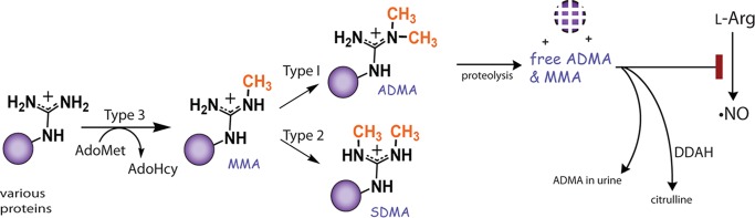
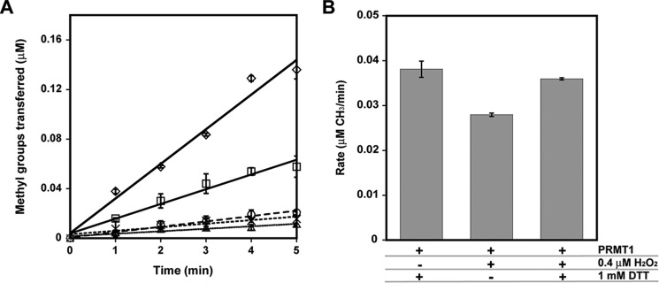
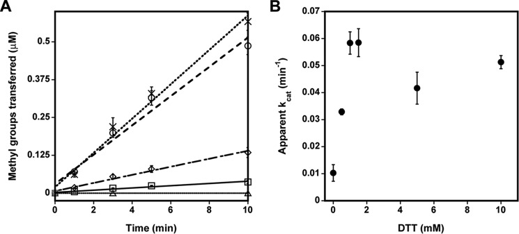
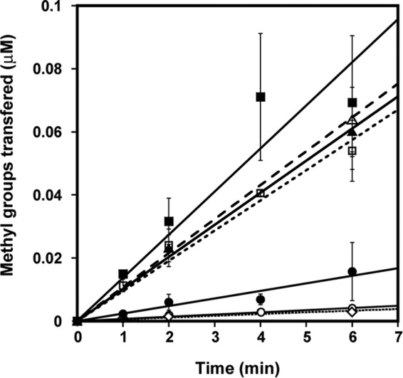
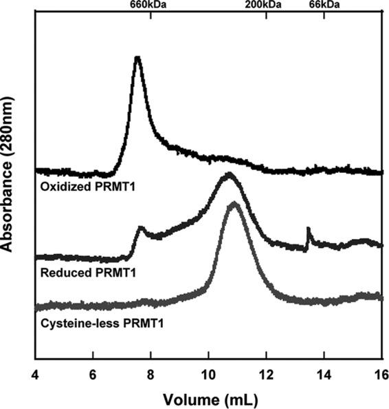
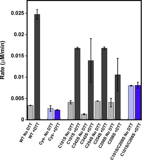
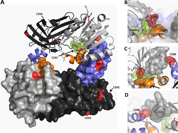
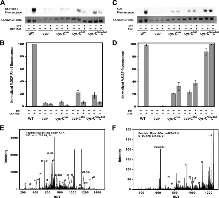
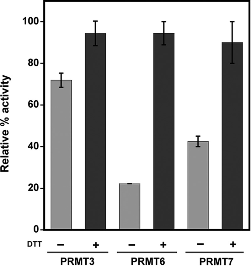
Elevated levels of asymmetric dimethylarginine (ADMA) correlate with risk factors for cardiovascular disease. ADMA is generated by the catabolism of proteins methylated on arginine residues by protein arginine methyltransferases (PRMTs) and is degraded by dimethylarginine dimethylaminohydrolase. Reports have shown that dimethylarginine dimethylaminohydrolase activity is down-regulated and PRMT1 protein expression is up-regulated under oxidative stress conditions, leading many to conclude that ADMA accumulation occurs via increased synthesis by PRMTs and decreased degradation. However, we now report that the methyltransferase activity of PRMT1, the major PRMT isoform in humans, is impaired under oxidative conditions. Oxidized PRMT1 displays decreased activity, which can be rescued by reduction. This oxidation event involves one or more cysteine residues that become oxidized to sulfenic acid (-SOH). We demonstrate a hydrogen peroxide concentration-dependent inhibition of PRMT1 activity that is readily reversed under physiological H2O2 concentrations. Our results challenge the unilateral view that increased PRMT1 expression necessarily results in increased ADMA synthesis and demonstrate that enzymatic activity can be regulated in a redox-sensitive manner.

Keywords: ADMA; PRMT1; arginine methylation; endothelial dysfunction; hydrogen peroxide; oxidative stress; posttranslational modification (PTM); protein arginine methyltransferase; redox regulation; sulfenic acid.


Figures

FIGURE 1.
FIGURE 1.
ADMA formation and degradation. PRMTs transfer a methyl group from donor AdoMet to arginine residues in substrate proteins. Type 1 PRMTs can transfer one or two methyl groups to the same-terminal guanidino nitrogen-producing MMA or ADMA, respectively. Upon degradation of the methylated proteins, free MMA and ADMA inhibit NO synthesis by acting as competitive inhibitors of nitric oxide synthase. Free ADMA is catabolized by DDAH to citrulline and dimethylamine or excreted in urine.
FIGURE 2.
FIGURE 2.
A and B, PRMT1 activity is inhibited by H2O2 in a concentration-dependent manner (A), and activity lost can be recovered by reduction (B). A, reduced PRMT1 was incubated with 0 (♢), 40 (□), 200 (○), 400 (×), or 800 (▵) μm H2O2 for 10 min at 37 °C, followed by the addition of catalase. Methyltransferase activity of the treated PRMT1 was measured with 200 μm R3 peptide as a substrate, as described under “Experimental Procedures.” B, reduced PRMT1 was treated with a physiologically relevant concentration of hydrogen peroxide or PBS and subsequently treated with 1 mm DTT or PBS prior to methyltransferase assays.
FIGURE 3.
FIGURE 3.
The effect of reducing agents on PRMT1 enzymatic activity. A, PRMT1 methyltransferase activity was measured without substrate (▵) or with 200 μm R3 peptide in the absence (□) and presence of DTT (○) or Tris(2-carboxyethyl)phosphine (×) or after desalting following a 10 min preincubation with DTT (♢). B, methyltransferase activity was measured as a function of DTT concentration.
FIGURE 4.
FIGURE 4.
The enhancing effect of DTT on PRMT1 methyltransferase activity is independent of the His6 tag. Enzymatic activity of HisTevPRMT1 (○), cleaved PRMT1 (▵), and dialyzed HisTevPRMT1 (□) measured with 200 μm R3 peptide in the absence (open symbols) and presence (closed symbols) of DTT, respectively, as well as HisTevPRMT1 treated with EDTA only as a control (♢).
FIGURE 5.
FIGURE 5.
Oligomeric state of PRMT1 proteins assessed by size exclusion chromatography. PRMT1 without DTT treatment (top trace) or incubated overnight with 1 mm DTT and 2 mm EDTA (center trace) compared with cysteineless PRMT1, which migrates at the same position regardless of treatment with DTT or EDTA. Reduction of PRMT1 or removal of all cysteine residues results in a shift toward a smaller oligomeric state.
FIGURE 6.
FIGURE 6.
Methyltransferase activity of PRMT1 cysteine variants in the absence or presence of DTT. WT PRMT1, cysteineless PRMT1, C101S PRMT1, C342S PRMT1, C254S PRMT1, C208S PRMT1, and C101S/C208S PRMT1 methylated 200 μm R3 peptide in the absence (light gray/purple) or presence (dark gray/purple) of DTT. The results shown correspond to the average of at least two biological replicates. Removing all cysteine residues from PRMT1 or making Cys101 and Cys208 unavailable for oxidation resulted in abolished redox control over PRMT1 catalytic activity.
FIGURE 7.
FIGURE 7.
Cysteine residues in rPRMT1. Shown is the PRMT1 dimer (PDB code 1OR8) colored as described in the text (AdoMet binding domain in light gray, β barrel domain in dark gray, dimerization arm in blue, αγ-loop-αZ in orange, AdoHcy in green, and cysteine residues in red). Residues 26–39 were modeled on the basis of the position of this helix in the PRMT3 structure (PDB code 1F3L). A, PRMT1 dimer. B, surface representation showing close active site interactions between AdoHcy, Cys101, and Phe36. C, top view of the dimerization arm in one monomer interacting with the AdoMet binding site in the other monomer. D, back view showing the packing of Cys208 in one monomer with the α helix in the AdoMet binding domain of the other monomer.
FIGURE 8.
FIGURE 8.
Sulfenic acid detection and free thiol content in PRMT1. A–D, air-oxidized wild-type PRMT1 (WT), cys-, cys-Cys101, cys-Cys208, and cys-Cys101Cys208 were denatured in 6 m urea and incubated with 1 mm DTT or buffer prior to addition of 10 μm DCP-Rho1 or 2.5 mm 5IAF. Labeled samples were resolved by SDS-PAGE. A, representative image of the rhodamine fluorescence signal and the corresponding Coomassie bands. B, graphical representation of triplicate gel analysis. Normalized percent DCP-Rho1 fluorescence represents the percentage of fluorescent signal divided by the amount of protein observed in the Coomassie bands, which is interpreted as the relative amount of sulfenic acid present. C, representative image of the 5IAF fluorescence signal and the corresponding Coomassie bands. D, graphical representation of triplicate 5IAF gel analysis, interpreted as relative amount of free thiols present. E and F, representative MS/MS fragmentation spectra for peptides containing dimedone-modified sulfenic acids in PRMT1. E, the CID fragments of peptide IECSSISDYAVK labeled with dimedone at Cys101. F, ETD fragments of peptide MCSIKDVAIK labeled with dimedone at Cys208. The mass shift by the modification is 138.068 (exact number), as denoted in the peptide sequence.
FIGURE 9.
FIGURE 9.
Redox control is conserved among PRMT family members. Human PRMT3 (residues 211–531), human PRMT6, and TbPRMT7 activities were tested with R3 peptide, bulk histones, or Histone 4, respectively, in the absence or presence of DTT. The average of three activity measurements are shown as relative percent activity for each isoform with its corresponding substrate.

References

    1. Go A. S., Mozaffarian D., Roger V. L., Benjamin E. J., Berry J. D., Blaha M. J., Dai S., Ford E. S., Fox C. S., Franco S., Fullerton H. J., Gillespie C., Hailpern S. M., Heit J. A., Howard V. J., Huffman M. D., Judd S. E., Kissela B. M., Kittner S. J., Lackland D. T., Lichtman J. H., Lisabeth L. D., Mackey R. H., Magid D. J., Marcus G. M., Marelli A., Matchar D. B., McGuire D. K., Mohler E. R., 3rd, Moy C. S., Mussolino M. E., Neumar R. W., Nichol G., Pandey D. K., Paynter N. P., Reeves M. J., Sorlie P. D., Stein J., Towfighi A., Turan T. N., Virani S. S., Wong N. D., Woo D., Turner M. B., American Heart Association Statistics Committee and Stroke Statistics Subcommittee (2014) Heart Disease and Stroke Statistics-2014 Update A Report From the American Heart Association. Circulation 129, e28–e292 - PMC - PubMed
    1. Lu T. M., Ding Y. A., Charng M. J., Lin S. J. (2003) Asymmetrical dimethylarginine: a novel risk factor for coronary artery disease. Clin. Cardiol. 26, 458–464 - PMC - PubMed
    1. Chen X., Niroomand F., Liu Z., Zankl A., Katus H. A., Jahn L., Tiefenbacher C. P. (2006) Expression of nitric oxide related enzymes in coronary heart disease. Basic Res. Cardiol. 101, 346–353 - PubMed
    1. Antoniades C., Shirodaria C., Leeson P., Antonopoulos A., Warrick N., Van-Assche T., Cunnington C., Tousoulis D., Pillai R., Ratnatunga C., Stefanadis C., Channon K. M. (2009) Association of plasma asymmetrical dimethylarginine (ADMA) with elevated vascular superoxide production and endothelial nitric oxide synthase uncoupling: implications for endothelial function in human atherosclerosis. Eur. Heart J. 30, 1142–1150 - PubMed
    1. Böger R. H., Bode-Böger S. M., Szuba A., Tsao P. S., Chan J. R., Tangphao O., Blaschke T. F., Cooke J. P. (1998) Asymmetric dimethylarginine (ADMA): a novel risk factor for endothelial dysfunction: its role in hypercholesterolemia. Circulation 98, 1842–1847 - PubMed

Publication types

MeSH terms

LinkOut - more resources