. 2015 Jun 1;194(11):5305-11.
doi: 10.4049/jimmunol.1402517. Epub 2015 Apr 24.

B7H6-Specific Bispecific T Cell Engagers Lead to Tumor Elimination and Host Antitumor Immunity

Affiliations

B7H6-Specific Bispecific T Cell Engagers Lead to Tumor Elimination and Host Antitumor Immunity

Ming-Ru Wu et al. J Immunol. .

Abstract

Substantial evidence showed that T cells are the key effectors in immune-mediated tumor eradication; however, most T cells do not exhibit antitumor specificity. In this study, a bispecific T cell engager (BiTE) approach was used to direct T cells to recognize B7H6(+) tumor cells. B7H6 is a specific ligand for the NK cell-activating receptor NKp30. B7H6 is expressed on various types of primary human tumors, including leukemia, lymphoma, and gastrointestinal stromal tumors, but it is not constitutively expressed on normal tissues. Data from this study showed that B7H6-specific BiTEs direct T cells to mediate cellular cytotoxicity and IFN-γ secretion upon coculturing with B7H6(+) tumors. Furthermore, B7H6-specific BiTE exhibited no self-reactivity to proinflammatory monocytes. In vivo, B7H6-specific BiTE greatly enhanced the survival benefit of RMA/B7H6 lymphoma-bearing mice through perforin and IFN-γ effector mechanisms. In addition, long-term survivor mice were protected against an RMA lymphoma tumor rechallenge. The B7H6-specific BiTE therapy also decreased tumor burden in murine melanoma and ovarian cancer models. In conclusion, B7H6-specific BiTE activates host T cells and has the potential to treat various B7H6(+) hematological and solid tumors.


Figures

Figure 1
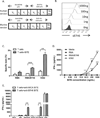
Figure 1. Design, production, and functionality of B7H6-specific BiTEs
(A) A schematic diagram of the human and murine B7H6-specific BiTE proteins. (B) Human T cells (CD3ε+, NKp30−, B7H6−) were stained with B7H6-specific BiTE protein (1ng-1000ng) followed by soluble B7H6-DyLight650. Data shown are representative histograms of staining intensity. (C) OKT3 activated human T cells were co-cultured with tumor cell lines at an E:T ratio of 5:1 (105:2×10⁴) with or without human B7H6-specific BiTE (1ug/mL). Six hours after co-culturing, culture supernatant was harvested and specific lysis was determined by LDH release assay. The data shown are mean +SD of triplicate wells, and are representative data from 3 different human donors. (D) OKT3 activated human T cells were co-cultured with B7H6− (RMA) or B7H6+ (RMA/B7H6, K562) tumor cells at an E:T of 1:1 (105:105) at the indicated concentrations of human B7H6-specific BiTE protein. Cell-free medium was collected after 24h and IFN-γ concentration determined by ELISA. The data shown are mean +SD of triplicate wells, and are representative of data from 3 different human T cell donors. (E) OKT3 activated human T cells were cultured with B7H6 negative (RMA, B16F10), B7H6 positive (RMA/B7H6, B16F10/B7H6), and B7H6 & MICA positive (K562) tumor cell lines at an E:T of 4:1 (105:2.5×10⁴) with B7H6-specific BiTE or a MICA-specific BiTE. Cell-free medium was collected after 24h and IFN-γ concentration determined by ELISA. Statistical analysis compares the IFN-γ value of B7H6 positive tumor cells (RMA/B7H6 or K562) to B7H6 negative tumor cells (RMA) at each BiTE concentration. An ** indicates p < 0.005; *** indicates p < 0.0001.
Figure 2
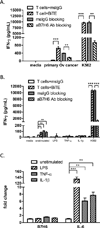
Figure 2. B7H6-specific BiTEs trigger T cells to produce IFN-γ to primary human ovarian cancer but not pro-inflammatory monocytes
(A) Activated human T cells were co-cultured with primary human ovarian cancer cells or K562 at an E:T ratio of 1:1 in the presence of murine IgG or B7H6-specific BiTE (250ng/mL). Blocking experiments were done by pre-incubating tumor samples with 10ug of murine IgG or anti-B7H6 mAbs for 45min before co-culturing. Cell-free medium was harvested after 24h and IFN-γ concentration determined by ELISA. Data shown are mean +SD of triplicate wells, and are representative of two independent experiments with different T cell donors. (B) Activated human T cells were co-cultured with unstimulated PBMCs or LPS, TNF-α, or IL-1β stimulated autologous PBMCs in the presence of murine IgG or B7H6-specific BiTE protein. Blocking experiments were done by pre-incubating target cells with 5ug of murine IgG or anti-B7H6 mAbs for 45min before co-culturing. Cell-free medium was harvested after 24h and IFN-γ concentration determined by ELISA. Data shown are representative of four different PBMC donors. (C) Unstimulated, LPS, TNF-α, or IL-1β stimulated PBMCs were harvested after 4h of stimulation. The amounts of IL-6 and B7H6 mRNA in each sample were measured by quantitative real-time PCR. The amount of mRNA in the unstimulated PBMCs is set to 1. Data shown are representative of two different PBMC donors. An * indicates p < 0.05; ** indicates p < 0.005; *** indicates p < 0.0001.
Figure 3
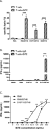
Figure 3. Murine B7H6-specific BiTE redirect T cells to specifically kill B7H6+ tumor cells and secrete IFN-γ
(A) Murine splenocytes were activated with concanavalin A (1µg/mL) and cultured with recombinant human IL-2 (25U/mL) for 4 to 6 days to generate murine T cells. Murine T cells were co-cultured with B7H6− tumor cells (RMA), or B7H6+ (RMA/B7H6, B16F10/B7H6, or ID8/B7H6) tumor cells at an E:T ratio of 5:1 for 6h. Specific lysis was quantified by LDH release assay. The data shown are mean +SD of quadruplicate wells and are representative of two independent experiments. (B) Murine T cells were co-cultured with B7H6− tumor cells (RMA, B16F10, ID8) or B7H6+ (B16F10/B7H6) tumor cells at an E:T ration of 4:1(105:2.5×10⁴) at a concentration of 500ng/mL B7H6-specific BiTE. Cell-free medium was harvested after 24h and IFN-γ concentration determined by ELISA. The data shown are mean +SD of triplicate wells and are representative of two independent experiments. (C) Murine T cells were co-cultured with indicated tumors at an E:T ratio of 5:1 (B16F10/B7H6) or 1:1 (RMA, RMA/B7H6) at various concentrations of murine B7H6-specific BiTE (7.8ng/mL–500ng/mL). Cell-free medium was harvested after 24h and IFN-γ concentration determined by ELISA. The data are shown as mean +SD of triplicate wells and are representative of two independent experiments. Statistical analysis compares the IFN-γ value of B7H6 positive tumor cells (RMA/B7H6 or B16F10/B7H6) to B7H6 negative tumor cells (RMA) in each BiTE concentration. An * indicates p < 0.05; ** indicates p < 0.005; *** indicates p < 0.0001.
Figure 4
Figure 4. Murine B7H6-specific BiTE mediate therapeutic efficacy against lymphoma in a perforin and IFN-γ dependent manner
(A) RMA/B7H6 cells (105 cells) were injected into mice i.v. on day 0. Murine IgG or B7H6-specific BiTE (10µg) was injected i.v. on days 3, 5, and 7. Kaplan-Meier survival curves are shown. Data shown are pooled results from two independent experiments (n = 12 for both groups). (B) RMA cells (105 cells) were injected into mice i.v. on day 0. Murine IgG or anti-B7H6 BiTE (10µg) was injected i.v. on days 3, 5, and 7. Kaplan-Meier survival curves are shown. (n = 6). (note the survival curves for both treatments were the same). (C) Long-term surviving mice from (A) were rechallenged with 2×10⁴ RMA (B7H6−) s.c. on the right shaved flank and tumor area (mm2) was measured. Data shown are representative of two independent experiments (n = 5). Error bars represent SEM. (D) Wild-type B6, Perforin-deficient, and Ifn-γ-deficient mice were injected with 105 RMA/B7H6 cells i.v. on day 0. Murine IgG or B7H6-specific BiTE (20µg) was injected i.v. on days 3, 5, and 7. Kaplan-Meier survival curves are shown. Data shown are pooled results from two independent experiments (n = 11 or 12). An * indicates p < 0.05; ** indicates p < 0.005; *** indicates p < 0.0001.
Figure 5
Figure 5. B7H6-specific BiTE protein mediates therapeutic efficacy in melanoma and ovarian cancer
(A) B16F10/B7H6 (106 cells) were injected into mice s.c. on the shaved right flank on day 0, and the mice were treated with 10µg murine IgG or B7H6-specific BiTE i.v. (days 5, 7, 9, and 11). Tumor area was measured. Error bars represent SEM. Data shown are pooled results from two independent experiments (n = 12). (B) B16F10 (106 cells) were injected into mice s.c. on the shaved right flank on day 0, and the mice were treated with 10µg murine IgG or anti-B7H6 BiTE i.v. (days 5, 7, 9, and 11). Tumor area was measured. Error bars represent SEM (n = 6). (C) ID8/B7H6 cells (5×10⁶ cells) were injected into mice i.p. on day 0, and the mice were treated with 10ug murine IgG or B7H6-specific BiTE i.p. (days 7, 9, and 11) with or without concanavalin A-activated and cultured T cells (5×10⁶ cells, i.p. on day 7) as an additional source of effector T cells. The number of solid tumors on the peritoneal wall and suspension tumors in the i.p. wash were quantified on day 50. Data shown are pooled results from three independent experiments. Dots represent values from individual mice (n = 9 to 16). Mann-Whitney U test was used for statistical analysis. An * indicates p < 0.05; ** indicates p < 0.005.

References

    1. Dudley ME, Rosenberg SA. Adoptive-cell-transfer therapy for the treatment of patients with cancer. Nat Rev Cancer. 2003;3:666–675. - PMC - PubMed
    1. Baeuerle PA, Reinhardt C. Bispecific T-cell engaging antibodies for cancer therapy. Cancer Res. 2009;69:4941–4944. - PubMed
    1. Hoffmann P, Hofmeister R, Brischwein K, Brandl C, Crommer S, Bargou R, Itin C, Prang N, Baeuerle PA. Serial killing of tumor cells by cytotoxic T cells redirected with a CD19-/CD3-bispecific single-chain antibody construct. Int J Cancer. 2005;115:98–104. - PubMed
    1. Offner S, Hofmeister R, Romaniuk A, Kufer P, Baeuerle PA. Induction of regular cytolytic T cell synapses by bispecific single-chain antibody constructs on MHC class I-negative tumor cells. Mol Immunol. 2006;43:763–771. - PubMed
    1. Bargou R, Leo E, Zugmaier G, Klinger M, Goebeler M, Knop S, Noppeney R, Viardot A, Hess G, Schuler M, Einsele H, Brandl C, Wolf A, Kirchinger P, Klappers P, Schmidt M, Riethmuller G, Reinhardt C, Baeuerle PA, Kufer P. Tumor regression in cancer patients by very low doses of a T cell-engaging antibody. Science. 2008;321:974–977. - PubMed

Publication types

MeSH terms