. 2015 Oct;17(10):1494-509.
doi: 10.1111/cmi.12451. Epub 2015 May 29.

Induced autoprocessing of the cytopathic Makes caterpillars floppy-like effector domain of the Vibrio vulnificus MARTX toxin

Affiliations

Induced autoprocessing of the cytopathic Makes caterpillars floppy-like effector domain of the Vibrio vulnificus MARTX toxin

Shivangi Agarwal et al. Cell Microbiol. 2015 Oct.

Abstract

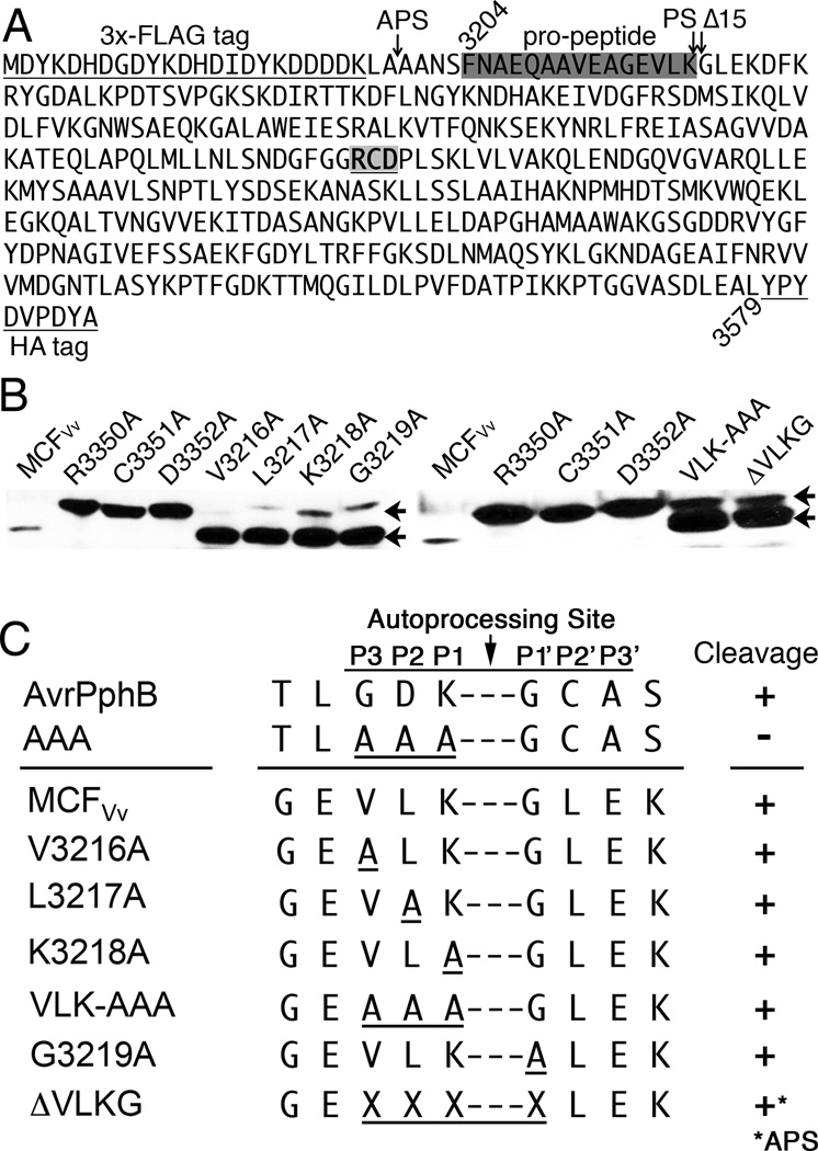
The multifunctional-autoprocessing repeats-in-toxin (MARTX(Vv)) toxin that harbours a varied repertoire of effector domains is the primary virulence factor of Vibrio vulnificus. Although ubiquitously present among Biotype I toxin variants, the 'Makes caterpillars floppy-like' effector domain (MCF(Vv)) is previously unstudied. Using transient expression and protein delivery, MCF(Vv) and MCF(Ah) from the Aeromonas hydrophila MARTX(Ah)) toxin are shown for the first time to induce cell rounding. Alanine mutagenesis across the C-terminal subdomain of MCF(Vv) identified an Arg-Cys-Asp (RCD) tripeptide motif shown to comprise a cysteine protease catalytic site essential for autoprocessing of MCF(Vv). The autoprocessing could be recapitulated in vitro by the addition of host cell lysate to recombinant MCF(Vv), indicating induced autoprocessing by cellular factors. The RCD motif is also essential for cytopathicity, suggesting autoprocessing is essential first to activate the toxin and then to process a cellular target protein resulting in cell rounding. Sequence homology places MCF(Vv) within the C58 cysteine protease family that includes the type III secretion effectors YopT from Yersinia spp. and AvrPphB from Pseudomonas syringae. However, the catalytic site RCD motif is unique compared with other C58 peptidases and is here proposed to represent a new subgroup of autopeptidase found within a number of putative large bacterial toxins.


Figures

Fig. 1
Fig. 1. Mcf homology domains in MARTX and other putative toxins
A. Schematic representation of MARTX toxins from V. vulnificus and A. hydrophila detailing cysteine protease domain (CPD) and distinct arrangement of effector domains in these two toxins (DUF1, domain of unknown function in first position; RID, Rho Inactivation Domain; ABH, alpha-beta hyrdrolase; DUF5; domain of unknown function in the 5th position; ACD, actin crosslinking domain). Enlarged top diagram of the MCFVv effector domain shows the locations of features revealed in this study including the autoprocesing site, the N-terminal helix (NTH), and the C58 peptidase homology (striped boxes). B. A C58 peptidase with conservation in the Photorhabdus Mcf toxins is found also in many other putative bacterial toxins of variable features as indicated, including the common toxin motif known as Mcf1-SHE, regions with homology to type III secretion effector HrmA (also known as HopA1), a membrane localization domain (MLD), glycosyl transferase (GTase) effector domain, and NCBI annotated conserved domain DUF3491. Many of these toxins also have a pore-forming domain similar to clostridial glucosylating toxins TcdA and TcdB (light striped boxes). C. Alignment of all amino acid sequences of proteins in panels A and B revealed only two short regions (I and II) of strong sequence conservation, indicated in panels A and B by white boxes inside the C58 peptidase grey striped boxes. These regions were aligned using the ClustalW algorithm with sequence identity (dark grey) and similarity (light grey) indicated. Asterisks indicates point mutations generated in MCFVv that did not affect cell rounding, black daggars indicate essential residues, and grey daggar is partially required. Sequences used were all from National Center for Biotechnology Information (NCBI) with accession numbers and references as follows: V. vulnificus CMCP6 MARTX toxin (Kim et al., 2011) (NP_762440.1); A. hydrophila 7966 MARTX toxin (Seshadri et al., 2006) (YP_855898), Ph. luminescens Mcf1 (Daborn et al., 2002) (AAM88787.1), Ph. luminescens Mcf2 (Waterfield et al., 2003) (AAR21118), Pseudomanas protegens FitD (Pechy-Tarr et al., 2008) (ABY91230), Providencia rettgeri YopT-type (WP_004260437, direct submission) Escherichia coli Toxin B (Hazen et al., 2013) (WP_024231151), Grimontia hollisae Toxin B (WP_005504930, Direct submission), Pseudomonas chlororaphis FitD (WP_009049649, direct submission), Yersina pestis YopT (Parkhill et al., 2001) (NP_395155), Pseudomonas syringae AvrPphB (Jenner et al., 1991) (also known as AvrPph3, AAA25727).
Fig. 2
Fig. 2. Ectopic expression of MCFVv induces cell rounding
A. Transient transfection of HeLa cells for 18 h with plasmids for expression of EGFP and MCFVv-EGFP (green) as indicated were stained with rhodamine phalloidin for F-actin (red) and DAPI for nucleus (blue). Percent cell rounding from 100 transfected cells from each of three independent transfections is noted in the inset. B. Western blot using anti-EGFP or anti-actin antibody on transfected cell lysates. C. Percent LDH release from transfected HeLa cells. D. Bright field or cells stained as above (inset) intoxicated with either 72 nM of LFN or LFN-MCFVv in combination with 168 nM of PA for 24 h. E. Coomassie-stained 12% SDS-PAGE gel showing the integrity of the LFN-MCFVv used for intoxication at two different concentrations as indicated. F. Percent LDH release from the intoxicated HeLa cells showing that there is no appreciable lysis when cells when MCFVv is delivered directly.
Fig. 3
Fig. 3. MCFVv mediated cell rounding is dependent on an RCD triad
A and B. Percent cell rounding from 100 transfected (A) or intoxicated (B) HeLa cells from each of three independent reactions was plotted. Data shown are mean ± standard deviation with mean labeled above the bar. C. Western blot using anti-EGFP antibody on indicated cell lysates. D. Migration of LFN-tagged mutant proteins used for intoxication on a Coomassie-stained 12% SDS-PAGE gel.
Fig. 4
Fig. 4. Asp-3352 can tolerate a tyrosine residue while other residues in the tripeptide motif are strictly conserved
A. Percent cell rounding from 100 transfected cells from each of three independent transfections was plotted. Data shown are mean ± standard deviation with mean labeled above the bar. B. Western blot using anti-EGFP and anti-actin antibodies on indicated cell lysates to demonstrate protein expression. Arrows on left blot indicate bands in same order as labeled on right blot.
Fig. 5
Fig. 5. MCF from A. hydrophila and from P. luminescens and P. asymbiotica show contrasting phenotypes
A and D. Confocal fluorescence microscopy of HeLa (A) or HEK293T (D) cells post-transfection of the plasmids as indicated. Bar=10 µm. B and E. Western blot detection of EGFP and EGFP-fusion proteins. For Western blot, actin was used as loading control. C and F. Percent cell rounding from 100 transfected cells from each of three independent transfections was plotted. Data shown are mean ± standard deviation.
Fig. 6
Fig. 6. MCFVv autoprocesses both in vivo and in vitro
A. Schematic representation of the FLAG and HA-tagged protein generated by transient expression in HEK293T cells. HEK293T cell lysates from cells transfected to express indicated protein were probed with anti-HA and anti-FLAG antibodies with actin as a loading control. B. Schematic representation of 6×-His tagged recombinant MCFVv with an HA tag (rMCFVv-HA). 20 µg purified protein was incubated in the presence or absence of 30 µg HEK293T cell lysate (CL) and both unprocessed and cleaved protein (cMCFVv-HA) were detected by Western blotting using anti-HA antibody. C. Schematic representation of the 6×-His tagged recombinant MCFVv (rMCFVv) used in panels C-G. Purified protein was incubated in the presence or absence of HEK293T cell lysate (CL) and reactions were resolved on 18% SDS-polyacrylamide gels and proteins visualized with coomassue blue. Cleaved protein is marked with small arrow and letter c. When indicated, rMCFVv was heat-treated prior to addition of CL (hkMCFVv) D–G. In vitro processing as in panel C except using (D) rMCFVv protien with amino acid changes as indicated, (E) rMCFVv protien pre-treated for 30 min with 1 mM NEM when indicated, (F) rMCFVv protein mixed with heat-treated (hk) CL when indicated, and (G) rMCFVv protein mixed with CL pre-treated with trypsin (ty) or proteinase K (pk) followed by boiling when indicated.
Fig. 7
Fig. 7. MCFVv processing of N-terminus is not sequence-specific
A. Protein sequence of MCFVv (aa 3204–3579) is shown with 3×FLAG and HA tags underlined. The pro-domain (boxed dark gray), the processing site (PS) between Lys-3218 and Gly-3219, the alternate processing site (APS) for ΔVLKG mutation, and position of Δ15 deletion for Fig. 8 are indicated with arrows. The RCD catalytic motif is bold and underlined. B. HEK293T cell lysates from cells transfected as in Fig. 6A to express indicated protein were probed with anti-HA antibody to detect both full-length (upper arrow) and cleaved MCFVv protein (lower arrow). C. Details of sequence of the various mutants in panel B with (+) indicating processing and (−) indicating failure to process. Data for AvrPphB is taken from Dowen et. al, 2009.
Fig. 8
Fig. 8. Ectopic expression of cleaved variant of MCFVv induces cell rounding
A. Schematic representation of the deletion variants of MCFVv and its catalytic mutants generated in fusion with EGFP; the small grey inverted triangle represents the processing site. Dashed lines indicate sequences deleted. Percent cell rounding for 100 transfected cells as done in panel B is represented as mean ± standard deviation. B. Confocal fluorescence microscopy images of HeLa cells transfected as indicated with inset for last panel magnified to highlight the punctae like structures indicative of subcellular localization. Figure is a composite of representative images from multiple experiments conducted over time as new deletions were tested, but all experiments were conducted with appropriate positive and negative controls. Bars = 10 µm. C. Western blot detection of EGFP and EGFP-fusion proteins in transiently transfected HeLa cells as indicated. Actin was used as loading control in Western blot.

References

    1. Ahrens S, Geissler B, Satchell KJ. Identification of a His-Asp-Cys catalytic triad essential for function of the Rho inactivation domain (RID) of Vibrio cholerae MARTX toxin. J Biol Chem. 2013;288:1397–1408. - PMC - PubMed
    1. Antic I, Biancucci M, Satchell KJ. Cytotoxicity of the Vibrio vulnificus MARTX toxin Effector DUF5 is linked to the C2A Subdomain. Proteins. 2014;82:2643–2656. - PMC - PubMed
    1. Chung KJ, Cho EJ, Kim MK, Kim YR, Kim SH, Yang HY, et al. RtxA1-induced expression of the small GTPase Rac2 plays a key role in the pathogenicity of Vibrio vulnificus. J Infect Dis. 2010;201:97–105. - PubMed
    1. Daborn PJ, Waterfield N, Silva CP, Au CP, Sharma S, Ffrench-Constant RH. A single Photorhabdus gene, makes caterpillars floppy (mcf), allows Escherichia coli to persist within and kill insects. Proc Nat Acad Sci USA. 2002;99:10742–10747. - PMC - PubMed
    1. Dowen RH, Engel JL, Shao F, Ecker JR, Dixon JE. A family of bacterial cysteine protease type III effectors utilizes acylation-dependent and -independent strategies to localize to plasma membranes. J Biol Chem. 2009;284:15867–15879. - PMC - PubMed

Publication types

MeSH terms