. 2015 Jul 1;403(1):101-13.
doi: 10.1016/j.ydbio.2015.04.014. Epub 2015 Apr 24.

A conserved MST1/2-YAP axis mediates Hippo signaling during lung growth

Affiliations

A conserved MST1/2-YAP axis mediates Hippo signaling during lung growth

Chuwen Lin et al. Dev Biol. .

Abstract

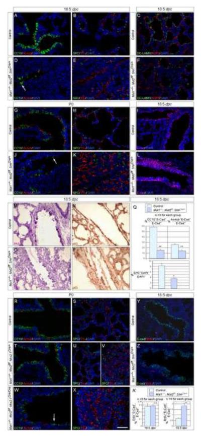
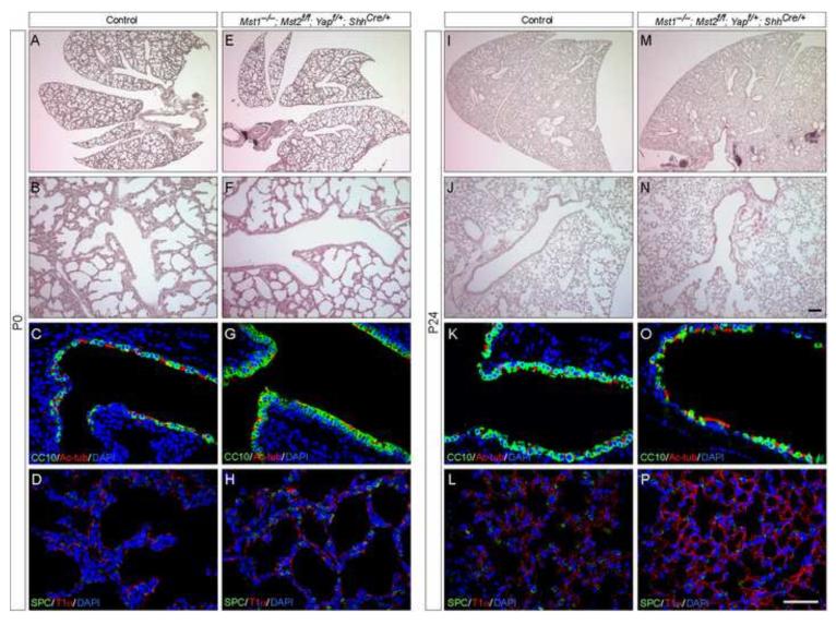
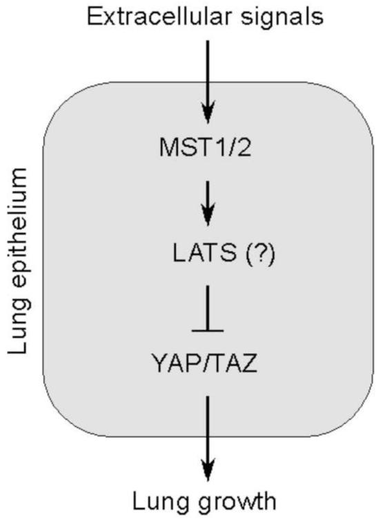
Hippo signaling is a critical player in controlling the growth of several tissues and organs in diverse species. The current model of Hippo signaling postulates a cascade of kinase activity initiated by the MST1/2 kinases in response to external stimuli. This leads to inactivation of the transcriptional coactivators, YAP/TAZ, due to their cytoplasmic retention and degradation that is correlated with YAP/TAZ phosphorylation. In most tissues examined, YAP plays a more dominant role than TAZ. Whether a conserved Hippo pathway is utilized during lung growth and development is unclear. In particular, the regulatory relationship between MST1/2 and YAP/TAZ in the lung remains controversial. By employing the Shh-Cre mouse line to efficiently inactivate genes in the lung epithelium, we show that loss of MST1/2 kinases in the epithelium can lead to neonatal lethality caused by lung defects. This is manifested by perturbation of lung epithelial cell proliferation and differentiation. These phenotypes are more severe than those produced by Nkx2.1-Cre, highlighting the effects of differential Cre activity on phenotypic outcomes. Importantly, expression of YAP targets is upregulated and the ratio of phospho-YAP to total YAP protein levels is reduced in Mst1/2-deficient lungs, all of which are consistent with a negative role of MST1/2 in controlling YAP function. This model gains further support from both in vivo and in vitro studies. Genetic removal of one allele of Yap or one copy of both Yap and Taz rescues neonatal lethality and lung phenotypes due to loss of Mst1/2. Moreover, knockdown of Yap in lung epithelial cell lines restores diminished alveolar marker expression caused by Mst1/2 inactivation. These results demonstrate that MST1/2 inhibit YAP/TAZ activity and establish a conserved MST1/2-YAP axis in coordinating lung growth during development.

Keywords: Development; Hippo signaling; Lung; Mst1/2; Yap.


Figures

Figure 1
Figure 1
Histological analysis of Mst1/2-deficient lungs generated by different mouse Cre lines (A-L) Hematoxylin-and-eosin (H&E)-stained lung sections collected from mouse strains that carry various combinations of Mst1 (null), Mst2f (conditional) and Cre alleles. (A-D) At postnatal (p) day 0 (P0), lungs from Mst1−/−; Mst2f/f; ShhCre/+ mice did not expand and appeared dense and compact compared to lungs from their wild-type littermates. By contrast, lungs from either Mst1−/−; Mst2f/f; Nkx2.1Cre/+ or Mst1−/−; Mst2f/f; Nkx2.1Cre/Cre mice were inflated although the upper regions of the lobes (arrows) were more compressed and contained less air. (E-H) Extra lung cells were detected in Mst1−/−; Mst2f/f; Nkx2.1Cre/+, Mst1−/−; Mst2f/f; Nkx2.1Cre/Cre and Mst1−/−; Mst2f/f; ShhCre/+ lungs and the severity of the lung phenotype increased in that order. While most Mst1−/−; Mst2f/f; ShhCre/+ mice died soon after birth, both Mst1−/−; Mst2f/f; Nkx2.1Cre/+ and Mst1−/−; Mst2f/f; Nkx2.1Cre/Cre mice survived postnatally. (I-L) Lungs from Mst1−/−; Mst2f/f; Nkx2.1Cre/Cre mice at various time points after birth showed morphological abnormalities, including extra lung cells and inflammatory responses. In some Mst1−/−; Mst2f/f; Nkx2.1Cre/Cre animals, collapsed lung lobes could be observed, likely due to ongoing destruction of the lungs. Rare Mst1−/−; Mst2f/f; ShhCre/+ mice escaped neonatal lethality and some of them displayed varying degrees of lung defects. Scale bar = 500 μm for A-D; 100 μm for E-L.
Figure 2
Figure 2
Phenotypic analysis of Mst1/2 mutant lungs produced by different mouse Cre lines (A-P) Immunostaining of lung sections collected from mouse strains that carry various combinations of Mst1 (null), Mst2f (conditional) and Cre alleles at 18.5 dpc or postnatal day 0 (P0). (A-L) Major lung cell types including Clara cells (CC10+), ciliated cells (Ac-tubulin+), alveolar type II cells (SPC+, DC-LAMP+) and alveolar type I cells (T1α+, AQP5+) were specified in Mst1−/−; Mst2f/f; ShhCre/+ lungs at 18.5 dpc or P0. However, fewer CC10+ and Ac-tubulin+ cells in the conducting airways (arrows) and fewer type II cells in the alveoli were detected in Mst1−/−; Mst2f/f; ShhCre compared to control lungs. Quantification of cell numbers is shown in (Q) in which the percentage of epithelial cells (E-cadherin+ [E-cad+]) that express CC10 or Ac-tubulin was calculated; the percentage of SPC+ cells on lung sections was enumerated by counting the total number of cells using DAPI. In many bronchioles, the simple columnar epithelium in wild-type lungs (I) is replaced by pseudostratified epithelium (L) in Mst1/2-deficient lungs. Note that A–B and D–E are adjacent sections. (M-P) Immunohistochemistry of lung sections for detecting PAS+ mucin-secreting goblet cells and p63+ basal cells. Both cell types can be readily detected in Mst1−/−; Mst2f/f; ShhCre/+ lungs at 18.5 dpc. (R-X) Major lung cell types were also present in Mst1−/−; Mst2f/f; Nkx2.1Cre/+ and Mst1−/−; Mst2f/f; Nkx2.1Cre/Cre lungs at P0. Similarly, the number of CC10+ and Ac-tubulin+ cells in the conducting airways (arrow) was reduced in Mst1−/−; Mst2f/f; Nkx2.1Cre/Cre lungs compared to control lungs. Image in (U) was taken from the lower lobe and (V) from the upper lobe of Mst1−/−; Mst2f/f; Nkx2.1Cre/+ lungs. (Y, Z) Immunostaining of lung sections collected from Mst1−/−; Mst2f/f; ShhCre/+ mice injected with BrdU or EdU. Lung epithelial cells were distinguished by E-cad staining. Cell proliferation rate (judged by BrdU or EdU staining) in the lung epithelium was quantified in (A’). While the number of BrdU+ or EdU+ cells was increased in the epithelium of Mst1−/−; Mst2f/f; ShhCre/+ lungs at 18.5 dpc, no apparent difference in the number of epithelial EdU+ cells was detected between control and Mst1−/−; Mst2f/f; ShhCre/+ lungs at 16.5 dpc. This is correlated with the lack of overt lung phenotypes in Mst1−/−; Mst2f/f; ShhCre/+ mice at 16.5 dpc. It is possible that increased cell proliferation and phenotypic consequences only become apparent after 16.5 dpc. Scale bar = 50 μm. * P < 0.05; ** P < 0.01; NS, not significant (unpaired Student’s t-test).
Figure 3
Figure 3
Molecular analysis of lung cell type markers, cell cycle regulators, YAP targets and YAP/LATS proteins in the absence of Mst1/2 in the lung (A-F) qPCR analysis of RNA extracted from Mst1−/−; Mst2f/f; ShhCre/+ (16.5 or 18.5 dpc) and Mst1−/−; Mst2f/f; Nkx2.1Cre/Cre (P2) lungs. (A, B) Expression of several cell cycle regulators (Mycn and Ccnd2) and MYCN targets (Ndrg1 and Ndrg2) was increased in both Mst1−/−; Mst2f/f; ShhCre/+ and Mst1−/−; Mst2f/f; Nkx2.1Cre/Cre lungs. (C, D) Alterations in expression of various lung cell markers followed a similar trend in Mst1−/−; Mst2f/f; ShhCre/+ and Mst1−/−; Mst2f/f; Nkx2.1Cre/Cre lungs. While expression of alveolar type II cell markers (SPC, SPB, Abca3), Clara cell marker (CC10), ciliated cell marker (Foxj1) was reduced, expression of alveolar type I markers (Pdpn, Aqp5) was elevated. Basal cell marker (p63) did not seem to be altered. (E, F) Expression of YAP targets (Ctgf, Ajuba, Cyr61, Lamc2 and Prkci) was significantly induced in both Mst1−/−; Mst2f/f; ShhCre/+ and Mst1−/−; Mst2f/f; Nkx2.1Cre/Cre lungs albeit Ctgf induction was more modest in Mst1−/−; Mst2f/f; Nkx2.1Cre/Cre lungs. (G, H) Western blot analysis of YAP and phospho-YAP protein levels using lysates derived from Mst1−/−; Mst2f/f; ShhCre/+ and Mst1−/−; Mst2f/f; Nkx2.1Cre/Cre lungs at 16.5 dpc and P2 respectively. The fraction of phospho-YAP protein (relative to the total YAP) was decreased in Mst1−/−; Mst2f/f; ShhCre/+ or Mst1−/−; Mst2f/f; Nkx2.1Cre/Cre lungs. By contrast, protein levels of LATS and phospho-LATS did not seem to be altered in Mst1−/−; Mst2f/f; ShhCre/+ or Mst1−/−; Mst2f/f; Nkx2.1Cre/Cre lungs. Tubulin was used as the loading control. All values are means ± SEM. * P < 0.05; ** P < 0.01; NS, not significant (unpaired Student’s t-test).
Figure 4
Figure 4
The effects of Yap/Taz removal on the development of Mst1/2 mutant lungs and the survival of the animals (A, B, E, F, I, J, M, N) Hematoxylin-and-eosin (H&E)-stained lung sections collected from control and Mst1−/−; Mst2f/f; Yapf/+; Tazf/+; ShhCre/+ mice postnatal (p) day 4 (P4) and 24 (P24). Unlike Mst1−/−; Mst2f/f; ShhCre/+ mice, most of which died soon after birth, Mst1−/−; Mst2f/f; Yapf/+; Tazf/+; ShhCre/+ mice survived to adulthood without apparent gross abnormalities. In addition, lungs from Mst1−/−; Mst2f/f; Yapf/+; Tazf/+; ShhCre/+ mice were fully expanded and devoid of any dense or compact architecture observed in Mst1−/−; Mst2f/f; ShhCre/+ lungs. Lung histology was indistinguishable between control and Mst1−/−; Mst2f/f; Yapf/+; Tazf/+; ShhCre/+ mice (n > 20 analyzed). Note that some blood was present on lung sections (E) of Mst1−/−; Mst2f/f; Yapf/+; Tazf/+; ShhCre/+ mice due to dissection. (C, D, G, H, K, L, O, P) Immunostaining of lung sections collected from control and Mst1−/−; Mst2f/f; Yapf/+; Tazf/+; ShhCre/+ mice at P4 and P24. Lung cell types appeared to be properly specified in Mst1−/−; Mst2f/f; Yapf/+; Tazf/+; ShhCre/+ mice. Scale bar = 100 μm for A, B, E, F, I, J, M, N; 50 μm for C, D, G, H, K, L, O, P.
Figure 5
Figure 5
The effects of Yap removal on the development of Mst1/2 mutant lungs and the survival of the animals (A, B, E, F, I, J, M, N) Hematoxylin-and-eosin (H&E)-stained lung sections collected from control and Mst1−/−; Mst2f/f; Yapf/+; ShhCre/+ mice at postnatal (p) day 4 (P4) and 24 (P24). Unlike Mst1−/−; Mst2f/f; ShhCre/+ mice, most of which died soon after birth, Mst1−/−; Mst2f/f; Yapf/+; ShhCre/+ mice survived to adulthood without apparent gross abnormalities. In addition, lungs from Mst1−/−; Mst2f/f; Yapf/+; ShhCre/+ mice were fully expanded and devoid of any dense or compact architecture seen in Mst1−/−; Mst2f/f; ShhCre/+ lungs. Lung histology was indistinguishable between control and Mst1−/−; Mst2f/f; Yapf/+; ShhCre/+ mice (n > 8 analyzed). (C, D, G, H, K, L, O, P) Immunostaining of lung sections collected from control and Mst1−/−; Mst2f/f; Yapf/+; ShhCre/+ mice at P4 and P24. Lung cell types appeared to be properly specified in Mst1−/−; Mst2f/f; Yapf/+; ShhCre/+ mice. Scale bar = 100 μm for A, B, E, F, I, J, M, N; 50 μm for C, D, G, H, K, L, O, P.
Figure 6
Figure 6
The effects of Mst1/2 and Yap knockdown on alveolar marker expression in lung cell lines (A, B) qPCR analysis of mRNA transcripts and Western blot analysis of proteins extracted from human A549 or mouse MLE 12 lung cell lines. In these studies, Mst1/2 were knocked down individually or in combination with Yap inactivation by shRNA as indicated. shRNA against GFP served as a control. Knockdown of Mst1/2 resulted in reduced expression of SPB/SPC (alveolar type II cell markers), consistent with findings in Mst1/2-deficient lungs. Inactivation of Mst1/2 also led to increased CTGF (YAP target) expression and reduced levels of phospho-YAP, both of which are associated with increased YAP activity. Such alteration in gene expression was restored when Yap was simultaneously knocked down by shRNA in conjunction with Mst1/2. These results further support conclusions from analysis of Mst1/2 mutant lungs. Tubulin was used as the loading control.
Figure 7
Figure 7
A model of a conserved MST1/2-YAP axis in regulating lung development A model of MST and YAP interactions in the lung. Control of YAP/TAZ activity by MST1/2 regulates many aspects of cellular behavior during lung development. YAP plays a more dominant role than TAZ in Hippo signaling. In this model, loss of MST1/2 shifts YAP toward the non-phosphorylated state, which is no longer sequestered in the cytoplasm and also free from degradation. Instead, non-phosphorylated YAP is enriched in the nucleus to activate YAP targets, leading to cell proliferation and fate determination and other changes in cellular properties. This canonical relationship between MST and YAP is observed in many tissues. The mediator of Hippo signaling between MST1/2 and YAP in the lung could be LATS (another conserved feature of the Hippo pathway) or some other unknown kinases. Factors that activate MST1/2 have been inferred from cell-based studies but signals that control MST activity in the lung remain unclear.

References

    1. Barry ER, Camargo FD. The Hippo superhighway: signaling crossroads converging on the Hippo/Yap pathway in stem cells and development. Current opinion in cell biology. 2013;25:247–253. - PubMed
    1. Borok Z, Whitsett JA, Bitterman PB, Thannickal VJ, Kotton DN, Reynolds SD, Krasnow MA, Bianchi DW, Morrisey EE, Hogan BL, Kurie JM, Walker DC, Radisky DC, Nishimura SL, Violette SM, Noble PW, Shapiro SD, Blaisdell CJ, Chapman HA, Kiley J, Gail D, Hoshizaki D. Cell plasticity in lung injury and repair: report from an NHLBI workshop, April 19-20, 2010. Proc Am Thorac Soc. 2011;8:215–222. - PMC - PubMed
    1. Chan EH, Nousiainen M, Chalamalasetty RB, Schafer A, Nigg EA, Sillje HH. The Ste20-like kinase Mst2 activates the human large tumor suppressor kinase Lats1. Oncogene. 2005;24:2076–2086. - PubMed
    1. Chan SW, Lim CJ, Chong YF, Pobbati AV, Huang C, Hong W. Hippo pathway-independent restriction of TAZ and YAP by angiomotin. The Journal of biological chemistry. 2011;286:7018–7026. - PMC - PubMed
    1. Chen MH, Wilson CW, Li YJ, Law KK, Lu CS, Gacayan R, Zhang X, Hui CC, Chuang PT. Cilium-independent regulation of Gli protein function by Sufu in Hedgehog signaling is evolutionarily conserved. Genes & development. 2009;23:1910–1928. - PMC - PubMed

Publication types

MeSH terms