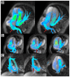
Improved quantification and mapping of anomalous pulmonary venous flow with four-dimensional phase-contrast MRI and interactive streamline rendering
- PMID: 25914149
- PMCID: PMC4843111
- DOI: 10.1002/jmri.24928
Improved quantification and mapping of anomalous pulmonary venous flow with four-dimensional phase-contrast MRI and interactive streamline rendering
Abstract
Background: Cardiac MRI is routinely performed for quantification of shunt flow in patients with anomalous pulmonary veins, but can be technically-challenging to perform. Four-dimensional phase-contrast (4D-PC) MRI has potential to simplify this exam. We sought to determine whether 4D-PC may be a viable clinical alternative to conventional 2D phase-contrast MR imaging.
Methods: With institutional review board approval and HIPAA-compliance, we retrospectively identified all patients with anomalous pulmonary veins who underwent cardiac MRI at either 1.5 Tesla (T) or 3T with parallel-imaging compressed-sensing (PI-CS) 4D-PC between April, 2011 and October, 2013. A total of 15 exams were included (10 male, 5 female). Algorithms for interactive streamline visualization were developed and integrated into in-house software. Blood flow was measured at the valves, pulmonary arteries and veins, cavae, and any associated shunts. Pulmonary veins were mapped to their receiving atrial chamber with streamlines. The intraobserver, interobserver, internal consistency of flow measurements, and consistency with conventional MRI were then evaluated with Pearson correlation and Bland-Altman analysis.
Results: Triplicate measurements of blood flow from 4D-PC were highly consistent, particularly at the aortic and pulmonary valves (cv 2-3%). Flow measurements were reproducible by a second observer (ρ = 0.986-0.999). Direct measurements of shunt volume from anomalous veins and intracardiac shunts matched indirect estimates from the outflow valves (ρ = 0.966). Measurements of shunt fraction using 4D-PC using any approach were more consistent with ventricular volumetric displacements than conventional 2D-PC (ρ = 0.972-0.991 versus 0.929).
Conclusion: Shunt flow may be reliably quantified with 4D-PC MRI, either indirectly or with detailed delineation of flow from multiple shunts. The 4D-PC may be a more accurate alternative to conventional MRI.
Keywords: flow; pulmonary; shunt; structural; veins.
© 2015 Wiley Periodicals, Inc.
Figures


References
-
- Festa P, Ait-Ali L, Cerillo AG, et al. [Accessed February 27, 2014];Magnetic resonance imaging is the diagnostic tool of choice in the preoperative evaluation of patients with partial anomalous pulmonary venous return. Int. J. Cardiovasc. Imaging. 2006 22(5):685–93. Available at: http://www.ncbi.nlm.nih.gov/pubmed/16547601. - PubMed
-
- Riesenkampff EM-C, Schmitt B, Schnackenburg B, et al. [Accessed February 27, 2014];Partial anomalous pulmonary venous drainage in young pediatric patients: the role of magnetic resonance imaging. Pediatr. Cardiol. 2009 30(4):458–64. Available at: http://www.ncbi.nlm.nih.gov/pubmed/19184180. - PubMed
-
- Robinson BL, Kwong RY, Varma PK, et al. [Accessed May 5, 2014];Magnetic resonance imaging of complex partial anomalous pulmonary venous return in adults. Circulation. 2014 129(1):e1–2. Available at: http://www.ncbi.nlm.nih.gov/pubmed/24396018. - PubMed
-
- Vyas HV, Greenberg SB, Krishnamurthy R. MR imaging and CT evaluation of congenital pulmonary vein abnormalities in neonates and infants. Radiographics. 2012;32(1):87–98. Available at: http://www.ncbi.nlm.nih.gov/pubmed/22236895. - PubMed
-
- Fisher MR, Hricak H, Higgins CB. Magnetic resonance imaging of developmental venous anomalies. AJR. Am. J. Roentgenol. 1985;145(4):705–9. Available at: http://www.ncbi.nlm.nih.gov/pubmed/3875985. - PubMed
Publication types
MeSH terms
Grants and funding
LinkOut - more resources
Full Text Sources
Other Literature Sources
Research Materials
Miscellaneous

