

All-trans retinoic acid induces oxidative phosphorylation and mitochondria biogenesis in adipocytes
- PMID: 25914170
- PMCID: PMC4442868
- DOI: 10.1194/jlr.M053652
All-trans retinoic acid induces oxidative phosphorylation and mitochondria biogenesis in adipocytes
Abstract
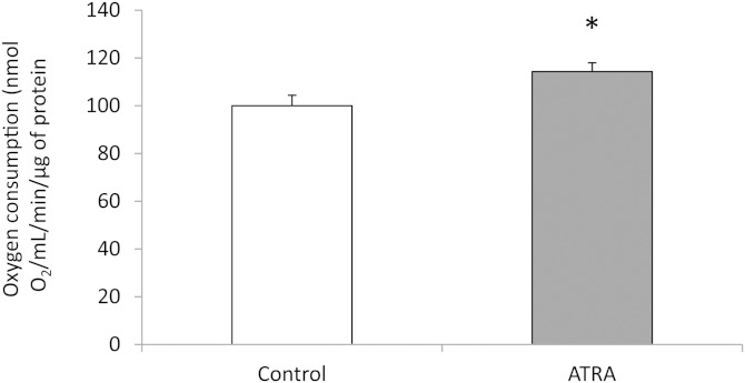
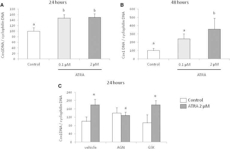
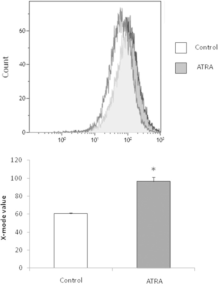
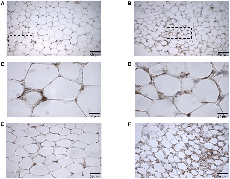
A positive effect of all-trans retinoic acid (ATRA) on white adipose tissue (WAT) oxidative and thermogenic capacity has been described and linked to an in vivo fat-lowering effect of ATRA in mice. However, little is known about the effects of ATRA on mitochondria in white fat. Our objective has been to characterize the effect of ATRA on mitochondria biogenesis and oxidative phosphorylation (OXPHOS) capacity in mature white adipocytes. Transcriptome analysis, oxygraphy, analysis of mitochondrial DNA (mtDNA), and flow cytometry-based analysis of mitochondria density were performed in mature 3T3-L1 adipocytes after 24 h incubation with ATRA (2 µM) or vehicle. Selected genes linked to mitochondria biogenesis and function and mitochondria immunostaining were analyzed in WAT tissues of ATRA-treated as compared with vehicle-treated mice. ATRA upregulated the expression of a large set of genes linked to mtDNA replication and transcription, mitochondrial biogenesis, and OXPHOS in adipocytes, as indicated by transcriptome analysis. Oxygen consumption rate, mtDNA content, and staining of mitochondria were increased in the ATRA-treated adipocytes. Similar results were obtained in WAT depots of ATRA-treated mice. We conclude that ATRA impacts mitochondria in adipocytes, leading to increased OXPHOS capacity and mitochondrial content in these cells.
Keywords: adipose tissue; browning; vitamin A.
Copyright © 2015 by the American Society for Biochemistry and Molecular Biology, Inc.
Figures
Similar articles
-
Stimulation of mitochondrial oxidative capacity in white fat independent of UCP1: a key to lean phenotype.Biochim Biophys Acta. 2013 May;1831(5):986-1003. doi: 10.1016/j.bbalip.2013.02.003. Epub 2013 Feb 20. Biochim Biophys Acta. 2013. PMID: 23454373
-
Peroxiredoxin 3 is a key molecule regulating adipocyte oxidative stress, mitochondrial biogenesis, and adipokine expression.Antioxid Redox Signal. 2012 Feb 1;16(3):229-43. doi: 10.1089/ars.2010.3766. Epub 2011 Oct 17. Antioxid Redox Signal. 2012. PMID: 21902452 Free PMC article.
-
Remodeling of white adipose tissue after retinoic acid administration in mice.Endocrinology. 2006 Nov;147(11):5325-32. doi: 10.1210/en.2006-0760. Epub 2006 Jul 13. Endocrinology. 2006. PMID: 16840543
-
Biochemical adaptations in white adipose tissue following aerobic exercise: from mitochondrial biogenesis to browning.Biochem J. 2020 Mar 27;477(6):1061-1081. doi: 10.1042/BCJ20190466. Biochem J. 2020. PMID: 32187350 Review.
-
Stress turns on the heat: Regulation of mitochondrial biogenesis and UCP1 by ROS in adipocytes.Adipocyte. 2017 Jan 2;6(1):56-61. doi: 10.1080/21623945.2016.1273298. Epub 2016 Dec 16. Adipocyte. 2017. PMID: 28452586 Free PMC article. Review.
Cited by
-
Inhibition of retinoic acid receptor β signaling confers glycolytic dependence and sensitization to dichloroacetate in melanoma cells.Oncotarget. 2017 Aug 24;8(48):84210-84223. doi: 10.18632/oncotarget.20476. eCollection 2017 Oct 13. Oncotarget. 2017. PMID: 29137417 Free PMC article.
-
The brown fat-specific overexpression of RBP4 improves thermoregulation and systemic metabolism by activating the canonical adrenergic signaling pathway.Exp Mol Med. 2025 Mar;57(3):554-566. doi: 10.1038/s12276-025-01411-6. Epub 2025 Mar 3. Exp Mol Med. 2025. PMID: 40025173 Free PMC article.
-
Reduced adiponectin expression after high-fat diet is associated with selective up-regulation of ALDH1A1 and further retinoic acid receptor signaling in adipose tissue.FASEB J. 2017 Jan;31(1):203-211. doi: 10.1096/fj.201600263RR. Epub 2016 Oct 11. FASEB J. 2017. PMID: 27729412 Free PMC article.
-
Maternal genetics and diet modulate vitamin A homeostasis of the offspring and affect the susceptibility to obesity in adulthood in mice.Am J Physiol Endocrinol Metab. 2024 Sep 1;327(3):E258-E270. doi: 10.1152/ajpendo.00116.2024. Epub 2024 Jul 17. Am J Physiol Endocrinol Metab. 2024. PMID: 39017681 Free PMC article.
-
DNA Methylation Changes are Associated with the Programming of White Adipose Tissue Browning Features by Resveratrol and Nicotinamide Riboside Neonatal Supplementations in Mice.Nutrients. 2020 Feb 12;12(2):461. doi: 10.3390/nu12020461. Nutrients. 2020. PMID: 32059412 Free PMC article.
References
-
- Flachs P., Rossmeisl M., Kuda O., Kopecky J. 2013. Stimulation of mitochondrial oxidative capacity in white fat independent of UCP1: a key to lean phenotype. Biochim. Biophys. Acta. 1831: 986–1003. - PubMed
-
- Blomhoff R., Blomhoff H. K. 2006. Overview of retinoid metabolism and function. J. Neurobiol. 66: 606–630. - PubMed
Publication types
MeSH terms
Substances
LinkOut - more resources
Full Text Sources
