

Intestinal genetic inactivation of caspase-8 diminishes migration of enterocytes
- PMID: 25914458
- PMCID: PMC4402296
- DOI: 10.3748/wjg.v21.i15.4499
Intestinal genetic inactivation of caspase-8 diminishes migration of enterocytes
Abstract
Aim: To verify the hypothesis that caspase-8 (Casp8), which regulates cellular apoptosis and necroptosis, is critically involved in enterocyte migration.
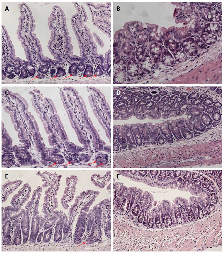
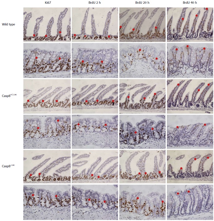
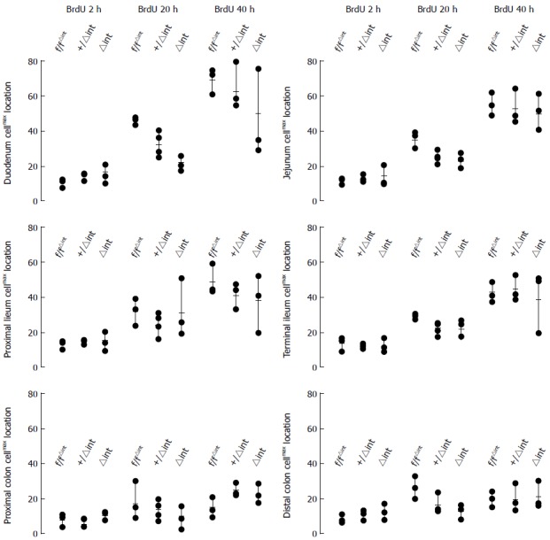
Methods: Casp8-silenced Caco2 cells were used in migration assays. In addition, enterocyte-specific Casp8 heterozygous (Casp8(+/∆int)) or homozygous knockout mice (Casp8(∆int)) were generated by crossing genetically modified mice carrying loxP recombination sites in intron 2 and 4 of the murine Casp8 gene with transgenic animals expressing a cre-transgene under control of the villin promoter in a pure C57/BL6 genetic background. The nucleoside analog BrdU was injected i.p. in male Casp8(+/∆int) and Casp8(∆int) animals 4 h, 20 h, or 40 h before performing morphometric studies. Locations of anti-BrdU-immunostained cells (cell(max)) in at least 50 hemi-crypts of 6 histoanatomically distinct intestinal mucosal regions were numbered and extracted for statistical procedures. For the mice cohort (n = 28), the walking distance of enterocytes was evaluated from cell(max) within crypt (n = 57), plateau (n = 19), and villus (n = 172) positions, resulting in a total of 6838 observations. Data analysis was performed by fitting a three-level mixed effects model to the data.
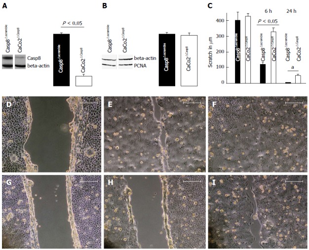
Results: In cell culture experiments with Caco2 cells, Casp8 knockdown efficiency mediated by RNA interference on Casp8 transcripts was 80% controlled as determined by Western blotting. In the scratch assay, migration of Casp8-deleted Caco2 cells was significantly diminished when compared with controls (Casp8(∆scramble) and Caco2). In BrdU-labeled Casp8(∆int) mice, cell(max) locations were found along the hemi-crypts in a lower position than it was for Casp8(+/∆int) or control (cre-negative) animals. Statistical data analysis with a three-level mixed effects model revealed that in the six different intestinal locations (distinct segments of the small and large intestine), cell movement between the three mice groups differed widely. Especially in duodenal hemi-crypts, enterocyte movement was different between the groups. At 20 h, duodenal cell(max) location was significantly lower in Casp8(∆int) (25.67 ± 2.49) than in Casp8(+/∆int) (35.67 ± 4.78; P < 0.05) or control littermates (44.33 ± 0.94; P < 0.01).
Conclusion: Casp8-dependent migration of enterocytes is likely involved in intestinal physiology and inflammation-related pathophysiology.
Keywords: Barrier function; Caspase 8; Cell migration; Inflammatory bowel disease; Intestinal morphogenesis.
Figures
Similar articles
-
Regulators of Intestinal Epithelial Migration in Sepsis.Shock. 2019 Jan;51(1):88-96. doi: 10.1097/SHK.0000000000001117. Shock. 2019. PMID: 29424793 Free PMC article.
-
Beta-7 integrin controls enterocyte migration in the small intestine.World J Gastroenterol. 2015 Feb 14;21(6):1759-64. doi: 10.3748/wjg.v21.i6.1759. World J Gastroenterol. 2015. PMID: 25684940 Free PMC article.
-
Deletion of the Casp8 gene in mice results in ileocolitis, gut barrier dysfunction, and malassimilation, which can be partially attenuated by inulin or sodium butyrate.Am J Physiol Gastrointest Liver Physiol. 2019 Oct 1;317(4):G493-G507. doi: 10.1152/ajpgi.00297.2018. Epub 2019 Aug 14. Am J Physiol Gastrointest Liver Physiol. 2019. PMID: 31411503
-
Regulation of enterocyte apoptosis by acyl-CoA synthetase 5 splicing.Gastroenterology. 2007 Aug;133(2):587-98. doi: 10.1053/j.gastro.2007.06.005. Epub 2007 Jun 8. Gastroenterology. 2007. PMID: 17681178
-
SGLT-1-mediated glucose uptake protects intestinal epithelial cells against LPS-induced apoptosis and barrier defects: a novel cellular rescue mechanism?FASEB J. 2005 Nov;19(13):1822-35. doi: 10.1096/fj.05-4226com. FASEB J. 2005. PMID: 16260652
Cited by
-
Regulators of Intestinal Epithelial Migration in Sepsis.Shock. 2019 Jan;51(1):88-96. doi: 10.1097/SHK.0000000000001117. Shock. 2019. PMID: 29424793 Free PMC article.
-
Notch inhibition counteracts Paneth cell death in absence of caspase-8.Virchows Arch. 2018 Jul;473(1):71-83. doi: 10.1007/s00428-018-2368-3. Epub 2018 May 16. Virchows Arch. 2018. PMID: 29770852
-
Targeting Wnt Signaling via Notch in Intestinal Carcinogenesis.Cancers (Basel). 2019 Apr 18;11(4):555. doi: 10.3390/cancers11040555. Cancers (Basel). 2019. PMID: 31003440 Free PMC article. Review.
-
The roles and functions of Paneth cells in Crohn's disease: A critical review.Cell Prolif. 2021 Jan;54(1):e12958. doi: 10.1111/cpr.12958. Epub 2020 Nov 11. Cell Prolif. 2021. PMID: 33174662 Free PMC article. Review.
-
Apoptotic cell death in disease-Current understanding of the NCCD 2023.Cell Death Differ. 2023 May;30(5):1097-1154. doi: 10.1038/s41418-023-01153-w. Epub 2023 Apr 26. Cell Death Differ. 2023. PMID: 37100955 Free PMC article. Review.
References
-
- van Es JH, van Gijn ME, Riccio O, van den Born M, Vooijs M, Begthel H, Cozijnsen M, Robine S, Winton DJ, Radtke F, et al. Notch/gamma-secretase inhibition turns proliferative cells in intestinal crypts and adenomas into goblet cells. Nature. 2005;435:959–963. - PubMed
-
- Stange DE, Clevers H. Concise review: the yin and yang of intestinal (cancer) stem cells and their progenitors. Stem Cells. 2013;31:2287–2295. - PubMed
-
- Potten CS, Loeffler M. Stem cells: attributes, cycles, spirals, pitfalls and uncertainties. Lessons for and from the crypt. Development. 1990;110:1001–1020. - PubMed
-
- Gribar SC, Anand RJ, Sodhi CP, Hackam DJ. The role of epithelial Toll-like receptor signaling in the pathogenesis of intestinal inflammation. J Leukoc Biol. 2008;83:493–498. - PubMed
Publication types
MeSH terms
Substances
LinkOut - more resources
Full Text Sources
Other Literature Sources
Miscellaneous
