

Endoscopic ultrasound guided radiofrequency ablation, for pancreatic cystic neoplasms and neuroendocrine tumors
- PMID: 25914783
- PMCID: PMC4390891
- DOI: 10.4240/wjgs.v7.i4.52
Endoscopic ultrasound guided radiofrequency ablation, for pancreatic cystic neoplasms and neuroendocrine tumors
Abstract
Aim: To outline the feasibility, safety, adverse events and early results of endoscopic ultrasound (EUS)-radiofrequency ablation (RFA) in pancreatic neoplasms using a novel probe.
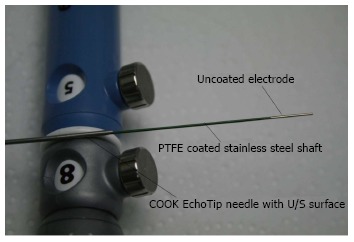
Methods: This is a multi-center, pilot safety feasibility study. The intervention described was radiofrequency ablation (RF) which was applied with an innovative monopolar RF probe (1.2 mm Habib EUS-RFA catheter) placed through a 19 or 22 gauge fine needle aspiration (FNA) needle once FNA was performed in patients with a tumor in the head of the pancreas. The Habib™ EUS-RFA is a 1 Fr wire (0.33 mm, 0.013") with a working length of 190 cm, which can be inserted through the biopsy channel of an echoendoscope. RF power is applied to the electrode at the end of the wire to coagulate tissue in the liver and pancreas.
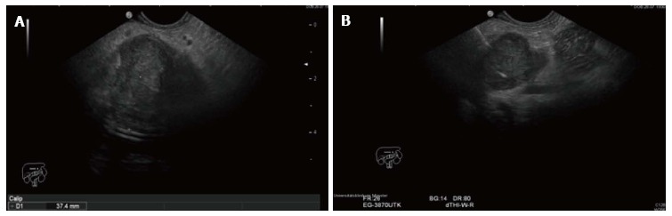
Results: Eight patients [median age of 65 (range 27-82) years; 7 female and 1 male] were recruited in a prospective multicenter trial. Six had a pancreatic cystic neoplasm (four a mucinous cyst, one had intraductal papillary mucinous neoplasm and one a microcystic adenoma) and two had a neuroendocrine tumors (NET) in the head of pancreas. The mean size of the cystic neoplasm and NET were 36.5 mm (SD ± 17.9 mm) and 27.5 mm (SD ± 17.7 mm) respectively. The EUS-RFA was successfully completed in all cases. Among the 6 patients with a cystic neoplasm, post procedure imaging in 3-6 mo showed complete resolution of the cysts in 2 cases, whilst in three more there was a 48.4% reduction [mean pre RF 38.8 mm (SD ± 21.7 mm) vs mean post RF 20 mm (SD ± 17.1 mm)] in size. In regards to the NET patients, there was a change in vascularity and central necrosis after EUS-RFA. No major complications were observed within 48 h of the procedure. Two patients had mild abdominal pain that resolved within 3 d.
Conclusion: EUS-RFA of pancreatic neoplasms with a novel monopolar RF probe was well tolerated in all cases. Our preliminary data suggest that the procedure is straightforward and safe. The response ranged from complete resolution to a 50% reduction in size.
Keywords: Cystic neoplasms; Endoscopic ultrasound; Neuroendocrine tumors; Pancreas; Radiofrequency ablation.
Figures
References
-
- Kitagawa Y, Unger TA, Taylor S, Kozarek RA, Traverso LW. Mucus is a predictor of better prognosis and survival in patients with intraductal papillary mucinous tumor of the pancreas. J Gastrointest Surg. 2003;7:12–18; discussion 18-19. - PubMed
LinkOut - more resources
Full Text Sources
Other Literature Sources
