

Autonomic and endocrine control of cardiovascular function
- PMID: 25914789
- PMCID: PMC4404375
- DOI: 10.4330/wjc.v7.i4.204
Autonomic and endocrine control of cardiovascular function
Abstract
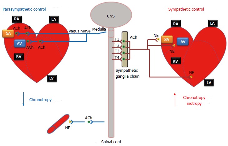
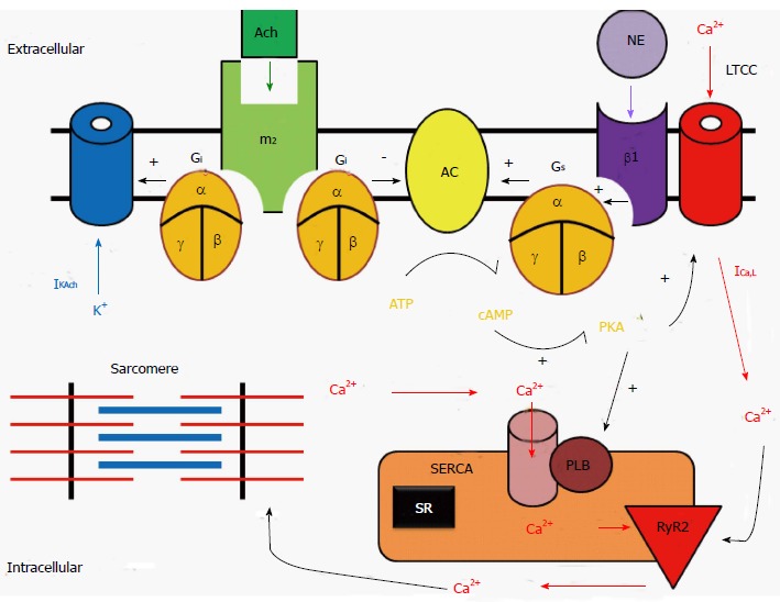
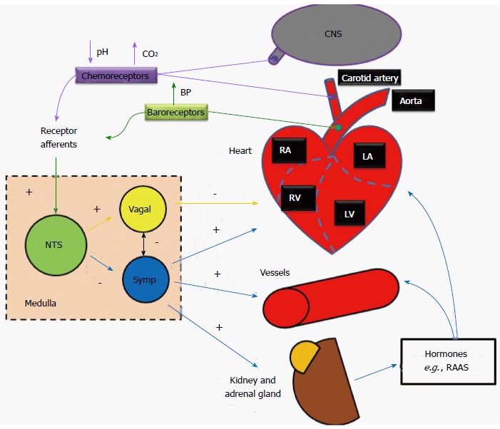
The function of the heart is to contract and pump oxygenated blood to the body and deoxygenated blood to the lungs. To achieve this goal, a normal human heart must beat regularly and continuously for one's entire life. Heartbeats originate from the rhythmic pacing discharge from the sinoatrial (SA) node within the heart itself. In the absence of extrinsic neural or hormonal influences, the SA node pacing rate would be about 100 beats per minute. Heart rate and cardiac output, however, must vary in response to the needs of the body's cells for oxygen and nutrients under varying conditions. In order to respond rapidly to the changing requirements of the body's tissues, the heart rate and contractility are regulated by the nervous system, hormones, and other factors. Here we review how the cardiovascular system is controlled and influenced by not only a unique intrinsic system, but is also heavily influenced by the autonomic nervous system as well as the endocrine system.
Keywords: Autonomic nervous system; Cardiovascular function; Endocrine system; Heart; Regulation.
Figures

References
-
- Boron W, Boulpaep E. Medical physiology: a cellular and molecular approach. 2nd ed. Philadelphia, PA: Elsevier Saunders; 2011.
-
- Gwathmey JK, Briggs GM, Allen PD. Heart Failure: Basic Science and Clinical Aspects. New York: Marcel Dekker Inc; 1994. pp. 282–283.
-
- Mann DL, Zipes DP, Libby P, Bonow RO. Braunwald’s Heart Disease: Textbook of Cardiovascular Medicine. 10th ed. Philadelphia, Pennsylvania: Elsevier - Health Sciences Division; 2014.
-
- Rhoadesand RA, Bell DR. Medical Physiology: Principles for Clinical Medicine. 3rd Ed. Philadelphia, Pennsylvania: Lippincott Williams and Wilkins, Wolters Kluwer Health; 2009.
-
- Kishi T. Heart failure as an autonomic nervous system dysfunction. J Cardiol. 2012;59:117–122. - PubMed
Publication types
Grants and funding
LinkOut - more resources
Full Text Sources
Other Literature Sources
