. 2015 May;17(5):615-626.
doi: 10.1038/ncb3160. Epub 2015 Apr 27.

TEAD and YAP regulate the enhancer network of human embryonic pancreatic progenitors

Affiliations

TEAD and YAP regulate the enhancer network of human embryonic pancreatic progenitors

Inês Cebola et al. Nat Cell Biol. 2015 May.

Abstract

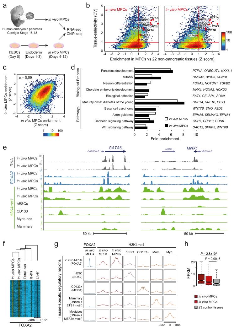
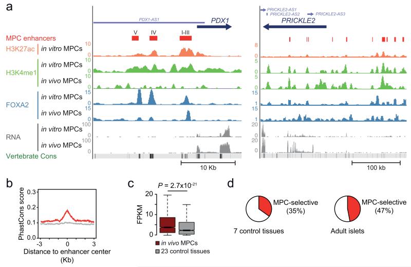
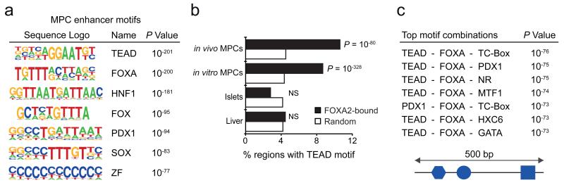
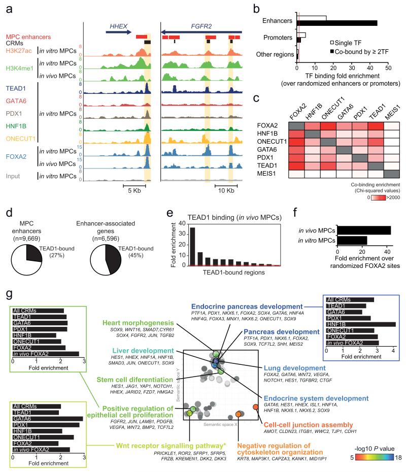
The genomic regulatory programmes that underlie human organogenesis are poorly understood. Pancreas development, in particular, has pivotal implications for pancreatic regeneration, cancer and diabetes. We have now characterized the regulatory landscape of embryonic multipotent progenitor cells that give rise to all pancreatic epithelial lineages. Using human embryonic pancreas and embryonic-stem-cell-derived progenitors we identify stage-specific transcripts and associated enhancers, many of which are co-occupied by transcription factors that are essential for pancreas development. We further show that TEAD1, a Hippo signalling effector, is an integral component of the transcription factor combinatorial code of pancreatic progenitor enhancers. TEAD and its coactivator YAP activate key pancreatic signalling mediators and transcription factors, and regulate the expansion of pancreatic progenitors. This work therefore uncovers a central role for TEAD and YAP as signal-responsive regulators of multipotent pancreatic progenitors, and provides a resource for the study of embryonic development of the human pancreas.


Figures

Figure 1
Figure 1
Human in vitro MPCs recapitulate transcriptional and epigenomic features of in vivo MPCs. (a) Experimental set-up. Pancreas was dissected from human Carnegie stage 16-18 embryos (in vivo MPCs). In vitro MPCs were derived from hESCs. (b) In vitro and in vivo MPCs share tissue-selective genes. Tissue-selectivity of RNAs was determined by the coefficient of variation (CV) across 25 embryonic and adult tissues or cell types. Enrichment of RNAs in MPCs relative to non-pancreatic tissues was quantified as a Z-score. Red lines define genes that are both tissue-selective and enriched in MPCs (CV>1, Z>1). Most known pancreatic regulatory TFs are in this quadrant in both sources of MPCs. Color scale depicts number of transcripts. (c) Z-scores of genes expressed in at least one source of MPCs were highly correlated for in vitro vs. in vivo MPCs (see also Supplementary Figure 1d for a comparison of unrelated tissues). Spearman's coefficient value is shown. Color scale depicts number of transcripts. (d) In vivo and in vitro MPC-enriched genes have common functional annotations. Shown are most significant terms for in vivo MPC-enriched genes, and their fold enrichment in both sources of MPCs. Representative genes from each category that are enriched in both MPCs are shown on the right. More extensive annotations are shown in Supplementary Table 3. (e) RNA, FOXA2 and H3K4me1 profiles of indicated samples in the GATA6 and MNX1 loci. (f) In vivo MPC FOXA2 occupancy is largely recapitulated by in vitro MPCs, but not by other tissues expressing FOXA2. Hierarchical clustering was performed on normalized FOXA2 ChIP-seq signal centered on all 5,760 in vivo MPC FOXA2 peaks. (g) In vitro MPCs recapitulate cell-specific H3K4me1 enrichment observed in chromatin from in vivo MPCs. Aggregation plots show H3K4me1 enrichment at occupancy sites of tissue-specific TFs. Mam.: Mammary Myo.: Myotubes. (h) Genes with ≥3 regions enriched in FOXA2 and H3K4me1 at in vivo MPCs are preferentially expressed in both in vivo and in vitro MPCs. Boxes show RNA interquartile range (IQR) and notches indicate median 95% confidence intervals (n=327 genes). P values were calculated with Wilcoxon rank-sum test.
Figure 2
Figure 2
A compendium of active enhancers in human pancreatic MPCs. (a) Predicted enhancers were defined by enrichment in H3K27ac and H3K4me1 (see schematic in Supplementary Fig. 2b). Shown are examples in the vicinity of PDX1, including a previously unannotated enhancer which we coin Area V, upstream of known enhancers (Areas I-IV),, and several enhancers near PRICKLE2, a non-canonical WNT signaling component (Supplementary Table 4). (b) MPC enhancer sequences are evolutionary conserved (17 species vertebrate PhastCons score). Conservation plots of random non-exonic sequences are shown as a light gray line. (c) Genes that are associated with 3 or more MPC enhancers show enriched expression in dissected in vivo MPCs relative to 23 other tissues. The boxes show interquartile range (IQR) of RNA levels, whiskers extend to 1.5 times the IQR or extreme values, and notches indicate 95% confidence intervals of the median. P value was calculated with Wilcoxon rank-sum test (n=2,093 genes). (d) Many MPC enhancers are tissue- and stage-selective. We defined enhancers of 8 control tissues using identical criteria as in MPCs (Supplementary Fig. 2c, Supplementary Table 8) and show the proportion of enhancers that are inactive in at least 6 out of 7 non-pancreatic tissues (left) or inactive in adult pancreatic islets (right).
Figure 3
Figure 3
MPC enhancers are enriched in DNA binding motifs for TEAD and known pancreatic transcription factors. (a) TEAD recognition motifs were strongly enriched in a de novo motif search in MPC enhancers. Other enriched matrices match binding sites of known pancreatic regulators. See Supplementary Tables 10 and 11 for a complete list of motifs enriched in MPC and MPC-selective enhancers, respectively. (b) TEAD motifs are highly enriched at genomic regions bound by FOXA2 in both in vivo and in vitro MPCs, but not at regions bound by FOXA2 in islets or liver. Binomial distribution P values were obtained using HOMER. NS: non-significant. (c) Combinations of recognition motifs for TEAD and other pancreatic regulators are specifically enriched in pancreatic MPC enhancers. We searched for combinations of 3 sequence motifs that were contained within 500 bp and were most enriched in pancreatic MPC enhancers relative to 8 other tissue enhancers. The top 50 most enriched motif combinations are shown in Supplementary Table 12.
Figure 4
Figure 4
TEAD1 is a core component of human pancreatic MPC cis-regulatory modules (CRMs). (a) ChIP-seq was used to locate binding sites of 6 TFs in MPCs, as illustrated in two loci encoding pancreatic TFs. CRMs were defined as enhancer regions with ≥2 overlapping TF-bound sites. Examples are highlighted in yellow. (b) TFs preferentially occupy MPC enhancers, and this is most pronounced for regions bound by ≥2 TFs. Binding enrichment was calculated over 1,000 permutations of enhancer or promoter genomic positions in the mappable genome. For comparison we analyzed all other genomic regions after exclusion of MPC enhancers or promoters. Red line indicates a fold enrichment of 1. (c) Pancreatic TFs co-occupy genomic regions, and TEAD1 shows a similar co-occupancy pattern as other known pancreatic TFs. Binding sites of MEIS1 in a non-pancreatic cell type were used as control. The heatmap depicts Chi-squared values for all pairwise comparisons of observed vs. expected co-binding. The latter was estimated by permuting each set of TF peaks independently 1,000 times. (d) Over 1/4 of MPC enhancers are bound by TEAD1, whereas 45% of genes associated with MPC enhancers include at least one TEAD1-bound enhancer. (e) ChIP-qPCR with in vivo MPCs confirms TEAD1 binding at in vitro MPC TEAD1-bound regions (regions and associated genes in Supplementary Table 15). (f) TEAD1 binding is enriched in regions bound by FOXA2 in either in vitro or in vivo MPCs. We calculated TEAD1-FOXA2 co-binding over the median expected value after generating 1,000 permutations of in vitro or in vivo FOXA2 peak positions. (g) CRMs underlie a pancreas developmental regulatory network. The 2,956 genes associated with CRMs were functionally annotated using GREAT, and REVIGO was used to visualize annotation clusters. The most significant terms from each cluster are highlighted according to the P value color scale. Bar graphs show that GO terms are similarly enriched in CRMs bound by different TFs. *Several WNT pathway related-terms were enriched, although manual annotation in this category revealed that most gene were either non-canonical WNT signaling mediators or antagonists of canonical WNT signaling (full annotations in Supplementary Table 17).
Figure 5
Figure 5
Functional validation of CRMs as transcriptional enhancers. (a) Thirty two CRMs were cloned into the pGL4.23 vector and tested in reporter assays, where 20 (62.5%) yielded significant activation of a minimal promoter driving luciferase in human pancreatic MPCs. Lines represent median with IQR. Two-tailed Mann-Whitney test P value is shown (n=4 replicate wells). (See also Supplementary Fig. 5a). (b) TEAD binding sites are essential for MPC enhancer activity. Mutation of one or more canonical TEAD binding sites in three CRMs abolished their activity in luciferase reporter assays in in vitro MPCs. Locations of the FGFR2 and MAP3K1 CRMs are highlighted in Figure 4a and Supplementary Figure 4c, respectively. Two-tailed t-test P values are listed in Supplementary Table 22 (n=3-4 transfections per construct, in 1-2 independent experiments). Error bars represent SEM. (c,d) A TEAD1-bound CRM near SOX9 (Fig. 7e) was fused to a minimal promoter and GFP, and injected into zebrafish embryos. In (c), a SOX9 CRM drove strong GFP expression in the pancreatic domain of 48 hpf zebrafish embryos (dotted circle, left panel), which was disrupted by a mutation in the TEAD recognition sequence (right). A midbrain-specific enhancer was used as internal control of transgenesis. Note that this experiment assessed activity of a single SOX9 CRM, which does not necessarily fully recapitulate the expression of endogenous sox9b. In the graph, +, +/− and − represent strong, weak and absent GFP expression in the pancreatic domain, respectively (n=110-140 embryos per condition, Chi-squared test P=1.37×10−83). (d) Immunofluorescence analysis of pancreatic MPCs in zebrafish embryos injected at one- to two-cell stage with constructs containing SOX9, MAP3K1 and FOXA2 CRMs driving GFP. Images show GFP in Pdx1+/Nkx6.1+ cells at 24/48 hpf, as indicated. In total, 8/10 CRMs yielded activity in Pdx1+/Nkx6.1+ progenitors (see also Supplementary Fig. 5b). The pancreatic progenitor domain is revealed by co-expression of Pdx1+ and Nkx6.1+ (dashed lines). Note that in zebrafish Nkx6.1 is specific to MPCs within embryonic pancreas. g: Pdx1+ gut cells, s: somites showing crossreactivity with anti-Pdx1 serum. (e) Percentage of transgenic embryos with CRM-driven GFP expression in MPCs, or in negative controls (neg.) (quantifications shown in Supplementary Table 21).
Figure 6
Figure 6
YAP is expressed in the nucleus of pancreatic MPCs, and shows co-occupancy with TEAD1 at MPC enhancers. (a) YAP is detected in the nucleus of PDX1+ in vivo MPCs from human Carnegie Stage 18 pancreas. (b) In 10 weeks post-conception (WPC) human pancreas YAP expression is strong in nuclei of PDX1+ progenitors, but shows markedly diminished signal intensity in NGN3+ progenitors (white arrow). Image depicts 5 cells in human embryonic pancreas 10 WPC. (c) Yap is detected in the nucleus of Sox9+ MPCs from mouse E12.5 embryonic pancreas (white arrow), whereas Yap is diffuse or absent in Ngn3+ endocrine progenitor cells (hollow arrowheads). (d) YAP is excluded from the nucleus in hESCs-derived pancreatic NGN3+ progenitor cells (hollow arrowheads). (e) ChIP-qPCR analysis of YAP occupancy in chromatin from in vitro MPCs shows that TEAD1-bound regions are often co-bound by YAP.
Figure 7
Figure 7
TEAD and YAP regulation of pancreas development. (a) Human in vitro MPCs were incubated with VP 24 hours to disrupt TEAD-YAP interactions, causing downregulation of genes associated with TEAD1-bound enhancers. Data was normalized by PBGD. Bars show mean values from 2 independent experiments, and points represent mean of 2 technical replicates. (b-d) VP treatment of E11.5 mouse pancreatic explants downregulated orthologs of TEAD1-bound genes, inhibited proliferation and reduced growth of pancreatic epithelial cells. Explants were treated with VP for 24 hours, washed, and incubated 24 hours before analysis. Data was normalized to Gapdh. *Two-tailed t test P<0.05 (individual values listed in Supplementary Table 22). Error bars represent SD from 3 independent experiments (each with n=2-4 embryos/condition). In (c) the percentage of proliferating epithelial cells was quantified with E-Cadherin and EdU immunolocalization. Two-tailed Mann-Whitney P value is shown for 3 experiments (each with n=2-3 pancreas/condition). In (d) GFP+ area in Sox9-EGFP transgenic embryo explants is shown at day 3 compared to day 1. Two-tailed Mann-Whitney test P values are shown for 3 experiments (each with n=2-4 buds/condition). In (c) and (d) boxes are IQR and median, whiskers 1.5 × IQR or extreme values. (e) Snapshot of the human SOX9 locus, encoding a regulator of MPC growth. The CRM tested in functional assays in Figure 5c and Figure 7f is highlighted. (f) yap1 inhibition decreased pancreatic sox9b expression. Injection of Mo-yap1 caused a reduction or absence of sox9b mRNA in the pancreatic domain (arrow) in 50/102 48 hpf embryos. Control embryos showed pancreatic sox9b expression in 100/100 embryos (Chi-squared P 2.61×10⁻¹⁵). Note that control and morphant embryos always showed sox9b expression in fin buds (fb). (g) Injection of Mo-yap1 (n=10 embryos) or the TEAD-EnR dominant negative (n=12 embryos) caused a decreased number of sox9b+/Pdx1+ pancreatic progenitors (dotted lines) in 24 hpf embryos vs. controls (n=9 embryos). Sox9b was detected by in situ hybridization and Pdx1 by immunofluorescence. The graph reflects the total number of pancreatic progenitors in each embryo. Mo-yap1 also increased ectopic expression of pancreatic markers (Supplementary Figure 7b). Student’s t test P values and SD are shown.
Figure 8
Figure 8
YAP/TEAD-dependent activation provides a regulatory switch for pancreatic MPC enhancers. A significant number of pancreatic MPC enhancers is co-bound by known stage-specific TFs along with TEAD and YAP. During pancreatic differentiation YAP is rapidly excluded from the nucleus and its expression is reduced, causing inactivation of MPC stage-specific enhancers. This simplified model depicts inhibition of YAP through Hippo kinase-induced phosphorylation or degradation, although additional non-mutually exclusive mechanisms for dynamic inhibition of YAP signaling are plausible. The model is supported by evidence showing that chemical or genetic inhibition of YAP and TEAD function causes inhibition of MPC enhancers.

References

    1. Fang H, et al. An organogenesis network-based comparative transcriptome analysis for understanding early human development in vivo and in vitro. BMC Syst Biol. 2011;5:108. - PMC - PubMed
    1. Fang H, et al. Transcriptome analysis of early organogenesis in human embryos. Developmental Cell. 2010;19:174–184. - PubMed
    1. Pan FC, Wright C. Pancreas organogenesis: From bud to plexus to gland. Developmental Dynamics. 2011;240:530–565. - PubMed
    1. Zaret KS, Grompe M. Generation and regeneration of cells of the liver and pancreas. Science. 2008 doi:10.1126/science.1161431. - PMC - PubMed
    1. Lango Allen H, et al. GATA6 haploinsufficiency causes pancreatic agenesis in humans. Nat Genet. 2012;44:20–22. - PMC - PubMed

METHODS REFERENCES

    1. O’Rahilly R, Müller F, Hutchins GM, Moore GW. Computer ranking of the sequence of appearance of 73 features of the brain and related structures in staged human embryos during the sixth week of development. Am. J. Anat. 1987;180:69–86. - PubMed
    1. Maestro MA, et al. Hnf6 and Tcf2 (MODY5) are linked in a gene network operating in a precursor cell domain of the embryonic pancreas. Human Molecular Genetics. 2003;12:3307–3314. - PubMed
    1. Piper K, et al. Beta cell differentiation during early human pancreas development. J. Endocrinol. 2004;181:11–23. - PubMed
    1. Petzold KM, Spagnoli FM. A system for ex vivo culturing of embryonic pancreas. J Vis Exp. 2012:e3979–e3979. doi:10.3791/3979. - PMC - PubMed
    1. Gaulton KJ, et al. A map of open chromatin in human pancreatic islets. Nat Genet. 2010;42:255–259. - PMC - PubMed

Publication types

MeSH terms