. 2015 Apr 27;10(4):e0125671.
doi: 10.1371/journal.pone.0125671. eCollection 2015.

STIM1 Is a Novel Component of ER-Chlamydia trachomatis Inclusion Membrane Contact Sites

Affiliations

STIM1 Is a Novel Component of ER-Chlamydia trachomatis Inclusion Membrane Contact Sites

Hervé Agaisse et al. PLoS One. .

Abstract

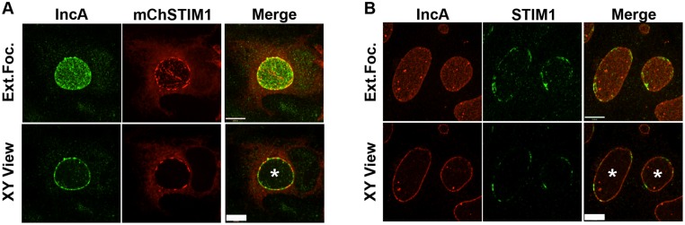
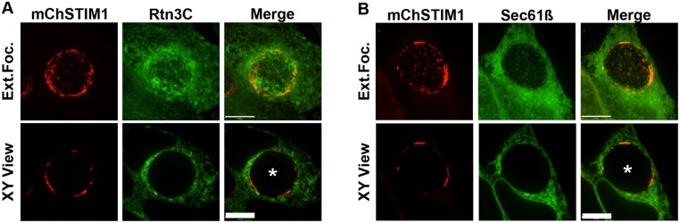
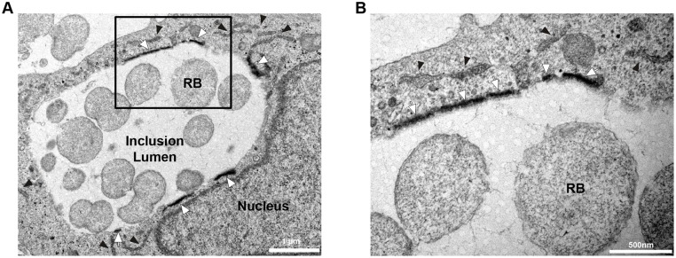
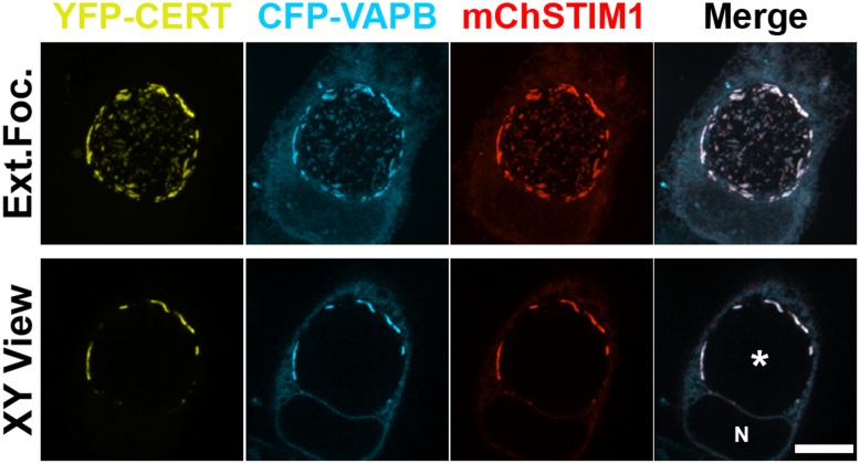
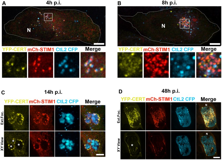
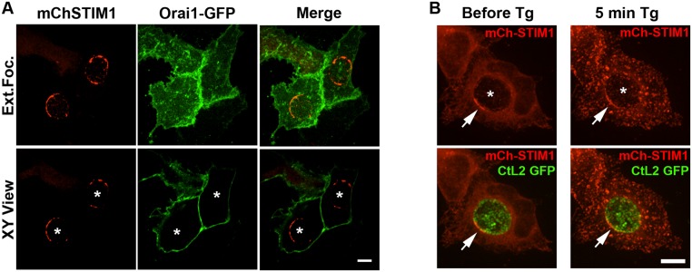
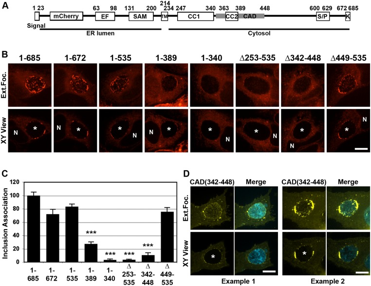
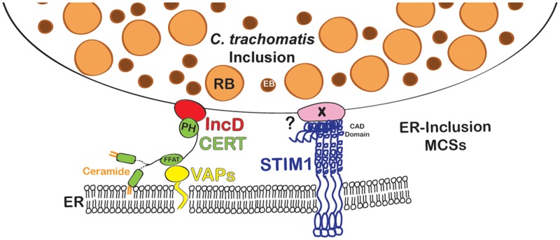
Productive developmental cycle of the obligate intracellular bacterial pathogen Chlamydia trachomatis depends on the interaction of the replicative vacuole, named the inclusion, with cellular organelles. We have recently reported the formation of ER-Inclusion membrane contact sites (MCSs), where the endoplasmic reticulum (ER) is in apposition to the inclusion membrane. These platforms contain the C. trachomatis inclusion membrane protein IncD, the mammalian ceramide transfer protein CERT and the ER resident proteins VAPA/B and were proposed to play a role in the non-vesicular trafficking of lipids to the inclusion. Here, we identify STIM1 as a novel component of ER-Inclusion MCSs. STIM1, an ER calcium (Ca2+) sensor that relocate to ER-Plasma Membrane (PM) MCSs upon Ca2+ store depletion, associated with C. trachomatis inclusion. STIM1, but not the general ER markers Rtn3C and Sec61ß, was enriched at the inclusion membrane. Ultra-structural studies demonstrated that STIM1 localized to ER-Inclusion MCSs. Time-course experiments showed that STIM1, CERT and VAPB co-localized throughout the developmental cycle. By contrast, Orai1, the PM Ca2+ channel that interacts with STIM1 at ER-PM MCSs, did not associate with C. trachomatis inclusion. Upon ER Ca2+ store depletion, a pool of STIM1 relocated to ER-PM MCSs, while the existing ER-Inclusion MCSs remained enriched in STIM1. Finally, we have identified the CAD domain, which mediates STIM1-Orai1 interaction, as the minimal domain required for STIM1 enrichment at ER-Inclusion MCSs. Altogether this study identifies STIM1 as a novel component of ER-C. trachomatis inclusion MCSs. We discuss the potential role(s) of STIM1 during the infection process.


Conflict of interest statement

Competing Interests: The authors have declared that no competing interests exist.

Figures

Fig 1
Fig 1. STIM1 is enriched in patches at the C. trachomatis inclusion membrane.
Confocal micrographs of HeLa cells infected with C. trachomatis for 24h and stained using antibodies against the C. trachomatis inclusion membrane protein IncA (A-B) (left panels, IncA, green (A) and red (B)). The cells were transfected with a mCherrySTIM1 construct 18h pre-infection (A) (middle panels, mChSTIM1, red) or stained using antibodies against STIM1 (B) (middle panels, STIM1, green). The merge is shown on the right. The top and bottom panels respectively correspond to the extended focus view combining all the confocal planes (Ext.Foc.) and a single plane crossing the middle of the inclusion (XY View). The asterisk in the XY View Merge panel indicates the inclusion. Scale bar: 10μm.
Fig 2
Fig 2. The ER markers Rtn3C and Sec61ß are not enriched at the C. trachomatis inclusion membrane.
Confocal micrographs of HeLa cells co-expressing mCherrySTIM1 (A-B) (left panels, mChSTIM1, red) and GFP-Rtn3C (A) (middle panels, Rtn3C, green) or GFP-Sec61ß (B) (middle panels, Sec61ß, green) and infected with C. trachomatis for 24h. The merge is shown on the right. The top and bottom panels respectively correspond to the extended focus view combining all the confocal planes (Ext.Foc.) and a single plane crossing the middle of the inclusion (XY View). The asterisk in the XY View Merge panel indicates the inclusion. Scale bar: 10μm.
Fig 3
Fig 3. STIM1 is enriched at ER-Inclusion MCSs.
Electron micrographs of HeLa cells expressing HRP-STIM1, infected with C. trachomatis for 24h and processed for conventional transmission electron microscopy coupled with peroxidase cytochemistry. White arrows and black arrowheads respectively indicate ER structures displaying high and low level of HRP-STIM1. The area outline by the box in A is shown at higher magnification in B. RB: Reticulate Body. Scale Bars: 1 μm (A), 500 nm (B).
Fig 4
Fig 4. STIM1 colocalizes with CERT and VAPB at the C. trachomatis inclusion membrane.
Confocal micrographs of HeLa cells co-expressing YFP-CERT (yellow), CFP-VAPB (cyan) and mCherrySTIM1 (red) and infected with C. trachomatis for 24h. The merge is shown on the right. The top and bottom panels respectively correspond to the extended focus view combining all the confocal planes (Ext.Foc.) and a single plane crossing the middle of the inclusion (XY View). The asterisk in the XY View Merge panel indicates the inclusion. N: Nucleus. Scale bar: 10μm.
Fig 5
Fig 5. STIM1 is associated with the C. trachomatis inclusion membrane throughout the developmental cycle.
A-B. Confocal micrographs of HeLa cells co-expressing YFP-CERT (yellow) and mCherrySTIM1 (mCh-STIM1, red) and infected with C. trachomatis expressing CFP (CtL2 CFP, cyan) for 4h (A), 8h (B). The extended focus view of 10 0.2 μm individual planes crossing the middle of the cell is shown. The boundary of the cell and the nucleus (N) are outlined and the area outlined by a box is shown at higher magnification below the whole cell micrograph. The merge is shown on the right. Scale bar: 10 μm. C-D. HeLa cells, as described in A and B, were infected for 14h (C) or 48h (D). Three small inclusions that have not yet fused are shown (C). The merge is shown on the right. The top and bottom panels respectively correspond to the extended focus view combining all the confocal planes (Ext.Foc.) and a single plane crossing the middle of the inclusion (XY View). The asterisk in the XY View Merge panel indicates the inclusions. N: Nucleus. Scale bars: 4μm (C), 20μm (D).
Fig 6
Fig 6. STIM1 cellular localization in response to Ca2+ store depletion.
A. Confocal micrographs of HeLa cells co-expressing mCherry-STIM1 (left panels, mChSTIM1, red) and Orai1-GFP (middle panels, Orai1-GFP, green) and infected with C. trachomatis for 24h. The merge is shown on the right. The top and bottom panels respectively correspond to the extended focus view combining all the confocal planes (Ext.Foc.) and a single plane crossing the middle of the inclusion (XY View). The asterisks in the XY View panels indicate the position of the inclusions. Scale bar: 10μm. B. Confocal micrographs of live HeLa cells expressing mCherry-STIM1 (mCh-STIM1, red) and infected for 24h with a strain of C. trachomatis that expresses GFP (CtL2 GFP, green). The images were acquired before (left panels, Before Tg) and 5 min after addition of Thapsigargin (Tg) (right panels, 5min Tg). The top and bottom panels respectively correspond to the mCh-STIM1 signal alone and the merge. An extended focus view combining all the confocal planes is shown. The arrow indicates STIM1 enrichment at ER-Inclusion MCSs. The asterisk indicates the position of the inclusion. Scale bar: 10μm.
Fig 7
Fig 7. The CAD domain of STIM1 is required for STIM1 localization to ER-Inclusion MCSs.
A. Schematic representation of the major domains of the STIM1 protein, their respective amino acid residue position and their respective cellular localization (ER lumen or Cytosol). Signal: signal peptide; EF: EF-hand; SAM: sterile alpha motif; TM: transmembrane domain; CC1: coiled-coil 1; CC2: coiled-coil 2; CAD: CRAC activation domain; S/P: Serine-proline-rich region; K: lysine-rich region. B. Confocal micrographs of HeLa cells expressing the indicated mCherry-STIM1 construct (red) and infected with C. trachomatis for 24h. The top and bottom panels respectively correspond to the extended focus view combining all the confocal planes (Ext.Foc.) and a single plane crossing the middle of the inclusion (XY View). The asterisk in the XY View Merge panel indicates the inclusion and N indicates the nucleus. Scale bar: 10μm. C. Quantification of inclusion association of the indicated mCh-STIM1 constructs compared to full-length mCh-STIM1. *** p value <0.001. D. Confocal micrographs of HeLa cells expressing YFP-CAD (CAD(342–448), yellow), and infected for 24h with a strain of C. trachomatis expressing CFP (cyan). The merge is shown on the right. Two representative examples of YFP-CAD pattern on the inclusion are shown. The top and bottom panels respectively correspond to the extended focus view combining all the confocal planes (Ext.Foc.) and a single plane crossing the middle of the inclusion (XY View). The asterisk in the XY View Merge panel indicates the inclusion. Scale bar: 10μm.
Fig 8
Fig 8. STIM1 depletion does not affect C. trachomatis growth.
HeLa cells were transfected with control siRNA (GFPsi) or a pool of CERT siRNA (CERTsi) or a pool of siRNA against STIM1 (STIM1si) for 3 days and infected with a strain of C. trachomatis expressing mCherry. (A) For each condition, the surface area of the inclusions was determined (Arbitrary Units). Each circle, square or triangle represents data from a single inclusion. (B) The number of infectious bacteria (IFUs/ml) was determined 48h p.i. (A-B) Data show the mean and standard deviation of biological triplicate of a representative experiment. ***P<0.0001 (student’s t test).
Fig 9
Fig 9. Schematic representation of ER-Inclusion MCSs.
CERT (green) localizes to ER-Inclusion MCSs. The PH domain of CERT binds to the C. trachomatis inclusion membrane protein IncD (red) and the FFAT motif of CERT allows CERT association with VAPA/B (Yellow) on the ER. In addition to CERT, VAPA/B and IncD, the ER Calcium sensor STIM1 (dark blue) also localizes to ER-Inclusion MCSs. The CAD domain of STIM1 is required for association with the inclusion. Future work will determine whether a Chlamydia factor X (light pink) is involved in this process.

References

    1. Friis RR. Interaction of L cells and Chlamydia psittaci: entry of the parasite and host responses to its development. J Bacteriol. 1972;110(2):706–721. - PMC - PubMed
    1. Moulder JW. Interaction of chlamydiae and host cells in vitro. Microbiol Rev. 1991;55(1):143–190. - PMC - PubMed
    1. Bastidas RJ, Elwell CA, Engel JN, Valdivia RH. Chlamydial intracellular survival strategies. Cold Spring Harb Perspect Med. 2013;3(5):a010256 10.1101/cshperspect.a010256 - DOI - PMC - PubMed
    1. Giles DK, Wyrick PB. Trafficking of chlamydial antigens to the endoplasmic reticulum of infected epithelial cells. Microbes Infect. 2008;10(14–15):1494–1503. 10.1016/j.micinf.2008.07.031 - DOI - PMC - PubMed
    1. Derré I, Swiss R, Agaisse H. The lipid transfer protein CERT interacts with the Chlamydia inclusion protein IncD and participates to ER-Chlamydia inclusion membrane contact sites. PLoS Pathog. 2011;7(6):e1002092 10.1371/journal.ppat.1002092 - DOI - PMC - PubMed

Publication types

MeSH terms