. 2015 Apr 27;10(4):e0125793.
doi: 10.1371/journal.pone.0125793. eCollection 2015.

Lipopolysaccharide (LPS)-Induced Biliary Epithelial Cell NRas Activation Requires Epidermal Growth Factor Receptor (EGFR)

Affiliations

Lipopolysaccharide (LPS)-Induced Biliary Epithelial Cell NRas Activation Requires Epidermal Growth Factor Receptor (EGFR)

Christy E Trussoni et al. PLoS One. .

Abstract

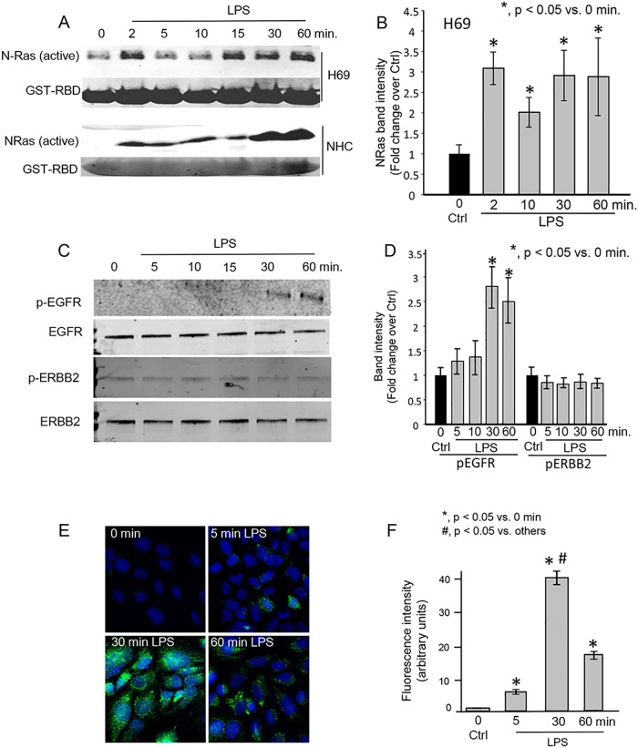
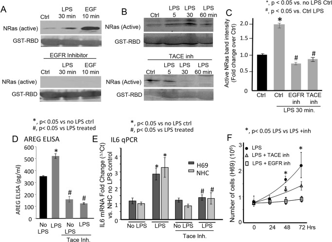
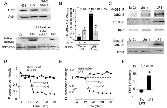
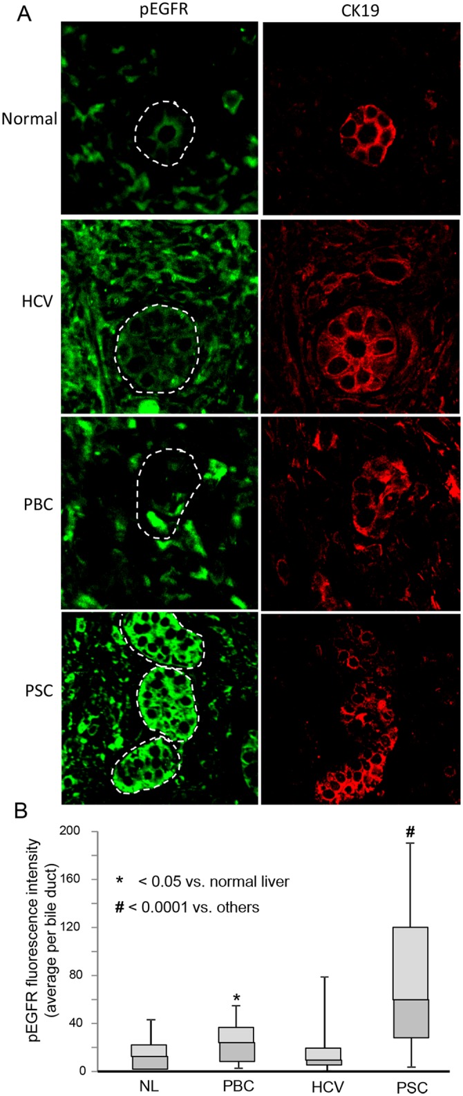
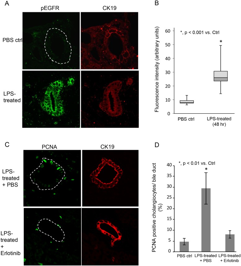
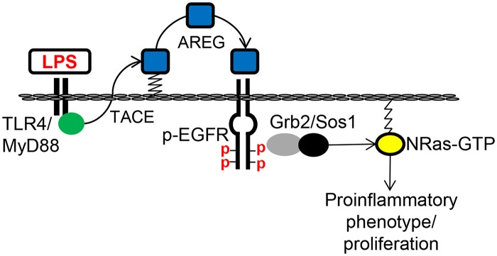
Cholangiocytes (biliary epithelial cells) actively participate in microbe-induced proinflammatory responses in the liver and contribute to inflammatory and infectious cholangiopathies. We previously demonstrated that cholangiocyte TLR-dependent NRas activation contributes to proinflammatory/ proliferative responses. We test the hypothesis that LPS-induced activation of NRas requires the EGFR. SV40-transformed human cholangiocytes (H69 cells), or low passage normal human cholangiocytes (NHC), were treated with LPS in the presence or absence of EGFR or ADAM metallopeptidase domain 17 (TACE) inhibitors. Ras activation assays, quantitative RT-PCR, and proliferation assays were performed in cells cultured with or without inhibitors or an siRNA to Grb2. Immunofluorescence for phospho-EGFR was performed on LPS-treated mouse samples and specimens from patients with primary sclerosing cholangitis, primary biliary cirrhosis, hepatitis C, and normal livers. LPS-treatment induced an association between the TLR/MyD88 and EGFR/Grb2 signaling apparatus, NRas activation, and EGFR phosphorylation. NRas activation was sensitive to EGFR and TACE inhibitors and correlated with EGFR phosphorylation. The TACE inhibitor and Grb2 depletion prevented LPS-induced IL6 expression (p<0.05) and proliferation (p<0.01). Additionally, cholangiocytes from LPS-treated mouse livers and human primary sclerosing cholangitis (PSC) livers exhibited increased phospho-EGFR (p<0.01). Moreover, LPS-induced mouse cholangiocyte proliferation was inhibited by concurrent treatment with the EGFR inhibitor, Erlotinib. Our results suggest that EGFR is essential for LPS-induced, TLR4/MyD88-mediated NRas activation and induction of a robust proinflammatory cholangiocyte response. These findings have implications not only for revealing the signaling potential of TLRs, but also implicate EGFR as an integral component of cholangiocyte TLR-induced proinflammatory processes.


Conflict of interest statement

Competing Interests: The authors have declared that no competing interests exist.

Figures

Fig 1
Fig 1. LPS induces NRas activation in cultured human cholangiocytes.
A. Confluent NHC and H69 cells were treated with LPS for 0, 2, 5, 10, 15, 30, and 60 minutes. LPS induced rapid and persistent NRas activation. B. Quantitation of NRas intensity. NRas band intensity was normalized to GST-RBD Ponceau red band intensity and is presented as fold change over the 0 time point +/- SEM from three independent experiments. C. Western blots for phospho-EGFR (pEGFR), total EGFR, phospho—ERBB2 (pERBB2), and total ERBB2 were also performed on lysates from the LPS-treated H69 cells. Phospho-EGFR was detected 30 and 60 minutes post-LPS treatment. D. Quantitation of pEGFR and pERBB2. Band intensity was normalized to total EGFR or ERBB2, respectively, and is presented as fold change over 0 time point control. E. Confocal immunofluorescence microscopy detected a progressive increase in phospho-EGFR following LPS treatment of H69 cells. F. Semi-quantitation of fluorescence intensity demonstrates an increase of phospho-EGFR fluorescence through the 30 minute time point. Data is presented as mean +/- SEM from three independent experiments and >100 cells per experiment.
Fig 2
Fig 2. Inhibition of ADAM metallopeptidase 17 (ADAM17 [TACE]) blocks LPS-induced NRas activation, AREG secretion, IL6 expression and cholangiocyte proliferation.
A. The EGFR inhibitor blocked LPS-induced NRas activation at the 30 minute post-LPS treatment time point. The EGFR inhibitor also blocked EGF-induced NRas activation. B. The TACE inhibitor, TAPI-1, blocked LPS-induced NRas activation at the 30 and 60 minute post-LPS treatment time point. C. Quantitation of NRas immunoblot band intensity for the 30 minute post-LPS timepoint for both the EGFR and TACE inhibitors. Data is presented as fold change +/- SEM from four independent experiments. D. An ELISA demonstrated that the TACE inhibitor (TAPI-1) blocked LPS-induced secretion of the EGFR ligand, AREG. Data is presented as pg/ml from three independent experiments. E. RT-PCR was performed and demonstrated that the TACE inhibitor blocked LPS-induced IL6 expression in both H69 and NHC cells. Data is presented as fold change (ΔΔCt) vs. NHC no LPS control. F. The TACE and EGFR inhibitors also block LPS-induced cholangiocyte proliferation. In the presence of LPS, H69 cells exhibited a doubling time of 46 hours, while TACE inhibitor treated cells exhibited a doubling time of approximately 80 hours (p < 0.05 at 48 and 72 hours control vs. inhibited cells). EGFR inhibitor treated cells exhibited minimal proliferation over the course of the experiment. Data is presented as mean number of cells +/- SEM from three independent experiments.
Fig 3
Fig 3. Depletion of the downstream EGFR molecular adaptor, Grb2 blocks LPS-induced NRas activation.
A. H69 cells were treated with either a scrambled control (Scr) or a Grb2 siRNA. Expression of Grb2 was normalized to actin to demonstrate efficient depletion. Untransfected (Ctrl), control scrambled (Scr) siRNA, and Grb2 siRNA transfected cells were treated with LPS. Grb2 siRNA blocked LPS-induced NRas activation. B. Quantitiative PCR demonstrated that Grb2 siRNA blocked LPS-induced IL6 expression. The experiment was performed in triplicate; *, p<0.05 compared to LPS treatment in the absence of the siRNA. LPS also induced an interaction between components of the TLR4 and EGFR signaling complex. C. Co-immunoprecipitations were performed on cells cultured in the presence or absence of LPS. Myd88 was immunoprecipitated from lysates of cholangiocytes cultured in the presence or absence of LPS. LPS induced an increase in the amount of Grb2 immunoprecipitated with MyD88, and an interaction between MyD88 and TLR4. Moreover, LPS induced an interaction between the well characterized Ras guanine exchange factor, Sos1, and its molecular adaptor, Grb2. Acceptor photobleaching quantitative FRET further demonstrated LPS-induced Grb2 and MyD88 interaction. D. In the absence of LPS, photobleaching of the acceptor Cy5 (at 16s) did not induce an increase in donor fluorescence, indicating that these molecules are greater than 10 nm apart. In contrast, LPS-treatment induced an increase in donor Cy3 fluorescence detection following Cy5 (acceptor) photobleaching (E), demonstrating the induced proximity (≤ 10 nm) of MyD88 and Grb2. F. FRET efficiency was calculated using the Donormax/Donormin method. The LPS-treated cells demonstrated energy transfer between the fluorophores (~10%), while the cells cultured in the absence of LPS did not.
Fig 4
Fig 4. Cholangiocytes in PSC diseased livers exhibit increased phospho-EGFR compared to normal and disease control livers.
A. Representative confocal immunofluorescence images for phospho-EGFR (pEGFR) and the cholangiocyte marker cytokeratin 19 (CK19) in normal (NL), HCV-infected, primary biliary cirrhosis (PBC), and primary sclerosing cholangitis (PSC) human livers. Normal livers exhibited minimal phospho-EGFR fluorescence. Cholangiocytes from both PBC and PSC diseased livers exhibited an increase in phospho-EGFR immunofluorescence detection. B. Semi-quantitative analyses of fluorescence intensity, using the Mann Whitney U test, revealed an increase in phospho-EGFR immunofluorescence in both PBC (p < 0.05) and PSC (p < 0.0001) compared to normal livers. Moreover, cholangiocytes from PSC diseased livers exhibited an increase in phospho-EGFR immunofluorescence compared to all other conditions (p < 0.0001). Data is presented as box plots (N = 4 for each condition; minimum of 5 ducts per sample) with mean (horizontal line between shaded boxes), 25th and 75th percentiles, with bars indicating maximum and minimum values.
Fig 5
Fig 5. LPS induces cholangiocyte EGFR phosphorylation and proliferation in a mouse model.
LPS (5mg/kg body weight) or saline (control) was injected into the tail vein of C57-black mice and the mice were sacrificed at 48 hours post-injection. A. Representative confocal immunofluorescence images for phospho-EGFR and cytokeratin 19 (CK19) in PBS control and mice treated with LPS. Control livers exhibited minimal phospho-EGFR fluorescence. Cholangiocytes from LPS-treated mouse livers exhibited an increase in phospho-EGFR immunofluorescence detection. B. Semi-quantitative analyses of fluorescence intensity, using the Mann Whitney U test (p < 0.001). Data is presented as box plots (N = 3 for each condition; minimum of 4 ducts per sample) with mean (horizontal line between shaded boxes), 25th and 75th percentiles, with bars indicating maximum and minimum values. C. Representative confocal immunofluorescence images for PCNA and CK19 in LPS treated and LPS treated with in the presence of the EGFR inhibitor Erlotinib. Cholangiocytes from LPS treated mice in the absence of Erlotinib exhibited increased PCNA positive nuclei. D. Quantitative analysis of PCNA positive nuclei. The number of PCNA positive cells per bile duct was counted (N = 3 for each condition; minimum of 4 ducts per sample) and is presented as a percentage of total cholangiocytes.
Fig 6
Fig 6. Working model of LPS-induced cholangiocyte NRas activation.
We propose that TLR4 activation by LPS promotes TACE-dependent cleavage of the EGFR ligand, AREG. Cleaved AREG engages the EGFR promoting EGFR phosphorylation and recruitment of the molecular adaptor, GRB2. GRB2 and associated guanine nucleotide exchange factor, SOS1, then promotes GTP-bound NRas, and subsequent signal transduction for the expression of proinflammatory mediators and cholangiocyte proliferation.

References

    1. Tabibian JH, Masyuk AI, Masyuk TV, O'Hara SP, LaRusso NF. Physiology of cholangiocytes. Compr Physiol. 2013;3: 541–565. 10.1002/cphy.c120019 - DOI - PMC - PubMed
    1. Chen XM, O'Hara SP, LaRusso NF. The immunobiology of cholangiocytes. Immunol Cell Biol. 2008;86: 497–505. 10.1038/icb.2008.37 - DOI - PMC - PubMed
    1. Harada K, Shimoda S, Sato Y, Isse K, Ikeda H, Nakanuma Y. Periductal interleukin-17 production in association with biliary innate immunity contributes to the pathogenesis of cholangiopathy in primary biliary cirrhosis. Clin Exp Immunol. 2009;157: 261–270. 10.1111/j.1365-2249.2009.03947.x - DOI - PMC - PubMed
    1. Karrar A, Broome U, Sodergren T, Jaksch M, Bergquist A, Bjornstedt M, et al. Biliary epithelial cell antibodies link adaptive and innate immune responses in primary sclerosing cholangitis. Gastroenterology. 2007;132: 1504–1514. - PubMed
    1. Mimura Y, Sakisaka S, Harada M, Sata M, Tanikawa K. Role of hepatocytes in direct clearance of lipopolysaccharide in rats. Gastroenterology. 1995;109: 1969–1976. - PubMed

Publication types

MeSH terms