. 2015 Jun 20;6(17):15235-49.
doi: 10.18632/oncotarget.3837.

Malignant T cells express lymphotoxin α and drive endothelial activation in cutaneous T cell lymphoma

Affiliations

Malignant T cells express lymphotoxin α and drive endothelial activation in cutaneous T cell lymphoma

Britt Lauenborg et al. Oncotarget. .

Abstract

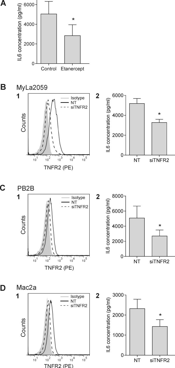
Lymphotoxin α (LTα) plays a key role in the formation of lymphatic vasculature and secondary lymphoid structures. Cutaneous T cell lymphoma (CTCL) is the most common primary lymphoma of the skin and in advanced stages, malignant T cells spreads through the lymphatic to regional lymph nodes to internal organs and blood. Yet, little is known about the mechanism of the CTCL dissemination. Here, we show that CTCL cells express LTα in situ and that LTα expression is driven by aberrantly activated JAK3/STAT5 pathway. Importantly, via TNF receptor 2, LTα functions as an autocrine factor by stimulating expression of IL-6 in the malignant cells. LTα and IL-6, together with VEGF promote angiogenesis by inducing endothelial cell sprouting and tube formation. Thus, we propose that LTα plays a role in malignant angiogenesis and disease progression in CTCL and may serve as a therapeutic target in this disease.

Keywords: CTCL; LTA; angiogenesis.


Conflict of interest statement

CONFLICTS OF INTEREST

The authors declare no competing interests.

Figures

Figure 1
Figure 1. LTα expression in CTCL
(A) Frozen biopsies of 10 patients diagnosed with CTCL (see Table 1) were subjected to IHC with an antibody directed against LTα showing strong cytoplasmic labelling of the neoplastic infiltrate in 8 of 10 cases (A × 400). (B) Shows an example of a patient staining negative for LTα (A × 400). (C) The expression of LTα and TNFα in non-malignant (MySi and MyLa1850) and malignant (MyLa2059 and PB2B) CTCL T cells lines were determined by ELISA. Bars represent mean values of three independent experiments.
Figure 2
Figure 2. JAK3/STAT5 drives the LTα expression
(A) LTα expression following inhibition of JAK activity in MyLa2059 with increasing concentrations of CP690,550 for 24 h *p < 0.05. (B) The phosphorylation status of STAT3 and 5 in MyLa2059 following incubation with CP690,550 for 24 h were analysed by WB. (C, D) MyLa2059 cells were transiently transfected with JAK1 and 3 (C) and STAT5a and b (D) or non-target (NT) control. 24h post-transfection, supernatants were harvested and LTα concentration measured by ELISA. Cells were lysed and the JAK3 (C) and STAT5 a and b (D) protein expression were analysed by WB. *p < 0.05 compared to NT control (paired t-test).
Figure 3
Figure 3. LTα is a transcriptional target of STAT5
(A) ChIP-seq reads from the LTA gene in malignant MyLa2059 cells. Reads (76 bases) obtained from immunoprecipitation of STAT5, STAT3, RELA, RELB and a negative control (rabbit IgG, bottom). The chromosomal position of LTA gene encoding LTα on chromosome 6 refers to hg19. Forward reads are indicated in green and reverse reads in red. (B) PCR analysis of ChIP samples using primers specific for the LTα promotor only detected the amplicon in the STAT5-precipitated sample, the 2% input, and the positive control (histone H3). (C) Pulldown assay using oligonucleotides representing a STAT binding site in the promoter region of the LTα gene. STAT5 binds to the sequence, while STAT3 do not.
Figure 4
Figure 4. Expression of TNF receptors in CTCL
Expression of TNFR1 and TNFR2 in malignant (MyLa2059, PB2B, Mac2a) CTCL T cells, Jurkat and Ramos were analysed by flowcytometry. Black line represents TNFR1 or TNFR2 expression, grey filled represent isotype control.
Figure 5
Figure 5. In situ TNFR2 expression in patients diagnosed with CTCL
Immunohistochemical (IHC) staining of cryostat sections of CTCL with an antibody directed against TNFR2 showed cytoplasmic labelling of the neoplastic cells (A × 400) in all cases (see Table 1).
Figure 6
Figure 6. LTα regulates the IL-6 expression in malignant CTCL T cells
(A) IL-6 expression in malignant CTCL cells (MyLa2059) were measured by ELISA following incubation with 100ug/ml Etanercept for 24 h. (B1, C1, D1) Selective down-regulation of TNFR2 with siRNA in malignant CTCL T cells (MyLa2059, PB2B and Mac2a). Filled grey represents isotype control, black line non-target (NT) control, and dashed line TNFR2 siRNA. (B2, C2, D2) IL-6 expression was measured after 24 h in MyLa2059, PB2B and Mac2a by ELISA following TNFR2 knock down. Bars represent mean values of three independent experiments. *p < 0.05 compared to NT (paired t-test).
Figure 7
Figure 7. Malignant LTα and IL-6 promote endothelial cells sprouting
Endothelial tube formation assays were performed on growth factor reduced matrigels in 24 well plates. (A) HUVEC cell sprouting when cultured in M200PRF alone (1), supplemented with VEGF (2) or malignant CTCL supernatant (1:2) (3). (B, C) Pictures were taken and number of branching points counted following treatment with human recombinant VEGF, LTα or IL-6 (B) or in the presence of malignant CTCL supernatant (1:2) either alone or supplemented with Avastin, Etanercept or Tocilizumab for 12 h. Bars represent mean values of three independent experiments. *p < 0.05 compared to control (paired t-test).

References

    1. Willemze R, Jaffe ES, Burg G, Cerroni L, Berti E, Swerdlow SH, Ralfkiaer E, Chimenti S, Diaz-Perez JL, Duncan LM, Grange F, Harris NL, Kempf W, et al. WHO-EORTC classification for cutaneous lymphomas. Blood. 2005;105:3768–3785. - PubMed
    1. Girardi M, Heald P, Wilson L. The pathogenesis of mycosis fungoides. N.Engl.J.Med. 2004;350:1978–1988. - PubMed
    1. Hwang ST, Janik JE, Jaffe ES, Wilson WH. Mycosis fungoides and Sezary syndrome. Lancet. 2008;371:945–957. - PubMed
    1. Kim E, Lin J, Junkins-Hopkins J, Vittorio C, Rook A. Mycosis fungoides and sezary syndrome: an update. Curr.Oncol.Rep. 2006;8:376–386. - PubMed
    1. Duvic M, Foss FM. Mycosis fungoides: Pathophysiology and emerging therapies. Semin. Oncol. 2007;34:21–27. - PubMed

Publication types

MeSH terms