. 2015 Jun;125(6):2335-46.
doi: 10.1172/JCI80297. Epub 2015 Apr 27.

Neurovascular crosstalk between interneurons and capillaries is required for vision

Neurovascular crosstalk between interneurons and capillaries is required for vision

Yoshihiko Usui et al. J Clin Invest. 2015 Jun.

Abstract

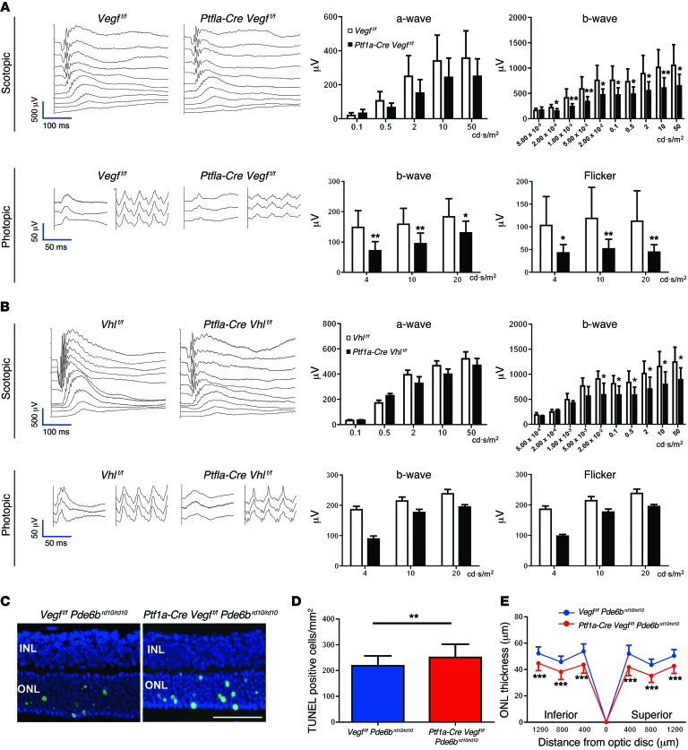
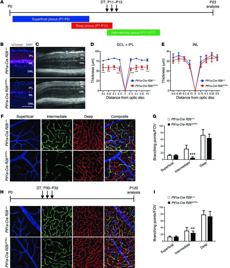
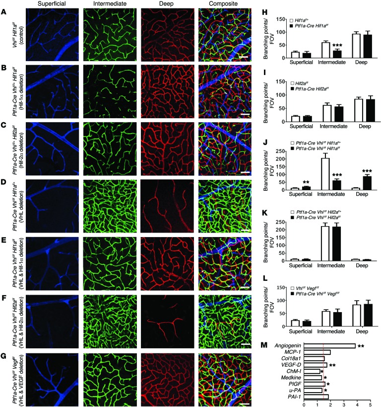
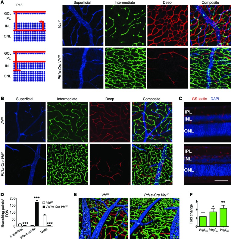
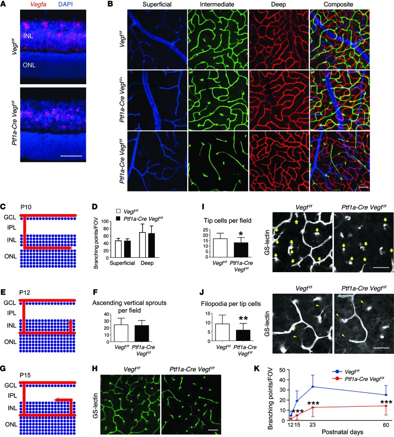
Functional interactions between neurons, vasculature, and glia within neurovascular units are critical for maintenance of the retina and other CNS tissues. For example, the architecture of the neurosensory retina is a highly organized structure with alternating layers of neurons and blood vessels that match the metabolic demand of neuronal activity with an appropriate supply of oxygen within perfused blood. Here, using murine genetic models and cell ablation strategies, we have demonstrated that a subset of retinal interneurons, the amacrine and horizontal cells, form neurovascular units with capillaries in 2 of the 3 retinal vascular plexuses. Moreover, we determined that these cells are required for generating and maintaining the intraretinal vasculature through precise regulation of hypoxia-inducible and proangiogenic factors, and that amacrine and horizontal cell dysfunction induces alterations to the intraretinal vasculature and substantial visual deficits. These findings demonstrate that specific retinal interneurons and the intraretinal vasculature are highly interdependent, and loss of either or both elicits profound effects on photoreceptor survival and function.


Figures

Figure 6
Figure 6. Intermediate plexus abnormalities are associated with visual dysfunction.
(A) Full-field ERGs in 2-month-old Ptf1a-Cre Vegff/f mice reveal significant defects in both rod- and cone-driven pathways (n = 8–10). (B) Full-field ERGs performed on 2-month-old Ptf1a-Cre Vhlf/f mice reveal that the rod-driven pathways are significantly impaired (n = 6). (CE) Photoreceptor atrophy is accelerated in a mouse model of spontaneous retinal degeneration (rd10 mice) with impaired intermediate plexuses, based on TUNEL staining in the ONL (C; green, quantified in D), and reduced ONL thickness values (E) in P21 Ptf1a-Cre Vegff/f Pde6brd10/rd10 mice and controls (Vegff/f Pde6brd10/rd10; n = 6 each). *P < 0.05, **P < 0.01, ***P < 0.001; 2-tailed Student’s t tests. Error bars indicate mean ± SD. Scale bar: 50 μm.
Figure 5
Figure 5. Genetic ablation of amacrine and horizontal cells phenocopies the defects in the intermediate plexus observed in Hif-1α and Vegfa mutants.
(A) DT was injected daily at the time points indicated. (BE) The reduced number of cells after ablation was examined by comparing cryosectioned retinas from P23 Ptf1a-Cre R26iDTR/+:tdTomato/+ and P23 Ptf1a-Cre R26+/+:tdTomato/+ mice after injecting DT (B), and by measuring the thickness of the GCL/IPL and INL (C; yellow brackets) from images captured in vivo using SD-OCT (D and E) (n = 4–6). (F and G) An attenuated intermediate plexus is observed (F; green) and quantified (G) in P23 Ptf1a-Cre R26iDTR/+ mice (n = 6). (H and I) An attenuated intermediate plexus is also seen when amacrine and horizontal cells are ablated well after intermediate plexus development (H; quantified in I), suggesting that amacrine cells are required for development and maintenance of the intermediate plexus (n = 4). *P < 0.05, **P < 0.01, ***P < 0.001; 2-tailed Student’s t tests. Error bars indicate mean ± SD. Scale bar: 50 μm (B, F, and H).
Figure 4
Figure 4. VHL/HIF-1α/VEGF signaling regulates angiogenesis in the intermediate plexus.
(AF) Compared with control (A), combinatorial conditional KO strategies were employed to show that the loss of HIF-1α (B; quantified in H) but not HIF-2α (C; quantified in I) in amacrine and horizontal cells interferes with intermediate plexus development in haplosufficient P23 Vhl+/- mutants. (DF) Homozygous deletion of P23 Vhl and Hif-1α (E) prevents the neovascularization observed in Vhl mutants (D and E; quantified in J), but deletion of Hif-2α elicits no effect (F; quantified in K) compared with controls (D). (G; quantified in L) Homozygous deletion of Vhl and Vegfa also rescues the Vhl neovascular phenotype. (All assays were performed in P23-staged mice; n = 4–6). (M) Relative mRNA expression values from qPCR gene-profiling analysis of 84 angiogenesis-related genes in Ptf1a-Cre Vhlf/f Vegff/f retinas at P12 compared with controls (harboring floxed alleles but no Cre); upregulated genes (P < 0.05 or fold change > 1.5 [marked by red dashed line]) are plotted (n = 4). *P < 0.05, **P < 0.01, ***P < 0.001; 2-tailed Student’s t tests. Error bars indicate mean ± SD. Scale bars: 50 μm (AG).
Figure 3
Figure 3. Vhl deletion in amacrine and horizontal cells induces formation of a dense and convoluted intermediate plexus at the expense of the deep plexus.
(A and B) Schematic of angiogenesis in Vhlf/f (control) or Ptf1a-Cre Vhlf/f retinas at P13. Note dramatic alterations in the intermediate plexus (green) and deep plexus (red) at P13 (A) and P23 (B) in flat-mounted retinas. (C) 100 μm sections from P23 Ptf1a-Cre Vhlf/f mice were stained with GS-lectin to highlight the extent of the neovascularization in the VHL mutants. Counterstained with DAPI. (D) The number of branching events in P13 Vhlf/f or Ptf1a-Cre Vhlf/f retinas was plotted (n = 4). (E) Three-dimensional reconstruction of 3 retinal plexuses in P23 Ptf1a-Cre Vhlf/f retina (superficial, blue; intermediate, green; deep plexus, red) highlighted the abnormally dense intermediate plexus. (F) qPCR analyses revealed that nondiffusible Vegf188 was the most abundant isoform expressed in Ptf1a-Cre Vhlf/f mice at P15 (n = 4). *P < 0.05, **P < 0.01, ***P < 0.001; 2-tailed Student’s t tests. Error bars indicate mean ± SD. Scale bars: 50 μm (AC and E).
Figure 2
Figure 2. Vegfa deletion in amacrine and horizontal cells severely impairs intraretinal vasculature development.
(A) In situ hybridization was performed on P12 Vegff/f or Ptf1a-Cre Vegff/f cryosectioned retinas with a Vegfa probe (counterstained with DAPI). (B) The intermediate plexus (green) is severely attenuated in P23 Ptf1a-Cre Vegff/f compared with controls. (C) Schematic of deep plexus development in a P10-staged mouse. Arrow illustrates the lateral growth of blood vessels in the OPL. (D) The number of branching events in the superficial and deep plexuses of P10 Vegff/f or Ptf1a-Cre Vegff/f retinas were counted and plotted (n = 4). (E) Schematic of vertical sprouting events from the deep plexus in P12 retinas. Arrow illustrates the direction of vascular sprouting from the deep plexus to the IPL. (F) There are no differences in the numbers of ascending vertical sprouts of Ptf1a-Cre Vegff/f in flat-mounted retinas compared with controls (Vegff/f) (n = 4). (G) Schematic of intermediate plexus development at P15. Arrow illustrates the lateral growth of blood vessels in the IPL. (HJ) GS-lectin–positive laterally expanding sprouts are fewer in number in P15 Ptf1a-Cre Vegff/f mice due to a reduced number of tip cells (I; arrows) and filopodia (J; arrowheads) (n = 4–5). (K) The number of branching points in the intermediate plexus were counted, quantified, and plotted at P12, P15, P23, and P60 (n = 4–6). *P < 0.05, **P < 0.01, ***P < 0.001; 2-tailed Student’s t tests. Error bars indicate mean ± SD. Scale bars: 50 μm (A, B, and H); 40 μm (I and J).
Figure 1
Figure 1. Amacrine and horizontal cells form neurovascular units with the intraretinal capillaries.
(A) Pseudocolored cross section of an adult murine retina. (B) Immunohistochemistry was used to identify putative neurovascular units between amacrine cells (arrows) or horizontal cells (arrowheads) with the vasculature using anti-calbindin (green), anti-CD31 (red), and DAPI (blue) in WT retinal cryosections at P23. (C) Cre recombination reporters label amacrine and horizontal cell nuclei in P23 Ptf1a-Cre R26tdTomato/+ mice (tomato signal was pseudocolored green). (DI) Amacrine (DF) and horizontal cell (D and G) neurites (NF-M labeled, green) associate with the intraretinal vasculature (GS-lectin, blue) as seen in thick cut (100 μm) sections (amacrine/horizontal nuclei, red). (E) Adjacent optical slices from the region of interest boxed in (D); arrows mark colocalization. (F and G) Flat-mounted P23 Ptf1a-Cre R26tdTomato/+ retinas colabeled with anti-neurofilament and GS-lectin (endothelial cell marker). (H and I) Amacrine cell neurites are decorated with GFP in Ptf1a-Cre R26GFP mice and can be observed in close proximity to GS-lectin–positive endothelial cells. Immunofluorescence for MAP2 in whole-mount retinas at P23 also reveals colocalization of amacrine and horizontal cell neurites with the intraretinal vasculature. Scale bars: 50 μm (AE, H, and I); 20 μm (F and G).

References

    1. Storkebaum E, Quaegebeur A, Vikkula M, Carmeliet P. Cerebrovascular disorders: molecular insights and therapeutic opportunities. Nat Neurosci. 2011;14(11):1390–1397. doi: 10.1038/nn.2947. - DOI - PubMed
    1. Stanimirovic DB, Friedman A. Pathophysiology of the neurovascular unit: disease cause or consequence? J Cereb Blood Flow Metab. 2012;32(7):1207–1221. doi: 10.1038/jcbfm.2012.25. - DOI - PMC - PubMed
    1. Sapieha P. Eyeing central neurons in vascular growth and reparative angiogenesis. Blood. 2012;120(11):2182–2194. doi: 10.1182/blood-2012-04-396846. - DOI - PubMed
    1. Fruttiger M. Development of the retinal vasculature. Angiogenesis. 2007;10(2):77–88. doi: 10.1007/s10456-007-9065-1. - DOI - PubMed
    1. Caprara C, Thiersch M, Lange C, Joly S, Samardzija M, Grimm C. HIF1A is essential for the development of the intermediate plexus of the retinal vasculature. Invest Ophthalmol Vis Sci. 2011;52(5):2109–2117. doi: 10.1167/iovs.10-6222. - DOI - PubMed

Publication types

MeSH terms