. 2015 Apr 27;10(4):e0125701.
doi: 10.1371/journal.pone.0125701. eCollection 2015.

Enhanced Sensitivity for Detection of HIV-1 p24 Antigen by a Novel Nuclease-Linked Fluorescence Oligonucleotide Assay

Affiliations

Enhanced Sensitivity for Detection of HIV-1 p24 Antigen by a Novel Nuclease-Linked Fluorescence Oligonucleotide Assay

Peihu Fan et al. PLoS One. .

Abstract

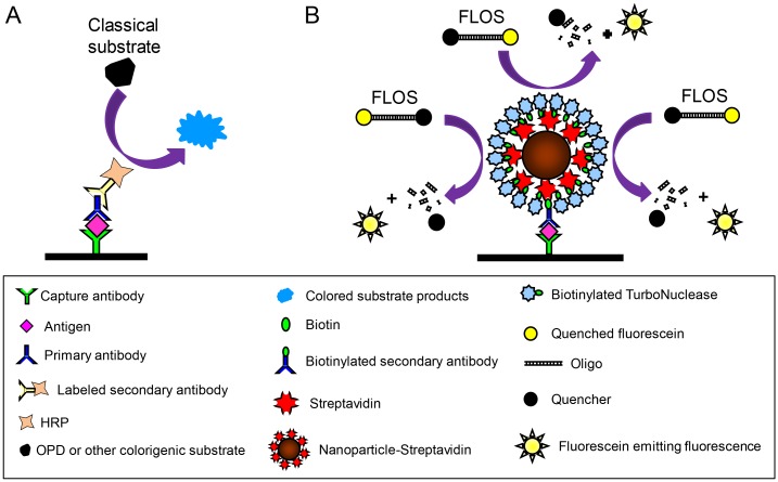
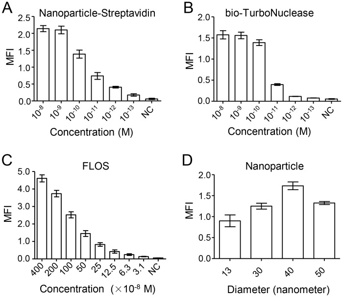
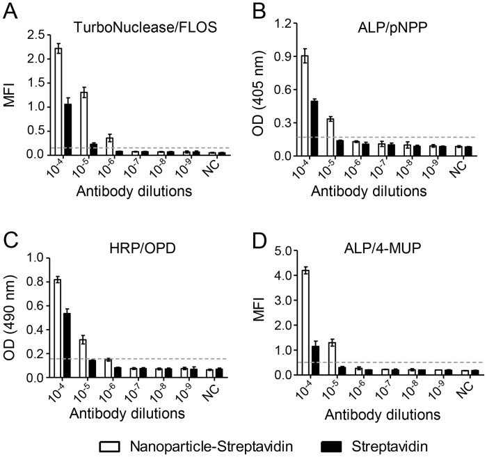
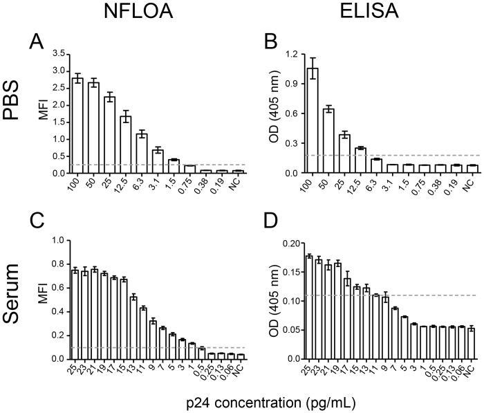
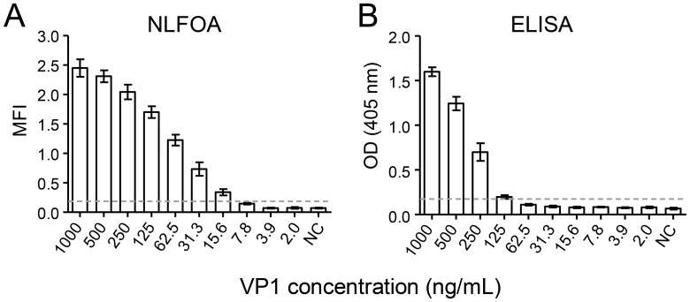
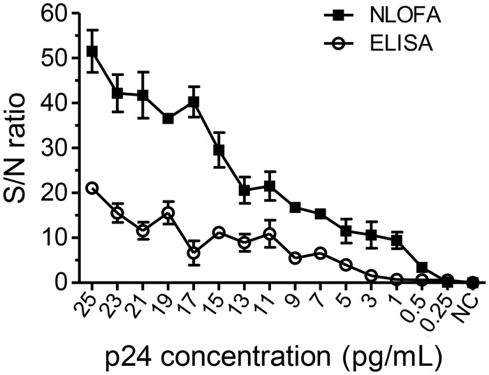
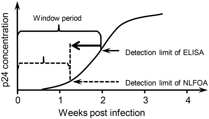
The relatively high detection limit of the Enzyme-linked immunosorbent assay (ELISA) prevents its application for detection of low concentrations of antigens. To increase the sensitivity for detection of HIV-1 p24 antigen, we developed a highly sensitive nuclease-linked fluorescence oligonucleotide assay (NLFOA). Two major improvements were incorporated in NLFOA to amplify antibody-antigen interaction signals and reduce the signal/noise ratio; a large number of nuclease molecules coupled to the gold nanoparticle/streptavidin complex and fluorescent signals generated from fluorescent-labeled oligonucleotides by the nuclease. The detection limit of p24 by NLFOA was 1 pg/mL, which was 10-fold more sensitive than the conventional ELISA (10 pg/mL). The specificity was 100% and the coefficient of variation (CV) was 7.8% at low p24 concentration (1.5 pg/mL) with various concentrations of spiked p24 in HIV-1 negative sera. Thus, NLFOA is highly sensitive, specific, reproducible and user-friendly. The more sensitive detection of low p24 concentrations in HIV-1-infected individuals by NLFOA could allow detection of HIV-1 infections that are missed by the conventional ELISA at the window period during acute infection to further reduce the risk for HIV-1 infection due to the undetected HIV-1 in the blood products. Moreover, NLFOA can be easily applied to more sensitive detection of other antigens.


Conflict of interest statement

Competing Interests: The authors have declared that no competing interests exist.

Figures

Fig 1
Fig 1. Schematic presentation of the nuclease-linked fluorescence oligonucleotide assay (NLFOA).
(A) In the conventional ELISA, the capture antibody-antigen complex binds to one primary antibody and subsequently to one enzyme-conjugated second antibody. The enzyme acts on the substrate to generate colored substrate products. The reaction is detected by measuring absorbance at a certain wavelength of the chemical reaction. (B) In NLFOA, the capture antibody-antigen complex binds to one biotinylated primary antibody, then to the nanoparticle-streptavidin complex containing multiple copies of streptavidins, and finally to multiple copies of biotinylated TurboNuclease. The fluorescent labeled oliogonucleotide substrate (FLOS), which does not emit fluorescence due to the Iowa Black FQ (quencher) at the other end of the oligo, can generate fluorescence after digestion by TurboNuclease.
Fig 2
Fig 2. Determination of relative activity of different nucleases.
(A) The nuclease activity was determined by mixing FLOS with serially diluted enzymes (1:10). The mean fluorescence intensity (MFI) was measured every 5 minutes. (B) Analysis of the enzyme contents in the commercial enzyme stocks by SDS-PAGE. Equal volume (10 μl) of each commercial nuclease was loaded onto a SDS-PAGE gel for electrophoresis. Exonuclease III and TurboNuclese were diluted at 1:100 and 1:10, respectively, due to the high protein concentration in the samples. (C) Determination of enzyme relative activity. The relative activity of each enzyme was defined by the positive MFI (2.1 folds above the background) at the lowest concentration of the enzyme (MFI/ng). All experiments were independently performed three times and results from one representative experiment are shown. Negative control (NC) contained all components except the enzyme. Means ± SD are shown.
Fig 3
Fig 3. Comparison of minimal required enzyme concentrations among different detection systems.
The lowest concentration of the enzyme required for detection was determined for four different enzyme-substrate systems: (A) TurboNuclease and FLOS system; (B) alkaline phosphatase (ALP) and p-nitrophenylphosphate (pNPP) system; (C) horseradish peroxidase (HRP) and o-Phenylenediamine (OPD) system; and (D) ALP and 4-methylumbelliferyl phosphate (4-MUP, a classical fluorogenic substrate) system. Each enzyme was prepared at 1:10 serial dilutions (starting at 10–7 M) and reacted with its corresponding substrate. Negative control (NC) contained all components except the enzyme. The cutoff (2.1 fold higher than the NC signal) is indicated by the dotted line. Means ± SD are shown.
Fig 4
Fig 4. Similar enzyme activity between TurboNuclease and bio-TurboNuclease.
The mean fluorescence intensity (MFI) was compared between TurboNuclease and bio-TurboNuclease at 1:10 serial dilutions (p>0.2, Student’s t test). Each assay was performed in five independent experiments. Means ± SD are shown.
Fig 5
Fig 5. Optimization of the components in NLFOA.
The NLFOA conditions were optimized for (A) nanoparticle-streptavidin, (B) bio-TurboNuclease, (C) FLOS and (D) diameters of the nanoparticles. Both nanoparticle-streptavidin and bio-TurboNuclease were prepared at 1:10 serial dilutions between 10–8 -10-13 M. The FLOS oligos were prepared at 1:2 serial dilutions between 400–3.1 ×10⁻⁸ M. Four different diameter sizes (13, 30, 40 and 50 nm) were analyzed for nanoparticles. Negative control (NC) contained all components except what was to be optimized. Each of the component was analyzed in five repeats and were performed in two independent experiments. Means ± SD are shown.
Fig 6
Fig 6. Increased sensitivity with nanoparticle-streptavidin and FLOS.
The signals generated by the nanoparticle-streptavidin and streptavidin systems were compared among four different enzyme-substrate systems: (A) TurboNuclease and FLOS; (B) alkaline phosphatase (ALP) and p-nitrophenylphosphate (pNPP); (C) horseradish peroxidase (HRP) and o-Phenylenediamine (OPD); and (D) alkaline phosphatase (ALP) and 4-methylumbelliferyl phosphate (4-MUP). Each enzyme was prepared at 1:10 serial dilutions (10–4–10–9 M). Negative control (NC) contained all components except the enzyme. The cutoff (2.1 fold higher than the NC signal) is indicated by the dotted line. Means ± SD are shown.
Fig 7
Fig 7. Comparison of the detection limit between NLFOA and the conventional ELISA.
The detection limit for HIV-1 p24 antigen diluted in PBS by NLFOA (A) and conventional ELISA (B). The detection limit of HIV-1 p24 antigen spiked in sera from a healthy human by NLFOA (C) and conventional ELISA (D). The dotted line indicates the cutoff value that is 2.1-fold of that of the negative control (NC). Experiments with p24 in PBS (A and B) were done in five repeats and experiments with p24 in sera (C and D) were done in eight repeats. Each assay was performed in three independent experiments. Means ± SD are shown.
Fig 8
Fig 8. Detection of the EV71 VP1 antigen by NLFOA.
The EV71 VP1 antigen was spiked in sera and anzlysed in parallel by NLFOA (A) and the conventional ELISA (B). The dotted line indicates the cutoff value that is 2.1-fold of that of the negative control (NC). Each assay was performed in three independent experiments. Means ± SD are shown.
Fig 9
Fig 9. Enhanced precision with NLFOA.
The signal/noise (S/N) ratios were determined for both NLFOA and ELISA assays at different p24 concentrations.
Fig 10
Fig 10. Shortened window period of HIV-1 infection by NLFOA.
The HIV-1 p24 antigen can only be detected after a period of time after HIV-1 infection (window period) by the conventional ELISA. The higher sensitivity of NLFOA can likely shorten this window period.

References

    1. Gan SD, Patel KR. Enzyme immunoassay and enzyme-linked immunosorbent assay. J Invest Dermatol. 2013;133: e12 10.1038/jid.2013.287 - DOI - PubMed
    1. Lequin RM. Enzyme immunoassay (EIA)/enzyme-linked immunosorbent assay (ELISA). Clin Chem. 2005;51: 2415–2418. - PubMed
    1. Crowther J. Enzyme Linked Immunosorbent Assay (ELISA) In: Walker JM, Rapley R, editors. Molecular Biomethods Handbook. 2nd ed New York: Humana Press; 2008. p. 657–682.
    1. Meng Y, High K, Antonello J, Washabaugh MW, Zhao Q. Enhanced sensitivity and precision in an enzyme-linked immunosorbent assay with fluorogenic substrates compared with commonly used chromogenic substrates. Anal Biochem. 2005;345: 227–236. - PubMed
    1. Yolken RH, Stopa PJ. Enzyme-linked fluorescence assay: Ultrasensitive solid-phase assay for detection of human rotavirus. J Clin Microbiol. 1979;10: 317–321. - PMC - PubMed

Publication types

MeSH terms