Review
. 2015 Jul;13(7):403-13.
doi: 10.1038/nrmicro3449. Epub 2015 Apr 27.

HIV-1 and interferons: who's interfering with whom?

Affiliations
Review

HIV-1 and interferons: who's interfering with whom?

Tomas Doyle et al. Nat Rev Microbiol. 2015 Jul.

Abstract

The ability of interferons (IFNs) to inhibit HIV-1 replication in cell culture models has long been recognized, and the therapeutic administration of IFNα to HIV-1-infected patients who are not receiving antiretroviral therapy produces a clear but transient decrease in plasma viral load. Conversely, studies of chronic HIV-1 infection in humans and SIV-infected animal models of AIDS show positive correlations between elevated plasma levels of IFNs, increased expression of IFN-stimulated genes (ISGs), biomarkers of inflammation and disease progression. In this Review, we discuss the evidence that IFNs can control HIV-1 replication in vivo and debate the controversial role of IFNs in promoting the pathological sequelae of chronic HIV-1 infection.


Conflict of interest statement

Competing interests statement

The authors declare no competing interests.

Figures
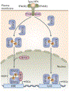
Figure 1 |
Figure 1 |. Induction of ISG expression.
Type I interferons (IFNs) bind to type I IFN receptor (IFNAR), which is composed of IFNAR1 and IFNAR2 subunits, leading to tyrosine kinase 2 (TYK2) and Janus kinase 1 (JAK1) activation. These kinases phosphorylate (P) signal transducer and activator of transcription 1 (STAT1) and STAT2 to allow homodimerization (for STAT1) and heterodimerization (STAT1 plus STAT2). These STAT dimers then translocate to the nucleus. STAT1–STAT2 dimers to bind interferon regulatory factor 9 (IRF9) to form the interferon-stimulated gene factor 3 (ISCF3) complex, which engages IFN-stimulated response elements (ISREs), whereas STAT1 homodimers engage gamma-activated seguences (GASs). Binding of the STAT dimers to ISREs and GASs activates transcription of IFN-stimulated genes (ISGs). Other STAT dimers, phosphoinositide 3-kinases and mitogen-activated protein kinases may also be activated downstream of type I IFNs.
Figure 2 |
Figure 2 |. Intracellular sensing of HIV-1 Infection.
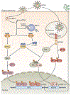
Following HIV-1 entry into the cell, viral RNA is reverse transcribed into cDNA, which is detected by the cytoplasmic receptors cyclic GMP–AMP (cGAMP) synthase (cGAS) and interferon-γ (IFNγ)-inducible protein 16 (IFI16). Following cDNA detection, IFI16 activates stimulator of IFN genes (STING), which leads to the activation of TANK-binding kinase 1 (TBK1) and the subseguent phosphorylation (P) of the IFN regulatory factor 3 (IRF3). Phosphorylated IRF3 can then engage IFN-stimulated response elements (ISREs), thereby inducing the expression of type I IFNs. When cGAS detects viral cDNA, the enzyme produces cGAMP, which leads to the activation of STING. STING then activates the inhibitor of NF-κB (IκB) kinase (IKK) complex and TBK1, leading to the activation of nuclear factor-κB (NF-κB) and IRF3, respectively. These transcription factors induce the expression of genes encoding IFNs and other pro-inflammatory cytokines. The cellular 3′-repair exonuclease 1 (TREX1) helps HIV-1 to evade cytosolic sensing by degrading viral cDNA in the cytoplasm. In addition to sensing cytoplasmic viral cDNA, cells can also sense HIV-1 single-stranded RNA (ssRNA) via Toll-like receptor 7 (TLR7) in endosomes, especially in plasmacytoid dendritic cells (pDCs). TLR7 activation by ssRNA in pDC endosomes results in the activation of myeloid differentiation primary response gene 88 (MYD88) and subseguent induction of IFN via activation of IRF7 and NF-κB.
Figure 3 |
Figure 3 |. HIV-1 restriction and resistance factors.
In the absence of viraLLy encoded antagonists (or viral escape), host cell proteins called HIV-1 restriction factors (yellow) inhibit various stages of the replication cycle. The tripartite motif-containing protein 5α (TRIM5α) promotes accelerated fragmentation of viral cores, preventing cDNA synthesis. SAM and HD domain-containing protein 1 (SAMHD1) depletes the cellular levels of 2′-deoxynucleoside 5′-triphosphates (dNTPs), which are reguired for efficient cDNA synthesis. APOBEC3 (apolipoprotein B mRNA-editing enzyme, catalytic polypeptide-like 3) proteins interfere with the processivity of HIV-1 reverse transcriptase and induce hypermutation of viral cDNA by cytidine deamination. Tetherin prevents the release of budded virions from the infected cell. Several viral proteins (blue) antagonize these cellular restriction factors. Viral infectivity factor (Vif) antagonizes APOBEC3 proteins, viral protein unique (Vpu) antagonizes tetherin, and the HIV-2 viral protein X (Vpx) antagonizes SAMHD1. HIV-1 resistance factors (brown) inhibit other stages of viral replication and are not counteracted by the virus. Myxovirus resistance 2 (MX2) prevents the nuclear import and integration of viral cDNA. Schlafen 11 (SLFN11) suppresses the translation of viral proteins. Interferon-induced transmembrane proteins (IFITMs) inhibit viral entry by interfering with membrane fusion. dsDNA, double-stranded DNA; gRNA, viral genomic RNA; LTR, long terminal repeat; ssDNA, single-stranded DNA.
Figure 4 |
Figure 4 |. The effect of IFNα treatment on plasma HIV-1 viral load.
The graph shows plasma HIV-1 viral load (PVL) responses in patients who are infected with HIV-1 and who have not received antiretroviral therapy, during 12 weeks of treatment with pegylated-interferon-α (IFNα). The thick dashed line indicates the median PVL. IFNα treatment induces a rapid decline in PVL in most patients, whereas a minority fail to respond. The PVL reaches a nadir at 2 weeks (median reduction of 1.3 log10 copies ml−1 from baseline), followed by partial reversal of the response. Adapted from Asmuth, D. M. et al., Safety, tolerability, and mechanisms of antiretroviral activity of pegylated interferon alfa-2a in HIV-l-monoinfected participants: a phase II clinical trial, J. Infect. Dis., 2010, 201, 11, 1686–1196, by permission of Oxford University Press.

References

    1. Faria NR et al. HIV epidemiology. The early spread and epidemic ignition of HIV-1 in human populations. Science 346, 56–61 (2014). - PMC - PubMed
    1. D’Arc M et al. Origin of the HIV-1 group O epidemic in western lowland gorillas. Proc. Natl Acad. Sci. USA 112, E1343–E1352 (2015). - PMC - PubMed
    1. Takeuchi O & Akira S Pattern recognition receptors and inflammation. Cell 140, 805–820 (2010). - PubMed
    1. Rotger M et al. Genome-wide mRNA expression correlates of viral control in CD4+ T-cells from HIV-1-infected individuals. PLoS Pathog. 6, e1000781 (2010). - PMC - PubMed
    2. This study characterizes the gene expression profile abnormalities of the CD4+ T cells of patients with HIV-1 infection according to plasma viral load.

    1. Hyrcza MD et al. Distinct transcriptional profiles in ex vivo CD4+ and CD8+ T cells are established early in human immunodeficiency virus type 1 infection and are characterized by a chronic interferon response as well as extensive transcriptional changes in CD8+ T cells. J. Virol. 81, 3477–3486 (2007). - PMC - PubMed

Publication types