. 2015 Apr 27;10(4):e0125293.
doi: 10.1371/journal.pone.0125293. eCollection 2015.

Computational Tools for Interpreting Ion Channel pH-Dependence

Affiliations

Computational Tools for Interpreting Ion Channel pH-Dependence

Ivan Sazanavets et al. PLoS One. .

Abstract

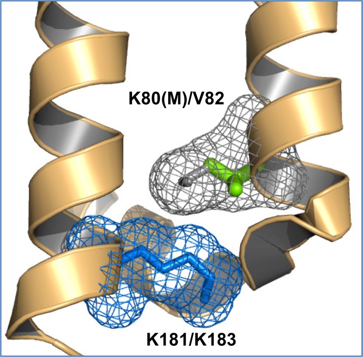
Activity in many biological systems is mediated by pH, involving proton titratable groups with pKas in the relevant pH range. Experimental analysis of pH-dependence in proteins focusses on particular sidechains, often with mutagenesis of histidine, due to its pKa near to neutral pH. The key question for algorithms that predict pKas is whether they are sufficiently accurate to effectively narrow the search for molecular determinants of pH-dependence. Through analysis of inwardly rectifying potassium (Kir) channels and acid-sensing ion channels (ASICs), mutational effects on pH-dependence are probed, distinguishing between groups described as pH-coupled or pH-sensor. Whereas mutation can lead to a shift in transition pH between open and closed forms for either type of group, only for pH-sensor groups does mutation modulate the amplitude of the transition. It is shown that a hybrid Finite Difference Poisson-Boltzmann (FDPB) - Debye-Hückel continuum electrostatic model can filter mutation candidates, providing enrichment for key pH-coupled and pH-sensor residues in both ASICs and Kir channels, in comparison with application of FDPB alone.


Conflict of interest statement

Competing Interests: This study was partly funded by Astra Zeneca. There are no patents, products in development or marketed products to declare. This does not alter the authors' adherence to all the PLOS ONE policies on sharing data and materials, as detailed online in the guide for authors.

Figures

Fig 1
Fig 1. Prediction of pH-sensing residues for Kir1.1.
(A) Cartoon of a Kir1.1 monomer (from the tetramer), with extracellular loops upper, intracellular domain lower and, the intervening helices marking the transmembrane regions. Residues identified as influencing the pH-dependent transition (see text) are marked with green surfaces. (B) and (C) Scatter plots of predicted pKas for closed and partially open channels, with the orange diagonal indicating equal pKas in these forms. Choosing a pH range of physiological interest between pH 6 and 8, the shaded regions (panel C) contain groups for which ionisation is predicted to change between closed and partially open forms, at physiological pH. Panel B is for FDPB calculations and panel C for FD/DH calculations, with groups K80 and K229 highlighted.
Fig 2
Fig 2. Predicted pH-sensor and pH-coupled residues in Kir1.1.
(A) pH-dependence of open-closed conformation energetics predicted for rat Kir1.1 wild type and R311Q mutant. A shift in the plots, rather than a change in slope, is shown, suggesting pH-coupling rather than pH-sensing. (B) A single monomer of rat Kir1.1 is shown, but with a positive potential contour (kT/2e) displayed (blue) for all four K80 amino sidechain groups in the tetramer. R311 and E302 lie outside of this contour, indicating low interaction with K80, consistent with pH-coupling. Other listed groups lie within the contour and therefore have potential to interact directly with the K80 pH sensor residue(s).
Fig 3
Fig 3. The environment of K181 in rat Kir1.1 compared with K183 of puffer fish Kir1.1.
Whereas the key pH-sensing residue K80 (grey stick and mesh) is a neighbour of K181 (bue mesh and stick) in rat Kir1.1, the presence of valine (green stick) at the equivalent position in puffer fish could allow an altered conformation of K183 (not drawn).
Fig 4
Fig 4. Prediction of pH-sensing residues for ASIC1a.
(A) Cartoon of an ASIC1a monomer (from the trimer), with the transmembrane helices evident in the lower part, and residues found to influence pH-dependence (see text) marked as green surfaces in the extracellular region. (B) and (C) Scatter plots of predicted pKas for closed (desensitised) and partially open channels, with the orange diagonal indicating equal pKas in these forms. With a pH range of physiological interest between pH 6 and 8, the red shaded regions (panel C) contain groups for which ionisation is predicted to change between closed and partially open forms. Panel B is for FDPB calculations and panel C for FD/DH calculations, with various groups highlighted.
Fig 5
Fig 5. Calcium binding and pH-dependence in ASIC1a.
Plots of predicted pH-dependence for the difference of partially open—closed (desensitised) channels are shown. The slope of a plot (blue) that takes no account of calcium binding is not in the right sense of relative open state stabilisation at lower pH, whilst that with carboxylic acid deletion to mimic calcium binding (red, see text) is consistent with channel opening. Finally, on the calcium-binding mimic background, the E412Q mutation (green) shifts the curve without substantially altering the slope.
Fig 6
Fig 6. Predicted ionisation differences for packing changes of partially buried groups in pH-sensitive and pH-insensitive ASICs.
(A) and (B) Scatter plots of ionisation changes (upon different repackings), calculated at pH 6.5, for the closed desensitised chicken ASIC1a structure (pH-sensitive, panel A), and a model of the equivalent structure for rat ASIC4 (pH-insensitive, panel B). Off-diagonal groups, those predicted to contribute to pH-dependence, are given with ASIC1a numbering.
Fig 7
Fig 7. Charge differences between pH-sensitive and pH-insensitive ASICs.
(A) Closed (desensitised) chicken ASIC1a structure and rat ASIC4 model, with electrostatic contours plotted (for physiological pH), at a relatively low contour (+/- kT/e, red is negative and blue shows positive potential). (B) The potential fields of (A) are differenced at a much higher level (+/- 16 kT/e) to identify the regions of most interest, which appear as the red mesh contour, indicating more negative contours in ASIC1a. These contours coincide with the groups indicated in the predictions of pH-sensitivity for ASIC1a (green surface) shown in Figs 4C and 6A. No calcium ions were included in these calculations.
Fig 8
Fig 8. Schematic representation of coupling to a pH-dependent transition.
In this example a salt-bridge is made in a channel closed form, but not in the open form, but this could be any type of interaction that varies between these forms. (A) General form of pH-dependent stability for a single folded conformation (F) relative to unfolded (U), ∆GFU. The standard pH titration ranges are marked for acidic and basic residues, omitting tyrosine, cysteine and amino acid termini, and ignoring groups with large ∆pKas. The effect of salt-bridge mutation has been exaggerated in scale, but importantly gives no change in slope of pH-dependence at neutral (physiological) pH. Wild type channel is shown schematically in panel B, together with schematic conformational energy curves for open and closed forms drawn such that they cross-over at pH50 around physiological pH. Upon salt-bridge mutation (loss of the acidic group, panel C), the open form curve remains the same as that for wild type, but the closed form is destabilised uniformly across physiological pH. As a result pH50 for the mutant form shifts, although there is no ionisation change at this pH between open and closed forms. The mutated interaction is therefore termed as coupled to pH-dependence, rather than underlying the pH-sensor itself.

Similar articles

Cited by

References

    1. Talley K, Alexov E (2010) On the pH-optimum of activity and stability of proteins. Proteins 78: 2699–2706. 10.1002/prot.22786 - DOI - PMC - PubMed
    1. Srivastava J, Barber DL, Jacobson MP (2007) Intracellular pH sensors: design principles and functional significance. Physiology (Bethesda) 22: 30–39. - PubMed
    1. Paynter JJ, Shang L, Bollepalli MK, Baukrowitz T, Tucker SJ (2010) Random mutagenesis screening indicates the absence of a separate H(+)-sensor in the pH-sensitive Kir channels. Channels (Austin) 4: 390–397. 10.4161/chan.4.5.13006 - DOI - PMC - PubMed
    1. Gonzalez W, Riedelsberger J, Morales-Navarro SE, Caballero J, Alzate-Morales JH, et al. (2012) The pH sensor of the plant K+-uptake channel KAT1 is built from a sensory cloud rather than from single key amino acids. Biochemical Journal 442: 57–63. 10.1042/BJ20111498 - DOI - PubMed
    1. Liechti LA, Berneche S, Bargeton B, Iwaszkiewicz J, Roy S, et al. (2010) A combined computational and functional approach identifies new residues involved in pH-dependent gating of ASIC1a. J Biol Chem 285: 16315–16329. 10.1074/jbc.M109.092015 - DOI - PMC - PubMed

Publication types

LinkOut - more resources