. 2015 Apr 27;10(4):e0125621.
doi: 10.1371/journal.pone.0125621. eCollection 2015.

REGULATOR OF BULB BIOGENESIS1 (RBB1) Is Involved in Vacuole Bulb Formation in Arabidopsis

Affiliations

REGULATOR OF BULB BIOGENESIS1 (RBB1) Is Involved in Vacuole Bulb Formation in Arabidopsis

Sang Won Han et al. PLoS One. .

Abstract

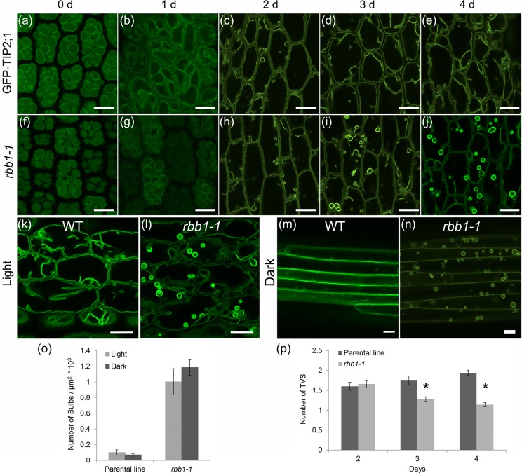
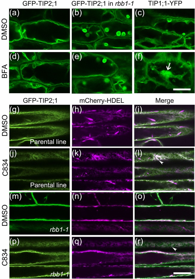
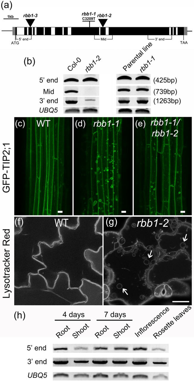
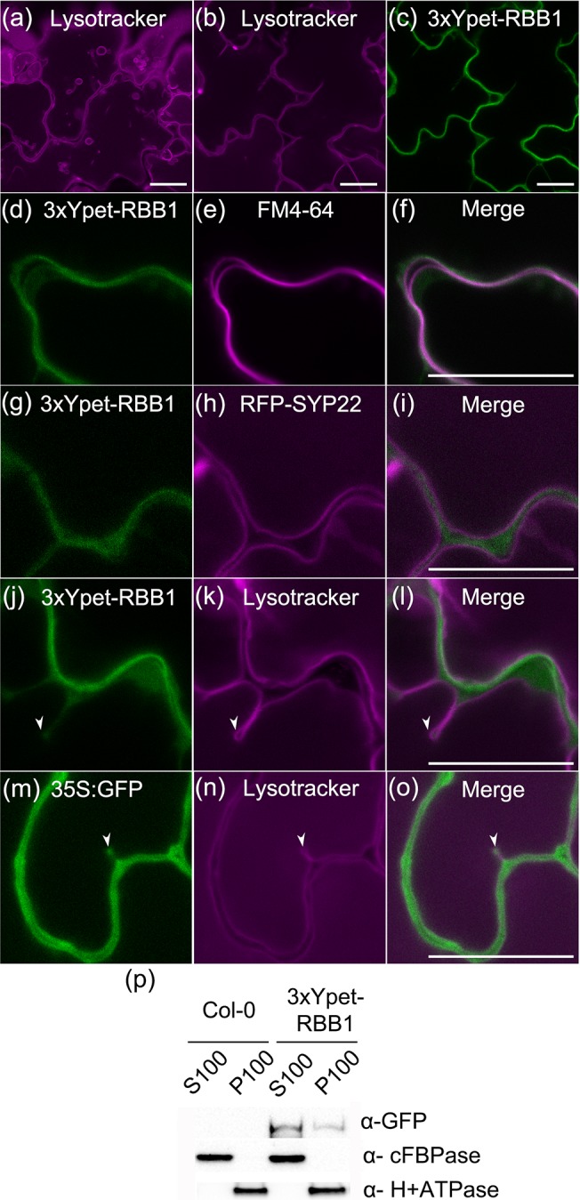
Vacuoles are dynamic compartments with constant fluctuations and transient structures such as trans-vacuolar strands and bulbs. Bulbs are highly dynamic spherical structures inside vacuoles that are formed by multiple layers of membranes and are continuous with the main tonoplast. We recently carried out a screen for mutants with abnormal trafficking to the vacuole or aberrant vacuole morphology. We characterized regulator of bulb biogenesis1-1 (rbb1-1), a mutant in Arabidopsis that contains increased numbers of bulbs when compared to the parental control. rbb1-1 mutants also contain fewer transvacuolar strands than the parental control, and we propose the hypothesis that the formation of transvacuolar strands and bulbs is functionally related. We propose that the bulbs may function transiently to accommodate membranes and proteins when transvacuolar strands fail to elongate. We show that RBB1 corresponds to a very large protein of unknown function that is specific to plants, is present in the cytosol, and may associate with cellular membranes. RBB1 is involved in the regulation of vacuole morphology and may be involved in the establishment or stability of trans-vacuolar strands and bulbs.


Conflict of interest statement

Competing Interests: The authors have declared that no competing interests exist.

Figures

Fig 1
Fig 1. regulator of bulb biogenesis1 contains many bulbs in many cell types.
(a-h) rbb1-1 mutants have more bulbs than the parental line. GFP-TIP2;1 localization in the parental line (WT, a-d) or rbb1-1 (e-h). Four-day-old seedlings were imaged by confocal microscopy to visualize morphology of the vacuole in the epidermis of hypocotyl (a, e) and cotyledon (b, f), and in root epidermis and cortex (c, g). The bulb phenotype was also observed in rosette leaves of 6-week-old plants (d, h). Scale bar = 20 μm. (i-n) GFP-TIP2;1 and RFP-TIP1;1 co-localize in rbb1-1 bulbs. The hypocotyls of 4-day-old dark-grown seedlings expressing GFP-TIP2;1 (green, i, l) and RFP-TIP1;1 (magenta, j, m) in the parental line (i-k) and rbb1-1 mutants (l-n) are shown. Merged images (k, n) are also shown. Scale bar: 20 μm. (o) rbb1-1 mutants do not accumulate higher levels of GFP-TIP2;1 in seedlings. Immunoblot of GFP-TIP2;1 accumulation in the parental line and rbb1-1 using antibodies against GFP (α-GFP) and Calreticulin (α-CRT) as loading control.
Fig 2
Fig 2. rbb1-1 mutants have fewer transvacuolar strands.
(a) The number of trans-vacuolar strands (TVS) per cell was counted in cotyledon cells from the parental line and rbb1-1 seedlings that were grown in the light. n = 48 cells from 8 seedlings. (b) rbb1-1 mutants have fewer TVS in hypocotyl from light- and dark-grown seedlings. Seedlings were germinated in the light or dark for 4 days and TVS were counted. n = 60 cells from 10 seedlings. Bars represent standard error. * Significantly different to the parental line in a t-test (P ≤ 0.05).
Fig 3
Fig 3. Developmental progression of the rbb1-1 phenotype and effects from light.
(a-j) The bulb phenotype is apparent in hypocotyls after 3 d of germination. Germinating seedlings from the parental line (GFP-TIP2;1, a-e) and rbb1-1 mutants (f-j) were imaged after 0–4 days of incubation. (k-n) The bulb phenotype of rbb1-1 is present in light- and dark-grown seedlings. GFP-TIP2;1 localization in hypocotyls was visualized in 4-d-old seedlings from the parental line (WT, k, m) or rbb1-1 (l, n) when grown under light (k, l) or dark (m, n) conditions. n = 36 cells from 6 seedlings. All scale bars = 20 μm. (o) Dark treatment does not alter the rbb1-1 phenotype. The number of bulbs was counted in hypocotyls from parental line and rbb1-1 seedlings that were grown in the light or in the dark as in k-n. n = 9–15 seedlings. Bars represent standard error. (p) rbb1-1 hypocotyls have fewer TVS after 3 d of germination. Seedlings were germinated in the light and TVS were counted after 2 days of incubation. n = 50 cells from 5 seedlings. Bars represent standard error. * Significantly different to the parental line in a t-test (P ≤ 0.01).
Fig 4
Fig 4. GFP-TIP2;1 trafficking to the vacuole is similar between rbb1-1 and the parental line.
(a-f) BFA inhibits trafficking of TIP1;1YFP but not GFP-TIP2;1 in the parental line or rbb1-1. Four-day-old seedlings from the parental (GFP-TIP2;1, a, d), rbb1-1 (b, e) or TIP1;1-YFP (c, f) were exposed to 0.1% DMSO (control, a, b, c) or 75 μM BFA (d, e, f) for 3 hours and hypocotyl cells were imaged. BFA compartments are indicated with arrows. Scale: 20 μm. (g-r) C834 inhibits GFP-TIP2;1 trafficking in the parental line and rbb1-1. Three-day-old seedlings from the parental line and rbb1-1 were exposed to 0.5% DMSO (g-i, m-o) or 55 μM C834 (j-l, p-r) for 48 h, and root cells were imaged in the microscope. Signal from GFP-TIP2;1 (green, g, j, m, p), the ER marker mCherry-HDEL (magenta, h, k, n, p) or the merged image is shown (i, l, o, r). Co-localization of GFP with mCherry-HDEL at the ER is indicated with arrowheads. Scale bar: 20 μm.
Fig 5
Fig 5. The RBB1 locus corresponds to At5g40450.
(a) Schematic structure of the RBB1 locus indicating the positions of rbb1 alleles. Exons are indicated as black boxes and introns are indicated as white boxes. 5' and 3' UTRs are indicated in gray. Triangles indicate T-DNA insertions of SALK lines. The position of three diagnostic RT-PCR amplicons at the 5’ end, middle and 3’ end are shown. (b) Accumulation of RBB1 transcripts as determined by RT-PCR with gene specific primers in seedlings from Col-0 (WT), rbb1-2, the GFP-TIP2;1 parental line and rbb1-1. Three pairs of gene specific primers were used to detect the transcript at the 5' end, middle, and 3' end of the RBB1 gene as indicated in (a). Numbers represent amplicon size. (c-e) Lack of genetic complementation between rbb1-1 and rbb1-2. Hypocotyls of four-day-old dark-grown seedlings from the parental control (WT, c), rbb1-1 (d) and the F1 progeny from a cross between rbb1-1 and rbb1-2 (rbb1-1/rbb1-2 double heterozygotes, e) were imaged by confocal microscopy. Scale bar: 20μm. (f-g) rbb1-2 contains bulbs in a similar manner as rbb1-1. Four-day-old seedlings were stained with 2 μM Lysotracker Red for 2 hours to label the vacuole in Col-0 (WT) and rbb1-2. Bulbs are indicated with arrows. Scale: 20 μm. (h) RBB1 is expressed in many developmental stages. The accumulation of RBB1 transcripts was determined by RT-PCR with gene specific primers in various tissues of 4 and 7-day old Col-0 (WT) plants. Two pairs of gene specific primers detected the 5' end and 3' end of RBB1 gene as indicated in (a).
Fig 6
Fig 6. 3xYpet-RBB1 is a cytoplasmic protein that associates with the tonoplast.
(a-c) A 3xYpet-RBB1 fusion complements the bulb phenotype of rbb1-2. Four-day-old seedlings from rbb1-2 (a) or rbb1-2 3xYpet-RBB1 (b, c) were stained with Lysotracker Red (magenta, a, b) to label the vacuole. The signal from 3xYpet-RBB1 (c) for the complemented plant is shown. (d-f) 3xYpet-RBB1 does not co-localize with FM4-64. Images show the cotyledon of 7-d-old seedlings expressing 3xYpet-RBB1 (d) stained with 5 μM FM4-64 (e) and the merged image (f). (g-i) 3xYpet-RBB1 and RFP-SYP22 do not co-localize. Cotyledons of 7-d-old seedlings expressing 3xYpet-RBB1 (g) and RFP-SYP22 (h) were imaged. Merged image (i) is shown. (j-l) 3xYpet-RBB1 and Lysotracker Red do not co-localize in rosette leaves. Seedlings from the 3xYpet-RBB1 line were stained with Lysotracker Red. Signal from 3xYpet-RBB1 (j), Lysotracker Red (k) and the merged image (l) are shown. Arrowheads indicate the localization of 3xYpet-RBB1 at the tip of elongating trans-vacuolar strands. (m-o) GFP molecules can also label the tips of transvacuolar strands. Seedlings from a 35::GFP marker line were stained with Lysotracker Red. The GFP signal (m), Lysotracker Red (n) and the merged image (o) are shown. Arrowheads indicate the tip of TVS. All scale bars = 20 μm. (p) 3xYpet-RBB1 accumulates in the soluble (S100) and membrane pellet (P100) fractions from whole seedlings. Immunoblot of 3xYpet-RBB1 soluble (S100) and membrane (P100) fractions from Col-0 and 3xYpet-RBB1 transgenic plants using antibodies against GFP (α-GFP), cFBPase (α-cFBPase, control for soluble fraction), and Plasma Membrane H+ATPase (α-H+ATPase, control for membrane fraction).

References

    1. Bassham DC, Brandizzi F, Otegui MS, Sanderfoot AA. The secretory system of Arabidopsis. The Arabidopsis Book 2008. p. 1–29. - PMC - PubMed
    1. Muntz K. Protein dynamics and proteolysis in plant vacuoles. J Exp Bot. 2007;58(10):2391–407. 10.1093/jxb/erm089 - DOI - PubMed
    1. Xiang L, Etxeberria E, Van den Ende W. Vacuolar protein sorting mechanisms in plants. FEBS J. 2013;280(4):979–93. 10.1111/febs.12092 - DOI - PubMed
    1. Ruthardt N, Gulde N, Spiegel H, Fischer R, Emans N. Four-dimensional imaging of transvacuolar strand dynamics in tobacco BY-2 cells. Protoplasma. 2005;225(3–4):205–15. 10.1007/s00709-005-0093-7 - DOI - PubMed
    1. Uemura T, Yoshimura SH, Takeyasu K, Sato MH. Vacuolar membrane dynamics revealed by GFP-AtVam3 fusion protein. Genes to Cells. 2002;7(7):743–53. - PubMed

Publication types

Substances