. 2015 Sep;45(10):1515-21.
doi: 10.1007/s00247-015-3359-z. Epub 2015 Apr 28.

Atypical MRI features in soft-tissue arteriovenous malformation: a novel imaging appearance with radiologic-pathologic correlation

Affiliations

Atypical MRI features in soft-tissue arteriovenous malformation: a novel imaging appearance with radiologic-pathologic correlation

Anand S Patel et al. Pediatr Radiol. 2015 Sep.

Abstract

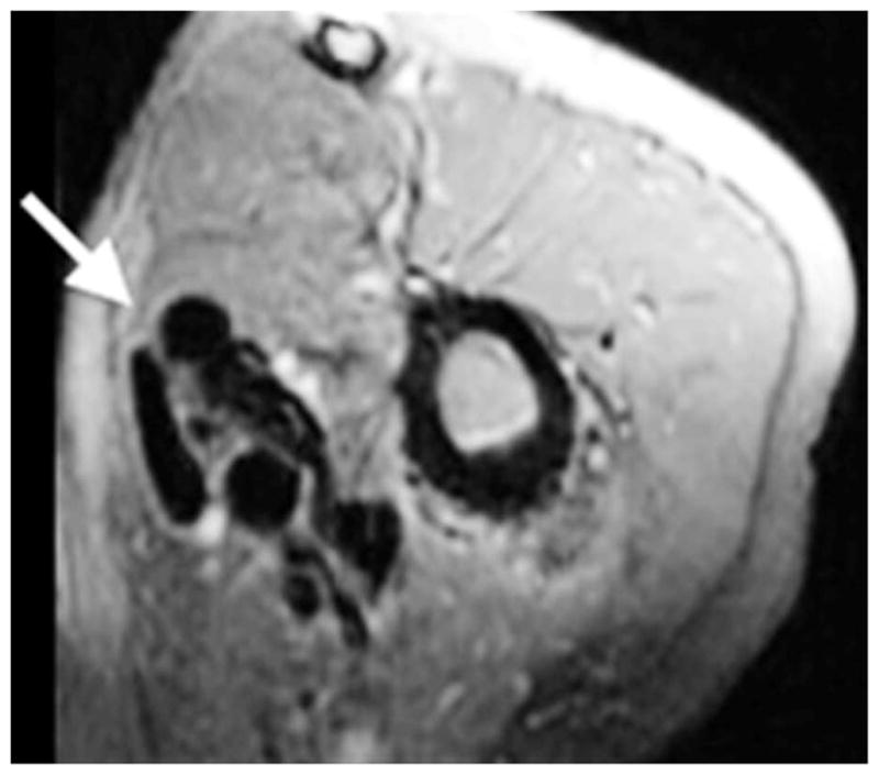
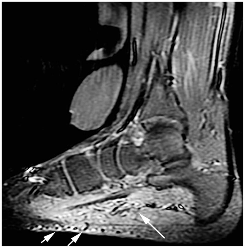
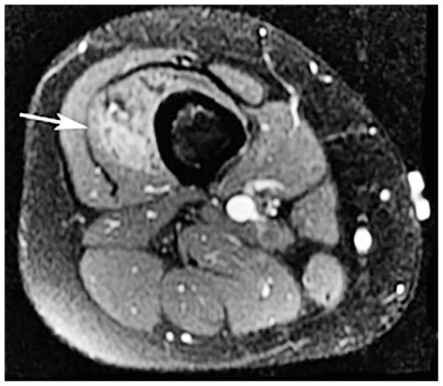
Background: The absence of a discrete mass, surrounding signal abnormality and solid enhancement are imaging features that have traditionally been used to differentiate soft-tissue arteriovenous malformations from vascular tumors on MRI. We have observed that these findings are not uncommon in arteriovenous malformations, which may lead to misdiagnosis or inappropriate treatment.

Objective: To estimate the frequency of atypical MRI features in soft-tissue arteriovenous malformations and assess their relationship to lesion size, location, tissue type involved and vascular architecture.

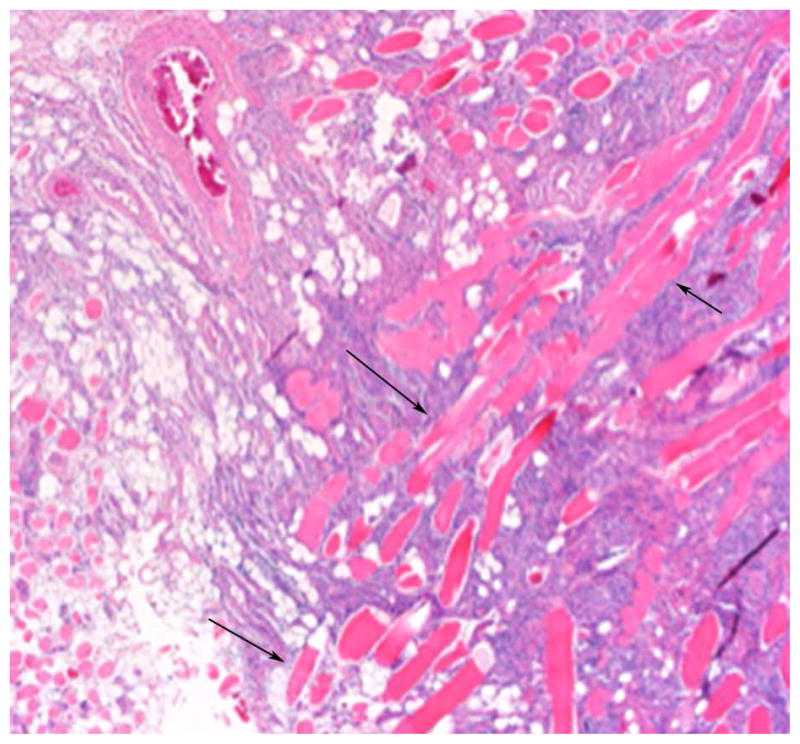
Materials and methods: Medical records, MRI and histopathology were reviewed in consecutive patients with soft-tissue arteriovenous malformations in a multidisciplinary vascular anomalies clinic. Arteriovenous malformations were divided into those with and without atypical MRI findings (perilesional T2 signal abnormality, enhancement and/or a soft-tissue mass). Lesion location, size, tissue involved and vascular architecture were also compared between groups. Tissue stains were reviewed in available biopsy or resection specimens to assess relationships between MRI findings and histopathology.

Results: Thirty patients with treatment-naïve arteriovenous malformations were included. Fifteen lesions demonstrated atypical MRI. There was no difference in age, gender, lesion size or involved body part between the groups. However, more than half of the atypical lesions demonstrated multicompartmental involvement, and tiny intralesional flow voids were more common in atypical arteriovenous malformations. Histopathology also differed in atypical cases, showing densely packed endothelial cells with connective tissue architectural distortion and edema.

Conclusion: Arteriovenous malformations may exhibit features of a vascular tumor on MRI, particularly when multicompartmental and/or containing tiny internal vessels. These features are important to consider in suspected fast-flow vascular malformations and may have implications with respect to their treatment.


Conflict of interest statement

Conflicts of interest Dr. Frieden is a consultant for Pierre Fabre. Drs. Patel, Schulman, Ruben, Hoffman, Dowd and Hess have no disclosures.

Figures

Fig. 1
Fig. 1
A 6-year-old boy with history of painful right forearm arteriovenous malformations. Axial fat-suppressed gadolinium-enhanced T1-weighted image demonstrates a typical arteriovenous malformation (arrows) lesion with prominent flow voids and no enhancement or soft-tissue abnormalities
Fig. 2
Fig. 2
A 27-year-old man with a history of painful left upper arm arteriovenous malformation. Axial fat-suppressed gadolinium-enhanced T1-weighted image demonstrates a typical arteriovenous malformation (arrow) lesion with prominent flow voids and no enhancement or soft-tissue abnormalities.
Fig. 3
Fig. 3
A 2-year-old girl with a history of painful pulsatile upper lip arteriovenous malformation. Axial fat-suppressed gadolinium-enhanced T1-weighted image demonstrates an atypical arteriovenous malformation (arrows) lesion with a discrete soft-tissue mass, enhancement and tiny intralesional flow voids
Fig. 4
Fig. 4
A 30-year-old woman with a history of painful right posterior neck arteriovenous malformations. Axial fat-suppressed gadolinium-enhanced T1-weighted image demonstrates an atypical arteriovenous malformation (arrow) lesion with a discrete soft-tissue mass, enhancement and tiny intralesional flow voids
Fig. 5
Fig. 5
An 11-year-old girl with a history of arteriovenous malformations within the plantar foot. Sagittal STIR images demonstrate an atypical arteriovenous malformation (arrows) with tiny indistinct flow voids, soft-tissue hyperintensity and mass-like characteristics of the lesion
Fig. 6
Fig. 6
A 23-year-old woman with a history of painful arteriovenous malformations within the right thigh. T2 fat-suppressed images demonstrate features of a typical and atypical arteriovenous malformation in the same patient. a. In the distal thigh, at the level of the nidus, there are tiny indistinct flow voids, soft-tissue hyperintensity and mass-like characteristics of the lesion. b. In the proximal thigh, there is a distinct large feeding vessel with flow void, and lack of associated soft-tissue mass
Fig. 6
Fig. 6
A 23-year-old woman with a history of painful arteriovenous malformations within the right thigh. T2 fat-suppressed images demonstrate features of a typical and atypical arteriovenous malformation in the same patient. a. In the distal thigh, at the level of the nidus, there are tiny indistinct flow voids, soft-tissue hyperintensity and mass-like characteristics of the lesion. b. In the proximal thigh, there is a distinct large feeding vessel with flow void, and lack of associated soft-tissue mass
Fig. 7
Fig. 7
A 27-year-old woman with chronic painful left forehead arteriovenous malformations status postsurgical excision. Hematoxylin and eosin stains of soft-tissue “atypical” arteriovenous malformation specimens. a Diffuse proliferation of small, crowded blood vessels permeates the connective tissue (40x). b Skeletal muscle fibers (arrows) are widely separated by small vessels and vascular lumina are not conspicuous (100x)
Fig. 7
Fig. 7
A 27-year-old woman with chronic painful left forehead arteriovenous malformations status postsurgical excision. Hematoxylin and eosin stains of soft-tissue “atypical” arteriovenous malformation specimens. a Diffuse proliferation of small, crowded blood vessels permeates the connective tissue (40x). b Skeletal muscle fibers (arrows) are widely separated by small vessels and vascular lumina are not conspicuous (100x)
Fig. 8
Fig. 8
A 30-year-old woman with chronic painful left temporal arteriovenous malformations status postsurgical excision. Hematoxylin and eosin stains of soft-tissue “typical” arteriovenous malformation specimens. a Variably sized vessels (arrows) throughout the subcutis (40x). b Individual, well-formed vessels (arrows) lie between skeletal muscle fibers, without disruption of the overall muscular architecture (100x)
Fig. 8
Fig. 8
A 30-year-old woman with chronic painful left temporal arteriovenous malformations status postsurgical excision. Hematoxylin and eosin stains of soft-tissue “typical” arteriovenous malformation specimens. a Variably sized vessels (arrows) throughout the subcutis (40x). b Individual, well-formed vessels (arrows) lie between skeletal muscle fibers, without disruption of the overall muscular architecture (100x)

References

    1. Donnelly LF, Adams DM, Bisset GS., 3rd Vascular malformations and hemangiomas: a practical approach in a multidisciplinary clinic. AJR Am J Roentgenol. 2000;174:597–608. - PubMed
    1. Dubois J, Alison M. Vascular anomalies: what a radiologist needs to know. Pediatr Radiol. 2010;40:895–905. - PubMed
    1. Konez O, Burrows PE. Magnetic resonance of vascular anomalies. Magn Reson Imaging Clin North Am. 2002;10:363–388. vii. - PubMed
    1. Enjolras O, Wassef M, Chapot R. Color atlas of vascular tumors and vascular malformations. Cambridge University Press; Cambridge, UK: 2007.
    1. Hovius SE, Borg DH, Paans PR, Pieterman H. The diagnostic value of magnetic resonance imaging in combination with angiography in patients with vascular malformations: a prospective study. Ann Plast Surg. 1996;37:278–285. - PubMed

MeSH terms