Review
. 2015 Jun;36(6):615-22.
doi: 10.1016/j.placenta.2015.04.003. Epub 2015 Apr 17.

Functional MRI of the placenta--From rodents to humans

Affiliations
Review

Functional MRI of the placenta--From rodents to humans

R Avni et al. Placenta. 2015 Jun.

Abstract

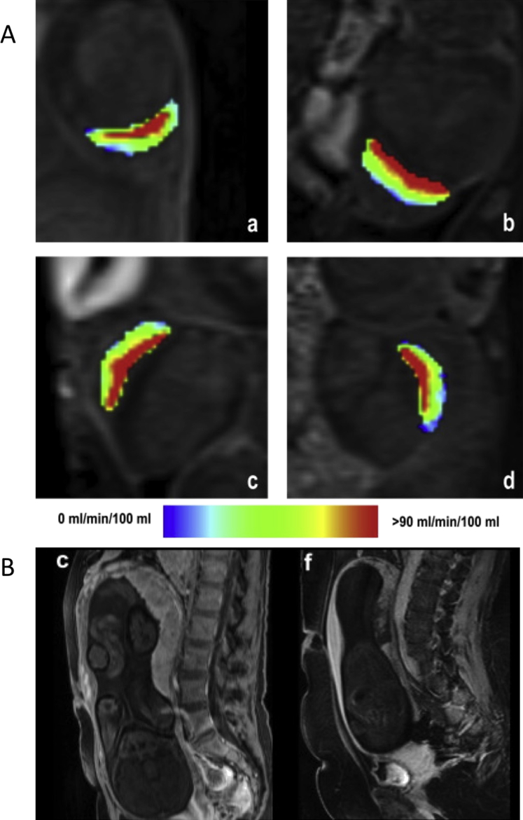
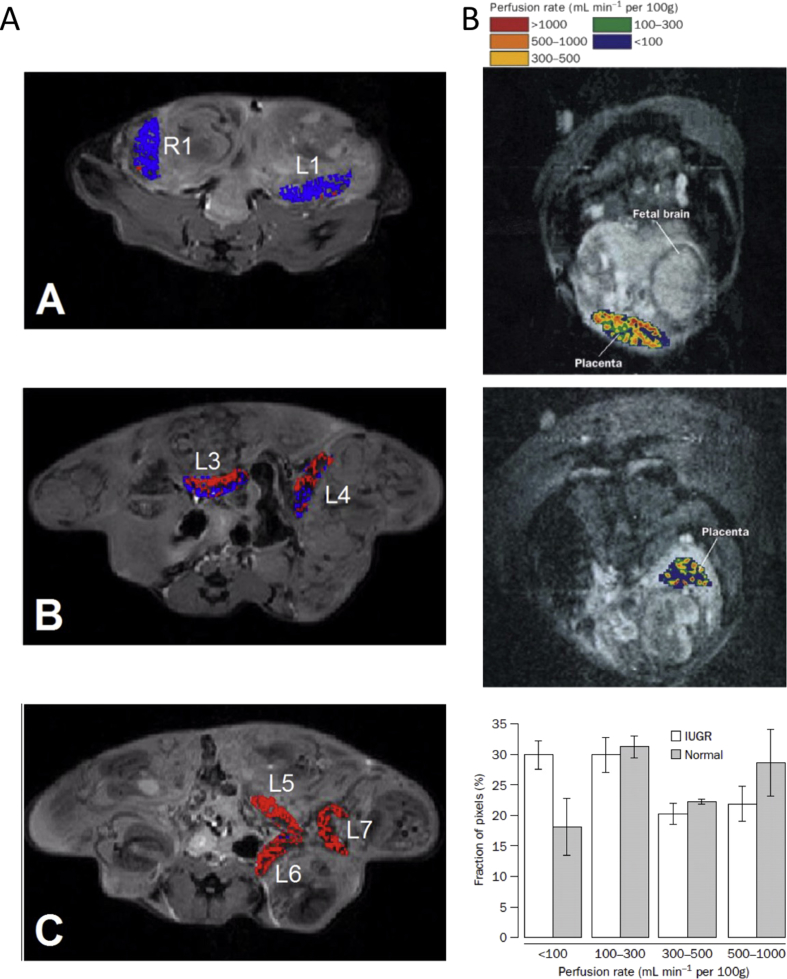
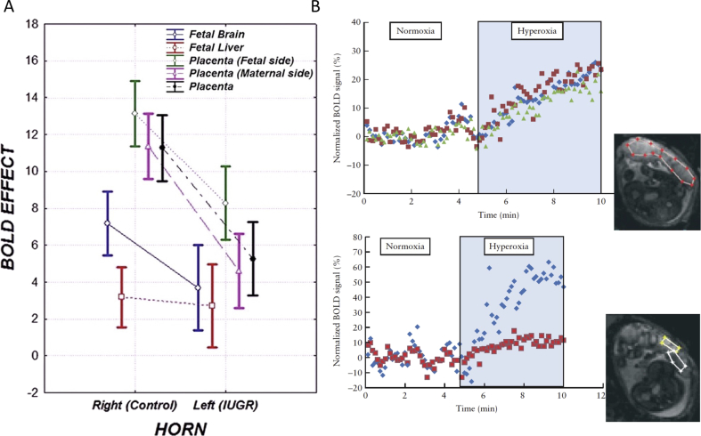
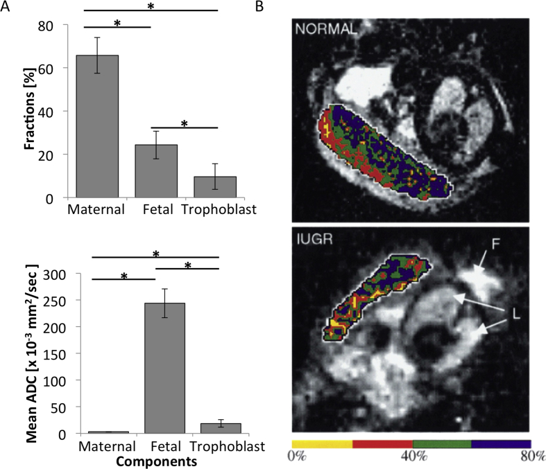
The placenta performs a wide range of physiological functions; insufficiencies in these functions may result in a variety of severe prenatal and postnatal syndromes with long-term negative impacts on human adult health. Recent advances in magnetic resonance imaging (MRI) studies of placental function, in both animal models and humans, have contributed significantly to our understanding of placental structure, blood flow, oxygenation status, and metabolic profile, and have provided important insights into pregnancy complications.

Keywords: Functional imaging; MRI; Placenta; Preclinical models.


Figures

Fig. 1
Fig. 1
Perfusion measurements using contrast agents. (A) Mouse placental perfusion maps calculated by the steepest slope model on embryonic day (E) 14.5 (a,b) and E16.5 (c,d), demonstrate, in all placentas, a high-flow compartment (red colored), which lies within the central labyrinth zone, and a low-flow compartment, which matches the peripheral labyrinth and the junctional zone. Reprinted from Ref. , with permission from Elsevier. (B) Human placental circulation in normal and IUGR pregnancies. 3D Contrast-enhanced, T1-weighted images acquired 2 min following administration of gadoterate meglumine in normal (c) and IUGR placentas (f). Normal placentas show a homogeneous signal increase, while IUGR-complicated placentas display many patchy, non-perfused areas. Reprinted from Ref. , with permission from Elsevier.
Fig. 2
Fig. 2
Perfusion and flow measurements using endogenous MRI contrast. (A) Assessment of the arterial blood supply to mouse placenta of an ICR pregnant mouse (E17.5) via bidirectional arterial spin labeling (BD-ASL) MRI. A–C: Color-coded BD-ASL maps (red and blue, positive and negative BD-ASL values, respectively) show that placentas near the cervix (Panel A: L1, R1) have negative BD-ASL values, whereas those near the ovary (Panel C: L5-7) have positive values. Placentas located in the middle of the uterine horn (Panel B, L3-4) have a dispersive pattern of positive and negative BD-ASL values, suggesting a dual blood supply with separate arterial inputs from each of the feeding arteries . (B) Left panel: Placental perfusion maps overlaid on an MRI image for: (top) a normal pregnancy and (bottom) a pregnancy complicated by IUGR. The placental perfusion rate in pregnancy complicated by IUGR is reduced, with many areas displaying infarction. Right panel: Histograms of placental perfusion fraction show a significant difference between IUGR and normal pregnancies in the distribution of perfusion categories. Reprinted from Ref. , with permission from Elsevier.
Fig. 3
Fig. 3
Oxygenation studies: (A) Effect of maternal hyperoxygenation on the placenta and different fetal organs in a rat ligation model of IUGR. Box-and-whisker plots summarize changes in T2 values between hyperoxygenation and ambient air in placentas, fetal livers and fetal brains, in the normal and ligated IUGR uterine horns. There is a significant effect of maternal hyperoxygenation on BOLD SI in all organs. IUGR placentas show a significant reduction in the BOLD response to hyperoxygenation. Reprinted from Ref. , with permission from RSNA®; (B) Changes in human placental oxygenation BOLD signals during a maternal hyperoxic respiration challenge. Upper panel: Normalized BOLD SI of the entire placenta, in three different slices, shows a significant increase during maternal hyperoxia. Lower panel: Maternal and fetal areas within the placenta display different dynamics in response to maternal changes in oxygenation. Placental region of interests (ROIs) are shown in the inset images. Reprinted from Ref. , with permission from John Wiley and Sons.
Fig. 4
Fig. 4
Microstructure: (A) Resolving mouse placental structures using a hybrid strategy, combining DW MRI, contrast-enhanced under SPEN acquisition. Upper panel: Estimated placental compartmental fraction. Lower panel: Corresponding ADC values in each compartment. According to this analysis, maternal, fetal, and trophoblastic contributions constitute 66%, 24%, and 10% of overall placental volume, respectively. ADC values reveal a freely diffusive maternal blood pool, a strongly perfused fetal blood flow, and intermediate behavior for the trophoblastic labyrinth cell layer . (B) Human placental perfusion fraction maps in normal and IUGR pregnancies, measured using IVIM. Perfusion fraction maps overlaid on MRI images for normal pregnancy (upper panel), indicating two zones of blood movement, and for pregnancy complicated by IUGR (lower panel), in which the placenta appears far more homogenous, with its outer zone containing a significantly reduced proportion of mobile blood. Reprinted from Ref. , with permission from Elsevier.
Fig. 5
Fig. 5
Metabolism: (A) 31P MRS in healthy and IUGR complicated pregnancies: Upper panel: representative 31P spectrum of a normal human placenta at 38 weeks gestation (a), fitted spectrum (b), and residual (c). Spectral intensities of the following metabolites were evaluated: phosphomonoesters (PME = phosphoethanolamine (PE) + phosphocholine (PC)); inorganic phosphate; phosphodiesters (PDE = glycerophosphoethanolamine (GPE) + glycerophosphocholine (GPC)); phosphocreatine (PCr); and adenosine triphosphate (ATP). Lower panel: Scatter plot displaying PDE/PME spectral intensity ratio in early onset (<34 weeks) PE versus normal pregnancy. Women with early onset preeclampsia have a higher PDE/PME spectral intensity ratio than women with normal pregnancies. Reprinted from Ref. , with permission from Elsevier. (B) 1H MRS: upper panel: placental 1H MRS spectrum acquired from normal human pregnancy. Choline and lipid spectral peaks appear at frequencies of 3.21, 1.3 and 0.9 parts per million (ppm), respectively. Lower panel: Placental 1H MRS spectrum acquired from IUGR-compromised pregnancy. The lipid spectral peak appears at a frequency of 1.42 ppm. The choline peak is below the level of reliable detection. Reprinted from Ref. , with permission from PLOS ONE.

References

    1. Salomon L.J., Siauve N., Balvay D., Cuenod C.A., Vayssettes C., Luciani A. Placental perfusion MR imaging with contrast agents in a mouse model. Radiology. 2005;235(1):73–80. - PubMed
    1. Taillieu F., Salomon L.J., Siauve N., Clement O., Faye N., Balvay D. Placental perfusion and permeability: simultaneous assessment with dual-echo contrast-enhanced MR imaging in mice. Radiology. 2006;241(3):737–745. - PubMed
    1. Salomon L.J., Siauve N., Taillieu F., Balvay D., Vayssettes C., Frija G. In vivo dynamic MRI measurement of the noradrenaline-induced reduction in placental blood flow in mice. Placenta. 2006;27(9–10):1007–1013. - PubMed
    1. Alison M., Quibel T., Balvay D., Autret G., Bourillon C., Chalouhi G.E. Measurement of placental perfusion by dynamic contrast-enhanced MRI at 4.7 T. Invest Radiol. 2013;48(7):535–542. - PubMed
    1. Tomlinson T.M., Garbow J.R., Anderson J.R., Engelbach J.A., Nelson D.M., Sadovsky Y. Magnetic resonance imaging of hypoxic injury to the murine placenta. Am J Physiol Regul Integr Comp Physiol. 2010;298(2):R312–R319. - PMC - PubMed

Publication types