. 2015 May 26;43(10):4950-61.
doi: 10.1093/nar/gkv336. Epub 2015 Apr 27.

Ectopic expression of RNF168 and 53BP1 increases mutagenic but not physiological non-homologous end joining

Affiliations

Ectopic expression of RNF168 and 53BP1 increases mutagenic but not physiological non-homologous end joining

Dali Zong et al. Nucleic Acids Res. .

Erratum in

Abstract

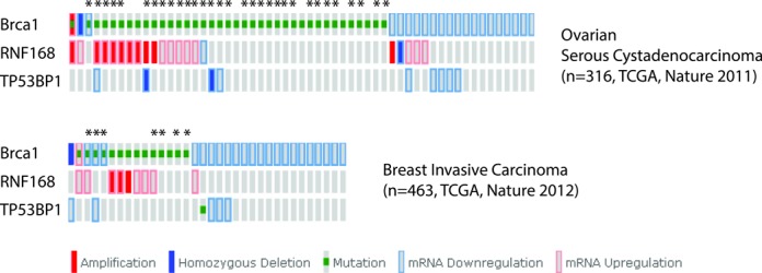
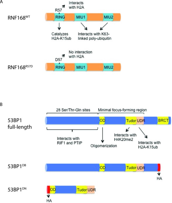
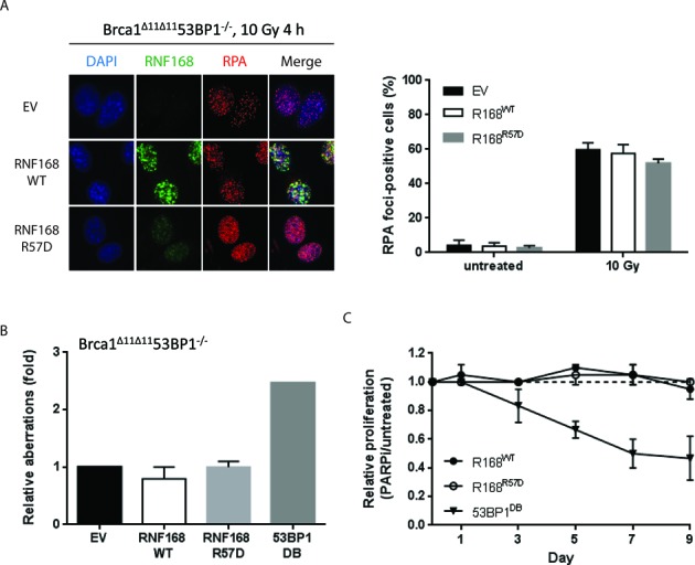
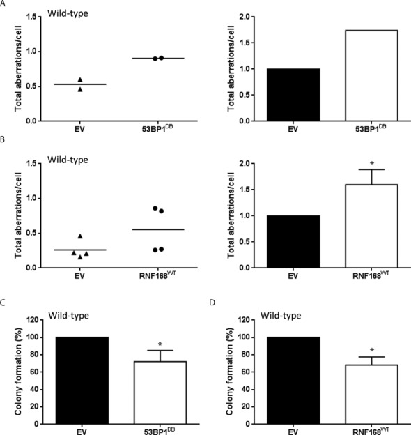
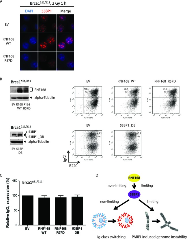
DNA double strand breaks (DSBs) formed during S phase are preferentially repaired by homologous recombination (HR), whereas G1 DSBs, such as those occurring during immunoglobulin class switch recombination (CSR), are repaired by non-homologous end joining (NHEJ). The DNA damage response proteins 53BP1 and BRCA1 regulate the balance between NHEJ and HR. 53BP1 promotes CSR in part by mediating synapsis of distal DNA ends, and in addition, inhibits 5' end resection. BRCA1 antagonizes 53BP1 dependent DNA end-blocking activity during S phase, which would otherwise promote mutagenic NHEJ and genome instability. Recently, it was shown that supra-physiological levels of the E3 ubiquitin ligase RNF168 results in the hyper-accumulation of 53BP1/BRCA1 which accelerates DSB repair. Here, we ask whether increased expression of RNF168 or 53BP1 impacts physiological versus mutagenic NHEJ. We find that the anti-resection activities of 53BP1 are rate-limiting for mutagenic NHEJ but not for physiological CSR. As heterogeneity in the expression of RNF168 and 53BP1 is found in human tumors, our results suggest that deregulation of the RNF168/53BP1 pathway could alter the chemosensitivity of BRCA1 deficient tumors.


Figures

Figure 1.
Figure 1.
The RNF168/53BP1 pathway is altered in a subset of BRCA1-deficient tumors. Mutation data for two TCGA studies were extracted from cBioPortal (www.cbioportal.org) and used to generate oncoprints showing examples of RNF168/53BP1 pathway heterogeneity in BRCA1-deficient tumors (–58). Asterisks denote tumors where BRCA1 is mutated in one allele and deleted in the other. All other BRCA1-mutated tumors contain heterozygous germline mutations.
Figure 2.
Figure 2.
Schematic depiction of the retroviral vectors used in this study. (A) The RNF168WT and RNF168R57D differ in their ability to specifically interact with and ubiquitylate histone H2A due to a point mutation in the RING domain. MIU, motif interacting with ubiquitin. (B) 53BP1DB lacks the C-terminal BRCT domain present in full-length 53BP1 while 53BP1DN consists only of the minimal region required for foci formation. 53BP1DB is functionally wild-type with respect to end-protection and CSR (28), whereas 53BP1DN acts as a dominant negative mutant. OD, oligomerization domain; UDR, ubiquitylation-dependent recruitment.
Figure 3.
Figure 3.
Overexpression of RNF168 or 53BP1 exacerbates PARP inhibitor-induced genomic instability and cytotoxicity. (A) BRCA1Δ11/Δ11 cells were treated with 1 μM PARPi for 24 h and processed for standard immunofluorescence. Left panel: cells were stained for 53BP1 (red) and imaged at 63× magnification. A representative experiment is shown. Right panel: the percentage of cells that contain >5 foci of 53BP1 from two independent experiments. At least 200 cells were scored for each sample and treatment condition. (B) BRCA1Δ11/Δ11 MEFs stably transduced with retroviral vectors encoding RNF168WT or RNF168R57D were treated with 1 μM PARPi (24 h) and harvested for preparation of metaphase spreads. Left panel: dot plots indicating the total amount of aberrations per cell in four independent experiments. At least 100 metaphases were analyzed for each condition. Right panel: histograms depicting PARPi-induced chromosomal aberration load relative to empty vector-transduced cells for the same experiments as shown in the corresponding left panels. (C) Similar to (B), except in BRCA1Δ11/Δ11 MEFs stably transduced with retroviral vectors encoding 53BP1DB or 53BP1DN. (D) Cells were treated with 1 μM PARPi for 24 h and then incubated in drug-free medium to allow formation of colonies. After 9 days, culture dishes were stained with crystal violet and colonies containing >50 cells were counted. Results are mean ± SD of three independent experiments. For (A)–(D), Statistical significance was determined with two-tailed unpaired Student's t-test; *, P < 0.05 compared to empty vector-transduced cells.
Figure 4.
Figure 4.
RNF168 and 53BP1 block RPA foci formation and RPA2 phosphorylation. (A) BRCA1Δ11/Δ11 MEFs stably transduced with retroviral vectors encoding RNF168WT or RNF168R57D were irradiated with 10 Gy and fixed 4 h later. Samples were processed for standard immunofluorescence. Left panel: cells were co-stained for RNF168 (green) and RPA2 (red) and imaged at 63× magnification. Note that the polyclonal anti-RNF168 antibody used in this study recognizes only the exogenously expressed human RNF168. A representative experiment is shown. Right panel: the percentage of cells that contain >10 RPA2 foci. (B) BRCA1Δ11/Δ11 MEFs stably transduced with retroviral vectors encoding 53BP1DB or 53BP1DN were irradiated and processed for standard immunofluorescence as in (A). Left panel: cells were co-stained for 53BP1 (green) and RPA2 (red) and imaged at 63x magnification. A representative experiment is shown. Right panel: the percentage of cells that contain >10 RPA2 foci. The right panels in A and B show mean ± SD of three independent experiments. At least 200 cells were scored for each sample and treatment condition. (C) Similar to (A), except cells were harvested at the indicated post-irradiation time points for western blot analysis. (D) Similar to (B), except cells were harvested at the indicated post-irradiation time points for western blot analysis. A representative blot is shown in (C) and (D). Experiments were repeated three times. For (A) and (B), statistical significance was determined with two-tailed unpaired Student's t-test; *, P < 0.05 compared to empty vector-transduced cells.
Figure 5.
Figure 5.
RNF168 uses 53BP1 to block RPA loading in BRCA1-deficient cells. (A) BRCA1Δ11/Δ1153BP1−/− MEFs stably transduced with retroviral vectors encoding RNF168WT or RNF168R57D were irradiated with 10 Gy and fixed 4 h later. Samples were processed for standard immunofluorescence. Left panel: cells were co-stained for RNF168 (green) and RPA2 (red) and imaged at 20x magnification (optovar: 1.25×). A representative experiment is shown. Right panel: the percentage of cells that contain >10 RPA2 foci. At least 100 cells were scored for each sample and treatment condition. (B) BRCA1Δ11/Δ1153BP1−/− MEFs stably transduced with retroviral vectors encoding RNF168WT, RNF168R57D or 53BP1DB were treated with 1 μM PARPi (24 h) and harvested for preparation of metaphase spreads. Histograms depict PARPi-induced chromosomal aberration load relative to empty vector-transduced cells for two-three independent experiments. At least 100 metaphases were analyzed for each condition. (C) BRCA1Δ11/Δ1153BP1−/− MEFs stably transduced with retroviral vectors encoding RNF168WT or RNF168R57D (GFP-positive) were plated with non-transduced BRCA1Δ11/Δ1153BP1−/− MEFs (GFP-negative) at a 1:1 ratio. Alternatively BRCA1Δ11/Δ1153BP1−/− MEFs stably transduced with retroviral vectors encoding 53BP1DB (GFP-negative) were mixed 1:1 with BRCA1Δ11/Δ1153BP1−/− MEFs stably transduced with the empty vector (GFP-positive). Cells were treated or not with 1 μM PARPi continuously for 9 days. Samples were collected before (day 0) as well as after PARPi treatment on days 1, 3, 5, 7 and 9 for flow cytometric analysis. Relative proliferation is calculated by normalizing the fraction of GFP-positive cells in PARPi-treated versus untreated samples collected on the same day. Data shown represents mean ± SD of three independent experiments.
Figure 6.
Figure 6.
53BP1 suppresses HR in wild-type cells. Wild-type cells were stably transduced with retroviral vectors encoding 53BP1DB or RNF168WT. (A, B) Cells were treated with 1 μM PARPi (24 h) and harvested for preparation of metaphase spreads. Left panels: dot plots indicating the total amount of aberrations per cell. Right panels: histograms depict PARPi-induced chromosomal aberration load relative to empty vector-transduced cells. At least 100 metaphases were scored for chromosomal aberrations for each condition. Results are mean ± SD of two (A) and four (B) independent experiments, respectively. (C, D) Cells were treated with 1 μM PARPi continuously for 10 days, after which culture dishes were stained with crystal violet and colonies containing >50 cells were counted. Results are mean ± SD of three independent experiments. For (B), (C) and (D), statistical significance was determined with two-tailed unpaired and paired Student's t-test, respectively; *P < 0.05 compared to empty vector-transduced cells.
Figure 7.
Figure 7.
RNF168 and 53BP1 are not limiting factors during immunoglobulin class switch recombination (CSR). (A) BRCA1Δ11/Δ11 splenic B cells transduced with retroviral vectors encoding RNF168WT or RNF168R57D were cultured in the presence of LPS/IL-4/RP105 to stimulate CSR. On day 3, cells were irradiated with 2 Gy and fixed 1 h later. Samples were stained for 53BP1 (red) and imaged at 63× magnification. (B) BRCA1Δ11/Δ11 splenic B cells were transduced with retroviral vectors encoding RNF168WT, RNF168R57D or 53BP1DB and cultured in the presence of LPS/IL-4/RP105 to stimulate CSR. Left panels: overexpression of RNF168 and 53BP1 were confirmed by western blotting. The upper band in the 53BP1 blot corresponds to the endogenous protein. Right panels: two-color flow cytometric analysis of IgG1 expression in B220-positive cells on day 4; scatter plots for B cells overexpressing RNF168 and its empty vector counterpart were gated on GFP. A representative experiment is shown. (C) Frequency of IgG1 expression in B220-positive BRCA1Δ11/Δ11 B cells from three-four independent experiments. (D) Model depicting the differential roles of 53BP1 and RNF168 in physiological and mutagenic NHEJ.

References

    1. Matsuoka S., Ballif B.A., Smogorzewska A., McDonald E.R. 3rd, Hurov K.E., Luo J., Bakalarski C.E., Zhao Z., Solimini N., Lerenthal Y., et al. ATM and ATR substrate analysis reveals extensive protein networks responsive to DNA damage. Science. 2007;316:1160–1166. - PubMed
    1. Doil C., Mailand N., Bekker-Jensen S., Menard P., Larsen D.H., Pepperkok R., Ellenberg J., Panier S., Durocher D., Bartek J., et al. RNF168 binds and amplifies ubiquitin conjugates on damaged chromosomes to allow accumulation of repair proteins. Cell. 2009;136:435–446. - PubMed
    1. Huen M.S., Grant R., Manke I., Minn K., Yu X., Yaffe M.B., Chen J. RNF8 transduces the DNA-damage signal via histone ubiquitylation and checkpoint protein assembly. Cell. 2007;131:901–914. - PMC - PubMed
    1. Kolas N.K., Chapman J.R., Nakada S., Ylanko J., Chahwan R., Sweeney F.D., Panier S., Mendez M., Wildenhain J., Thomson T.M., et al. Orchestration of the DNA-damage response by the RNF8 ubiquitin ligase. Science. 2007;318:1637–1640. - PMC - PubMed
    1. Mailand N., Bekker-Jensen S., Faustrup H., Melander F., Bartek J., Lukas C., Lukas J. RNF8 ubiquitylates histones at DNA double-strand breaks and promotes assembly of repair proteins. Cell. 2007;131:887–900. - PubMed

Publication types

MeSH terms

Substances