

Systematic comparison of MRI findings in pediatric ependymoblastoma with ependymoma and CNS primitive neuroectodermal tumor not otherwise specified
- PMID: 25916887
- PMCID: PMC4490876
- DOI: 10.1093/neuonc/nov063
Systematic comparison of MRI findings in pediatric ependymoblastoma with ependymoma and CNS primitive neuroectodermal tumor not otherwise specified
Abstract
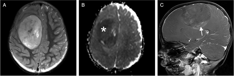
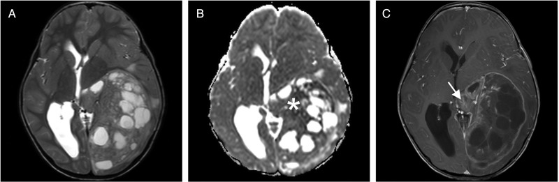
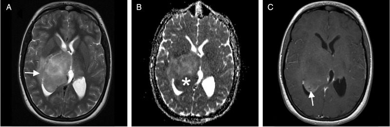
Background: Ependymoblastoma (EBL), ependymoma (EP), and primitive neuroectodermal tumors of the central nervous system not otherwise specified (CNS-PNET NOS) are pediatric brain tumors that can be differentiated by histopathology in the clinical setting. Recently, we described specific MRI features of EBL. In this study, we compare standardized MRI characteristics of EBL with EP and CNS-PNET NOS in a series comprising 22 patients in each group.
Methods: All 66 centrally reviewed cases were obtained from the database of the German multicenter HIT trials. We systematically analyzed the initial MRI scans at diagnosis according to standardized criteria, and paired comparison was performed for EBL and EP, as well as for EBL and CNS-PNET NOS.
Results: We found differences between EBL and EP regarding age at diagnosis, MR signal intensity, tumor margin and surrounding edema, presence and size of cysts, and contrast enhancement pattern. Although MRI appearance of EBL shares many features with CNS-PNET NOS, we revealed significant differences in terms of age at diagnosis, tumor volume and localization, tumor margins, edema, and contrast enhancement.
Conclusion: This is the first study that systematically compares multiple parameters of MRI in pediatric EBL with findings in EP and CNS-PNET NOS. Although a definite differentiation by means of MRI alone might not be feasible in the individual case, we identify significant differences between these tumor entities.
Keywords: CNS-PNET NOS; MRI; ependymoblastoma; ependymoma; magnetic resonance imaging.
© The Author(s) 2015. Published by Oxford University Press on behalf of the Society for Neuro-Oncology. All rights reserved. For permissions, please e-mail: journals.permissions@oup.com.
Figures
References
-
- Gerber NU, von Hoff K, von Bueren AO, et al. Outcome of 11 children with ependymoblastoma treated within the prospective HIT-trials between 1991 and 2006. J Neurooncol. 2011;102(3):459–469. - PubMed
-
- Morovic A, Damjanov I. Neuroectodermal ovarian tumors: a brief overview. Histol Histopathol. 2008;23(6):765–771. - PubMed
-
- Santi M, Bulas D, Fasano R, et al. Congenital ependymoblastoma arising in the sacrococcygeal soft tissue: a case study. Clin Neuropathol. 2008;27(2):78–82. - PubMed
-
- Bailey P, Cushing H. A Classification of the Tumors of the Glioma Group on a Histogenetic Basis With a Correlated Study of Prognosis. Philadelphia: J. B. Lippincott Co.; 1926.
Publication types
MeSH terms
LinkOut - more resources
Full Text Sources
Other Literature Sources
Medical
