. 2015 Jul;83(7):2762-70.
doi: 10.1128/IAI.00425-15. Epub 2015 Apr 27.

Enterococcus faecalis Gelatinase Mediates Intestinal Permeability via Protease-Activated Receptor 2

Affiliations

Enterococcus faecalis Gelatinase Mediates Intestinal Permeability via Protease-Activated Receptor 2

Nitsan Maharshak et al. Infect Immun. 2015 Jul.

Abstract

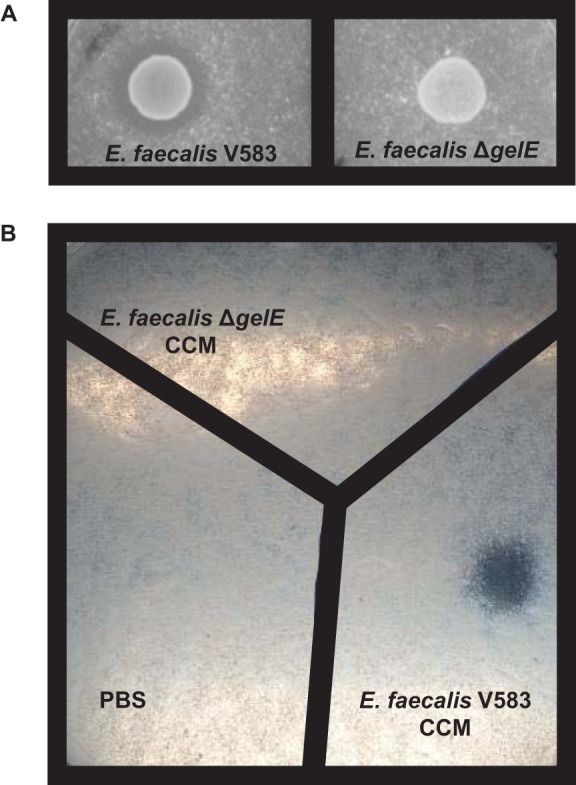
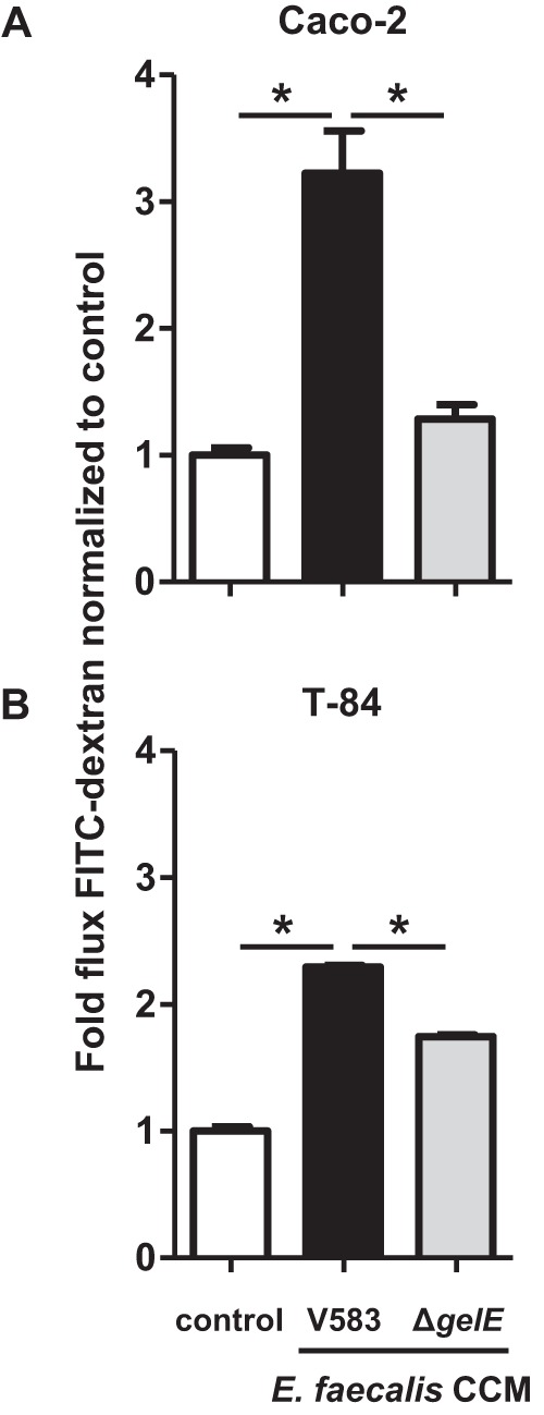
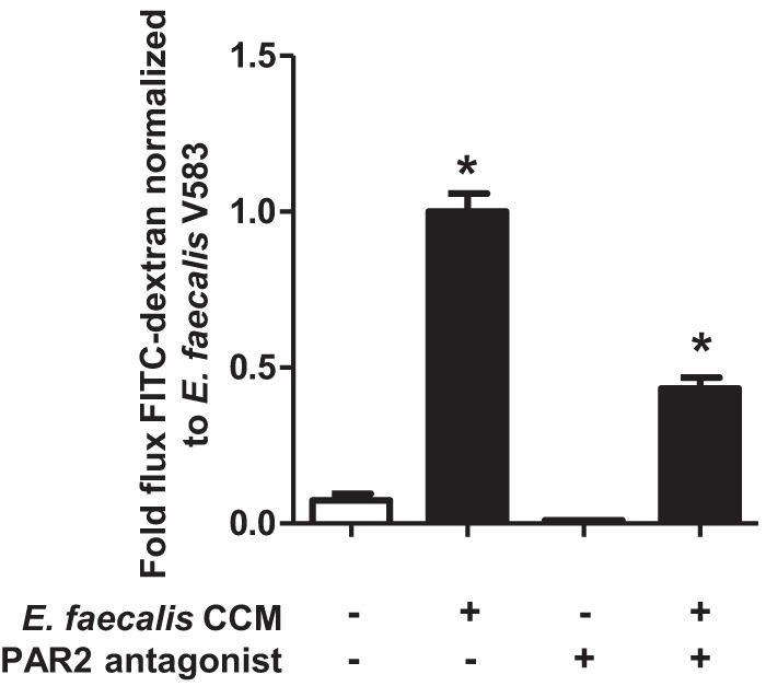
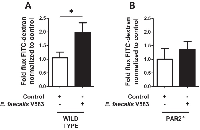
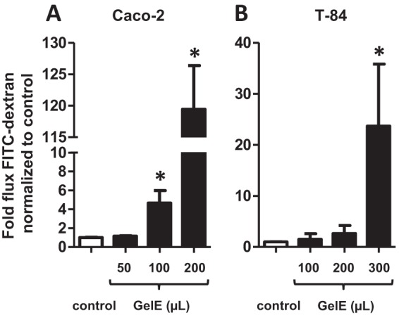
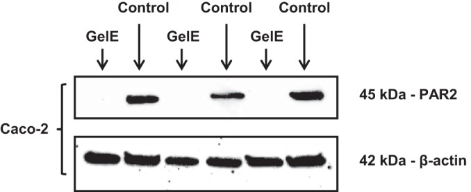
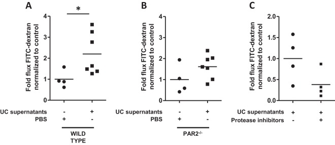
Microbial protease-mediated disruption of the intestinal epithelium is a potential mechanism whereby a dysbiotic enteric microbiota can lead to disease. This mechanism was investigated using the colitogenic, protease-secreting enteric microbe Enterococcus faecalis. Caco-2 and T-84 epithelial cell monolayers and the mouse colonic epithelium were exposed to concentrated conditioned media (CCM) from E. faecalis V583 and E. faecalis lacking the gelatinase gene (gelE). The flux of fluorescein isothiocyanate (FITC)-labeled dextran across monolayers or the mouse epithelium following exposure to CCM from parental or mutant E. faecalis strains indicated paracellular permeability. A protease-activated receptor 2 (PAR2) antagonist and PAR2-deficient (PAR2(-/-)) mice were used to investigate the role of this receptor in E. faecalis-induced permeability. Gelatinase (GelE) purified from E. faecalis V583 was used to confirm the ability of this protease to induce epithelial cell permeability and activate PAR2. The protease-mediated permeability of colonic epithelia from wild-type (WT) and PAR2(-/-) mice by fecal supernatants from ulcerative colitis patients was assessed. Secreted E. faecalis proteins induced permeability in epithelial cell monolayers, which was reduced in the absence of gelE or by blocking PAR2 activity. Secreted E. faecalis proteins induced permeability in the colonic epithelia of WT mice that was absent in tissues from PAR2(-/-) mice. Purified GelE confirmed the ability of this protease to induce epithelial cell permeability via PAR2 activation. Fecal supernatants from ulcerative colitis patients induced permeability in the colonic epithelia of WT mice that was reduced in tissues from PAR2(-/-) mice. Our investigations demonstrate that GelE from E. faecalis can regulate enteric epithelial permeability via PAR2.


Figures

FIG 1
FIG 1
Deletion of gelE from E. faecalis V583 results in the loss of casein degradation. E. faecalis V583, E. faecalis ΔgelE, and CCM from the growth of these strains were incubated overnight on agar plates containing 2.5% skim milk. (A) The parental E. faecalis strain V583 exhibits caseinolytic activity, while deletion of the gelE gene from E. faecalis V583 results in the loss of caseinolytic activity. (B) CCM from the parental E. faecalis strain V583 exhibits caseinolytic activity, while CCM from E. faecalis ΔgelE exhibits no caseinolytic activity. These observations were made consistently upon multiple incubations with parental and mutant E. faecalis cells and CCM on skim milk agar plates.
FIG 2
FIG 2
Deletion of gelE from E. faecalis V583 reduces the permeability of epithelial cell monolayers. (A) Caco-2 cell monolayers were incubated with CCM from E. faecalis V583 (filled bars) or from E. faecalis lacking the gelE gene (ΔgelE) (shaded bars). The flux of FITC-dextran across Caco-2 cell monolayers (indicative of paracellular permeability) was significantly greater when monolayers were apically cultured with E. faecalis V583 CCM for 24 h than with the medium control (open bars) (*, P < 0.0001). The flux of FITC-dextran across Caco-2 cell monolayers was significantly reduced to that of E. faecalis V583 CCM when cultured with CCM from E. faecalis ΔgelE for 24 h (*, P < 0.0001). (B) T-84 cell monolayers were incubated with CCM from E. faecalis V583 or E. faecalis ΔgelE. The flux of FITC-dextran across T-84 cell monolayers was significantly greater when monolayers were basally cultured with E. faecalis V583 CCM for 24 h than with the medium control (*, P < 0.0001). The flux of FITC-dextran across T-84 cell monolayers was significantly reduced to that of E. faecalis V583 CCM when cultured with CCM from E. faecalis ΔgelE for 24 h (*, P < 0.0001). Each experimental group represents at least 3 independent experiments with 2 technical replicates in each experiment. Data are expressed as mean values ± standard errors. Comparisons were made using one-way ANOVA.
FIG 3
FIG 3
E. faecalis V583 mediates monolayer permeability via PAR2. Caco-2 cell monolayers were incubated with or without a PAR2 antagonist (FSLLRY-NH2) for 24 h. The monolayers were then incubated with E. faecalis V583 CCM (filled bars) or a medium control (open bars) for a further 24 h. The flux of FITC-dextran across Caco-2 cell monolayers was significantly greater when monolayers were cultured with E. faecalis V583 CCM than with the control (*, P < 0.0001). The E. faecalis V583 CCM-mediated permeability of Caco-2 cell monolayers was significantly reduced when PAR2 activation was blocked with an antagonist (*, P < 0.0001). Each experimental group represents at least 3 independent experiments with 2 technical replicates in each experiment. Data are expressed as mean values ± standard errors. Comparisons were made using one-way ANOVA.
FIG 4
FIG 4
The E. faecalis V583 CCM-induced change in the permeability of the mouse colonic epithelium is absent in PAR2−/− mice. (A) The colonic epithelia of WT mice were exposed to CCM from E. faecalis V583 (filled bars), and the flux of FITC-dextran (paracellular permeability) was measured using Ussing chambers following 2 h of exposure. E. faecalis V583 CCM increased the flux of FITC-dextran across the epithelia from WT mice over that of the control (PBS) (open bars) (P < 0.04). (B) CCM from E. faecalis V583 could not induce a change in permeability in mice lacking the PAR2 receptor. Each bar represents experimental results for 10 WT or 5 PAR2−/− mice. Data are expressed as mean values ± standard errors. Comparisons were made using the nonparametric Mann-Whitney test.
FIG 5
FIG 5
GelE purified from E. faecalis V583 exhibits caseinolytic activity. (A) Putatively isolated GelE was run on an SDS gel to confirm its size and purity. The isolated protein had a molecular mass of approximately 38 kDa. (B) The putatively purified GelE exhibited caseinolytic activity following overnight incubation on agar plates containing 2.5% skim milk. These data demonstrate that purified GelE retained its functional activity. (C) Alignment of the 27 digested fragments of the putative GelE protein generated by LC–MS-MS. All digested fragments aligned with GelE from E. faecalis V583, confirming the identity of this protein. The shaded sequence indicates the areas homologous to digested fragments. Blue lines represent digested fragments.
FIG 6
FIG 6
GelE from E. faecalis V583 induces permeability in epithelial cell monolayers. Caco-2 (A) and T-84 (B) cell monolayers were incubated with GelE purified from E. faecalis V583 (filled bars). The flux of FITC-dextran across monolayers (indicative of paracellular permeability) was significantly greater when monolayers were apically (Caco-2) or basally (T-84) cultured with GelE from E. faecalis V583 for 24 h than when monolayers were cultured with the medium control (open bars) (*, P < 0.0001). Each experimental group represents at least 3 independent experiments with 2 technical replicates in each experiment. Data are expressed as mean values ± standard errors. Comparisons were made using the nonparametric Mann-Whitney test.
FIG 7
FIG 7
GelE from E. faecalis V583 activates PAR2 on epithelial cells. GelE was incubated with Caco-2 cells for 1 h. Antibodies could not detect PAR2 in epithelial cells exposed to GelE. Antibodies for β-actin demonstrated equal loading of proteins in each lane. This blot represents results from at least 3 independent experiments with 2 technical replicates in each experiment.
FIG 8
FIG 8
The permeability of the epithelium mediated by fecal supernatants from UC patients is lost in PAR2−/− mice. The colonic epithelia of mice were exposed to fecal supernatants generated from UC patients, and permeability was measured using Ussing chambers. (A) Exposure of the colonic epithelia of WT mice to supernatants from UC patients (n = 7) for 2 h induced a significant 2-fold increase in paracellular permeability over that of controls (P < 0.05). (B) Exposure of the colonic epithelia of PAR2−/− mice to supernatants from UC patients (n = 7) for 2 h did not exhibit a significant difference in paracellular permeability from controls. (C) Exposure of the colonic epithelia of WT mice to a subset of supernatants from UC patients (n = 4) for 2 h induced a reduced level of paracellular permeability when these supernatants were incubated with protease inhibitors. Comparisons were made using the nonparametric Mann-Whitney test.

References

    1. Arnott ID, Kingstone K, Ghosh S. 2000. Abnormal intestinal permeability predicts relapse in inactive Crohn disease. Scand J Gastroenterol 35:1163–1169. doi:10.1080/003655200750056637. - DOI - PubMed
    1. D'Incà R, Di Leo V, Corrao G, Martines D, D'Odorico A, Mestriner C, Venturi C, Longo G, Sturniolo GC. 1999. Intestinal permeability test as a predictor of clinical course in Crohn's disease. Am J Gastroenterol 94:2956–2960. doi:10.1111/j.1572-0241.1999.01444.x. - DOI - PubMed
    1. Dunlop SP, Hebden J, Campbell E, Naesdal J, Olbe L, Perkins AC, Spiller RC. 2006. Abnormal intestinal permeability in subgroups of diarrhea-predominant irritable bowel syndromes. Am J Gastroenterol 101:1288–1294. doi:10.1111/j.1572-0241.2006.00672.x. - DOI - PubMed
    1. Lee JW, Park JH, Park DI, Park JH, Kim HJ, Cho YK, Sohn CI, Jeon WK, Kim BI. 2010. Subjects with diarrhea-predominant IBS have increased rectal permeability responsive to tryptase. Dig Dis Sci 55:2922–2928. doi:10.1007/s10620-009-1094-8. - DOI - PubMed
    1. McOmber ME, Ou CN, Shulman RJ. 2010. Effects of timing, sex, and age on site-specific gastrointestinal permeability testing in children and adults. J Pediatr Gastroenterol Nutr 50:269–275. doi:10.1097/MPG.0b013e3181aa3aa9. - DOI - PMC - PubMed

Publication types

LinkOut - more resources