. 2015 Jun 1;194(11):5200-10.
doi: 10.4049/jimmunol.1500221. Epub 2015 Apr 27.

Collecting lymphatic vessel permeability facilitates adipose tissue inflammation and distribution of antigen to lymph node-homing adipose tissue dendritic cells

Affiliations

Collecting lymphatic vessel permeability facilitates adipose tissue inflammation and distribution of antigen to lymph node-homing adipose tissue dendritic cells

Emma L Kuan et al. J Immunol. .

Abstract

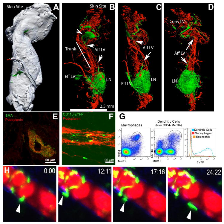
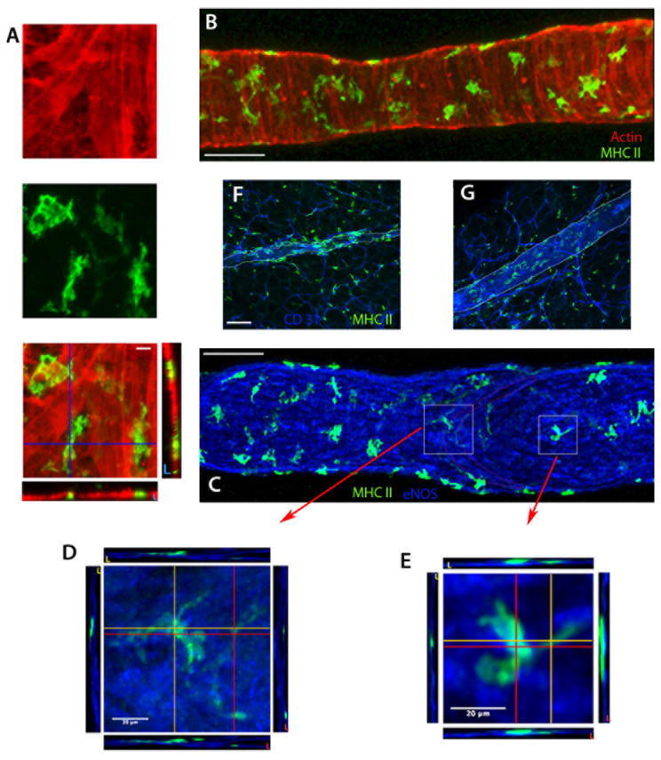
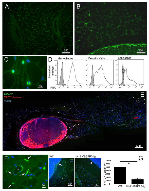
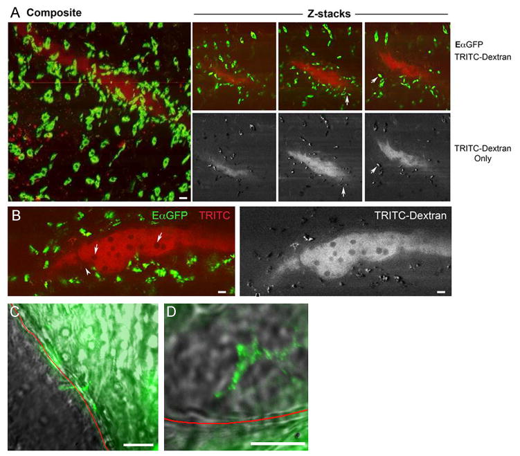
Collecting lymphatic vessels (CLVs), surrounded by fat and endowed with contractile muscle and valves, transport lymph from tissues after it is absorbed into lymphatic capillaries. CLVs are not known to participate in immune responses. In this study, we observed that the inherent permeability of CLVs allowed broad distribution of lymph components within surrounding fat for uptake by adjacent macrophages and dendritic cells (DCs) that actively interacted with CLVs. Endocytosis of lymph-derived Ags by these cells supported recall T cell responses in the fat and also generated Ag-bearing DCs for emigration into adjacent lymph nodes (LNs). Enhanced recruitment of DCs to inflammation-reactive LNs significantly relied on adipose tissue DCs to maintain sufficient numbers of Ag-bearing DCs as the LN expanded. Thus, CLVs coordinate inflammation and immunity within adipose depots and foster the generation of an unexpected pool of APCs for Ag transport into the adjacent LN.


Figures

Figure 1
Figure 1. Collecting lymphatic vessels in PAT and their interactions with DCs
A-D, Optical projection tomography of the brachial LN remove from scapular skin depicts the gross relationship between this LN and the surrounding lymphatic vessels and PAT. Green, lymphatic tracer; red, blood tracer. White color in Panel A shows the contour of PAT around LNs, with Panels B-D showing internal fluorescence in orientations identical to Panel A or partially rotated (B-C). Convergent lymphatics (Conv LVs) near the skin pool at a lymphatic sinus (arrowhead, B) and then a single major afferent lymphatic (Aff LV, arrows) moves along the truck of the PAT to the LN, with the efferent vessel following the blood supply emerging from the LN hilum. E) Cross-section of the afferent lymphatic in the PAT trunk showing smooth muscle actin and podoplanin costaining. F) CD11c-YFP+ cells visualized outside of podoplanin+ lymphatic vessels in PAT. G) Gating on macrophages, DCs, or eosinophils in PAT shows that only DCs are YFP+ in CD11c-YFP mice. H) Time-lapse images of CD11c-YFP DCs around popliteal PAT following injection of Evan's blue dye (fluoresces red) in the rear footpad. All images shown are representative of 2 independent experiments performed in triplicate.
Figure 2
Figure 2. MHC II+ cells within the wall of rat mesenteric collecting lymphatic vessels
A-E) Z-axis projections of isolated, rat mesenteric collecting lymphatics stained for MHC II (green), α-smooth muscle actin (red) and eNOS (blue). Panels A (higher magnification) and B (lower magnification) show the localization of MHC II+ cells within the α - smooth muscle actin-positive lymphatic wall. Panels C-E reveal MHC II+ cells within the lymphatic wall, here co-stained for eNOS. Boxed insets in Panel C show the downstream edge of valve leaflets, with Panels D and E are higher magnification images from C (white squares) depicting the orthogonal cross-sectional views at the locations marked by the red and orange lines revealing that the macrophages bodies are primarily ablumenal to the lymphatic endothelial cells. F-G) Z axis projections of rat mesenteric whole-mount preparations stained for MHC II+ cells (green) and endothelium by CD31 (blue). The collecting lymphatic wall is indicated by thin white lines. Panel F is a tissue section with an upstream lymphangion within the adipose near the gut, while G is a tissue section with a lymphangion within the adipose downstream of F, in the same lymphatic network near the lymph node. Scale bar is 100 μm unless otherwise indicated. Data in these panels are derived from 2-8 experiments, with n ≥ 2 replicates per condition.
Figure 3
Figure 3. Passage of soluble antigens from collecting lymphatic vessels to adipose tissue phagocytes
A, B) Low power cross-sections depicting PAT outside of the brachial LN 18 h after application to the skin surface of a contact sensitizer containing FITC (green) as a hapten (Panel B) compared to the same PAT without FITC painting in Panel A. C) Higher power view of FITC+ cells (green) in PAT as in B.D) Flow cytometric analysis to quantify FITC uptake 18 h after FITC skin painting in DCs (CD45+CD11c+MHCII+MERTK-CD64-), macrophages (CD45+MERTK+CD64+), and eosinophils (CD45+MERTK-SiglecF+). E) Low power tile-reconstructed view of a cross-section of PAT and brachial LN 20 h after EαGFP (green) was administered intradermally (i.d.) and 10 min. after 70 kDa TRITC-dextran (red) was injected i.d. F) Higher power view of PAT cross-section 10 min after FITC-dextran (green) i.d. shows a collecting vessel wall with dextran-enriched cells nearby (white arrow). G) Mice with defective lymphatics resulting from expression of VEGFR3-Ig from the K14 promoter and their littermate WT controls were subjected to FITC skin painting for 18 h. FITC+ cells in PAT cross-sections were counted (n=3 per group). Arrows point to the subscapsular sinus in each strain. Number above scale bars corresponds to number of microns. All images shown were representative of at least 3 animals per group.
Figure 4
Figure 4. Endocytic acquisition of tracers by phagocytes in PAT
A) Z stacks acquired from multiphoton microscopy on fixed PAT obtained from mice injected with EαGFP i.d. 12 h previously and then injected with TRITC-dextran (red) 10 min. prior to euthanasia. Red color identifies lymphatic lumen filled with TRITC dextran and cells acquiring TRITC dextran. Left panel, maximum projection of z-stacks compiled through 200 μm of PAT. Right panels show 3 individual z stack images; upper ones demonstrate both green (FITC) and red (TRITC) channels. Lower ones depict only the TRITC channel (in white contrast). Arrows indicate cells that are both FITC+ and TRITC. B) Higher power view of z-stack image. Dark circles (arrows) in the lumen are poorly endocytic T cells. Arrowhead indicates cells that took up EαGFP and extend pseudopods into the lumen. C, D) FITC-BSA was injected in the exteriorized rat mesentery. Live confocal imaging of the fluorescent tracer as it passed through mesenteric collecting vessel s began immediately. Red line delineates the border of the lymphatic vessel with surrounding adipose tissue and the lymphatic lumen as marked. Arrows depict cell bodies that appear to concentrate the tracer. Live imaging precluded immunostaining to identify these cells. Scale bar in all images is 30 μm. Data in each panel are representative of 2-3 independent experiments.
Figure 5
Figure 5. Inflammatory responses in PAT after skin immunization
A) Each line in the graphs represents an independent experiment in which the fold-increase in PAT CD11c+ inflammatory cells was measured 18 h after epicutaneous application of FITC sensitizer or 18 h after EαGFP was injected i.d. B) Quantitative PCR analysis to determine fold increase in mRNA for inflammatory mediators and key adipose-derived cytokines- TNFα, IL-6, iNOS, TLR4, CCR2, MCP-1, and CD36, 12 h after application of FITC sensitizer to skin. C, D) CD4+ T cells were purified from either naïve LNs (+Naïve T) or reactive LNs (+T) of CD45.1+ WT mice 4 days after FITC skin painting and transferred into CD45.2+ WT mice. These recipients were challenged 5 h later with FITC painting or an irrelevant antigen to which they were naïve (EαGFP) or not challenged (No Antigen). Accumulation of transferred CD45.1+ CD4+ and endogenous CD45.2+ CD4+ T cells in the site of skin challenge or the associated PAT was analyzed ∼36 h after challenge. Panel C shows the representative FACS plots of transferred T cells in skin or in PATs. Panel D charts the number of T cells accumulated in PAT of antigen challenged mice, expressed as fold-increase over the number of accumulated in the absence of antigen challenge. **, P < 0.01. All data were obtained from at least 2 independent experiments with more than 3 replicates per group in each experiment.
Figure 6
Figure 6. DCs from PAT use CCR7 to home to adjacent LNs
A) EαGFP or EαCherry (20 μg in 2 μl) was injected (intraPAT) into two sites of mouse PAT around brachial LN. The distribution of EαGFP+ cells was analyzed 18 h later. Arrowhead, injection site; arrow, EαGFP+ cells in LN. Dashed line delineates border of PAT and brachial LN on left, and the border of the LN B cell and T cell zone on right. B) Total number of EαGFP+YAe+CD11c+CD45+ cells in PAT and LN over time following intraPAT EαGFP delivery. At least 3 mice per time point were used. C) EαGFP and YAe expression in CD11c+ cells from brachial LNs 18 h after intraPAT (20 μg) or i.d. (40 μg in scapular skin) delivery of EαGFP. D) Sorted, CFSE-labeled naïve Tea transgenicT cellswere transferred i.v. into naïve recipient mice that 24 h later received intraPAT (20 μg/ PAT, solid bars) or i.d. (40 μg in upper scapular skin, open bars) injection of EαCherry. Total proliferated TEa+CD4+CFSElo T cells and CD69+ or CD44+ proportions in draining LNs were quantified. E) WT, CCR7 KO, or plt/plt mice received intraPAT EαGFP. After 18 h, percent EαGFP+YAe+ cells among CD11c+ LN cells was analyzed. F, G) Fluorescent bead-labeled bone marrow-derived DCs (green) from WT (F) or CCR7 KO (G) mice were injected directly into WT mice PAT for 24 h; 10 min before euthanasia, TRITC-dextran (red) was delivered i.d. to label lymphatics (white arrows). Scale bars in microns. Data show mean ± SEM. *, P < 0.05; **, P < 0.01; ***, P < 0.001. NS means non-statistical different. (A), and (C)-(G) were obtained from 3 independent experiments with at least 3 replicates per group in each experiment.
Figure 7
Figure 7. Inflammation increases mobilization of DCs from PAT to the adjacent LN
Mouse brachial LNs were inflamed using CFA (denoted as + CFA) or not (denoted as control). Then mice intraPAT injection of EαGFP (20 μg/ PAT) or FITC or TRITC painting on the skin. PAT and the adjacent LN were harvested 18 h later for imaging or flow cytometric analysis. A) Photomicrographs depicting distribution and density of EαGFP+ cells in PAT of control or CFA-treated mice. B) Total number of CD11c+EαGFP+YAe+ cells in PAT (upper panel) or LNs (lower panel) of control and CFA-treated mice. C) Images of PAT from mice painted with FITC or TRITC for 18 h. White arrows indicate the subcapsular sinus that feeds the conduit system in draining LNs. Scale bar, 50 μm. D) FITC (green) and TRITC (red) were painted together on mouse scapular back skin; 18 h later, the draining LN was collected for imaging analysis. Conduits (threadlike structures) in the LN are labeled with FITC but not TRITC. E) The total number of FITC+CD11c+ or TRITC+CD11c+ DCs in brachial LNs of control or CFA-treated mice. F) In control or CFA-treated mice, upper scapular skin was injected i.d. with EαGFP (40 μg/site) and the same skin site was painted with TRITC 9 h later. Draining LNs were collected 17 h after TRITC painting and the number of EαGFP+TRITC-CD11c+MHCIIhi (black bar), EαGFP+TRITC+CD11c+MHCIIhi (open bar), and EαGFP-TRITC+CD11c+MHCIIhi (gray bar) cells were quantified. Data depict mean ± SEM. *, P < 0.05; **, P < 0.01. All data were obtained from 3 independent experiments with more than 3 replicates per group in each experiment.

References

    1. Schmid-Schonbein GW. Microlymphatics and lymph flow. Physiol Rev. 1990;70:987–1028. - PubMed
    1. Baluk P, Fuxe J, Hashizume H, Romano T, Lashnits E, Butz S, Vestweber D, Corada M, Molendini C, Dejana E, McDonald DM. Functionally specialized junctions between endothelial cells of lymphatic vessels. The Journal of experimental medicine. 2007;204:2349–2362. - PMC - PubMed
    1. Muthuchamy M, Zawieja D. Molecular regulation of lymphatic contractility. Ann N Y Acad Sci. 2008;1131:89–99. - PubMed
    1. Scallan JP, Huxley VH. In vivo determination of collecting lymphatic vessel permeability to albumin: a role for lymphatics in exchange. The Journal of physiology. 2010;588:243–254. - PMC - PubMed
    1. Randolph GJ, Angeli V, Swartz MA. Dendritic-cell trafficking to lymph nodes through lymphatic vessels. Nature reviews Immunology. 2005;5:617–628. - PubMed

Publication types

MeSH terms

Substances