

CD8 T cells and Mycobacterium tuberculosis infection
- PMID: 25917388
- PMCID: PMC4439333
- DOI: 10.1007/s00281-015-0490-8
CD8 T cells and Mycobacterium tuberculosis infection
Abstract
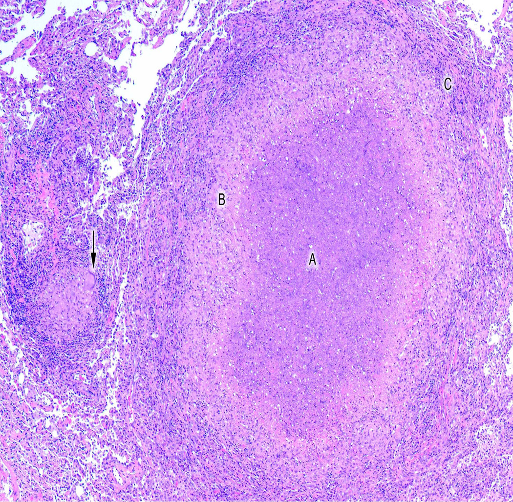
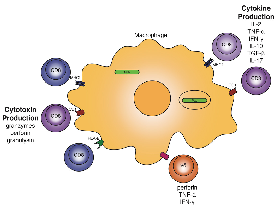
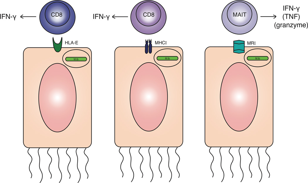
Tuberculosis is primarily a respiratory disease that is caused by Mycobacterium tuberculosis. M. tuberculosis can persist and replicate in macrophages in vivo, usually in organized cellular structures called granulomas. There is substantial evidence for the importance of CD4 T cells in control of tuberculosis, but the evidence for a requirement for CD8 T cells in this infection has not been proven in humans. However, animal model data support a non-redundant role for CD8 T cells in control of M. tuberculosis infection. In humans, infection with this pathogen leads to generation of specific CD8 T cell responses. These responses include classical (MHC Class I restricted) and non-classical CD8 T cells. Here, we discuss the potential roles of CD8 T cells in defense against tuberculosis, and our current understanding of the wide range of CD8 T cell types seen in M. tuberculosis infection.
Figures
References
-
- Organization WH. Global Tuberculosis Report 2014. World Health Organization; 2014.
-
- Gideon HP, Phuah J, Myers AJ, Bryson BD, Rodgers MA, Coleman MT, Maiello P, Rutledge T, Marino S, Fortune SM, Kirschner DE, Lin PL, Flynn JL. Variability in Tuberculosis Granuloma T Cell Responses Exists, but a Balance of Pro- and Anti-inflammatory Cytokines Is Associated with Sterilization. PLoS Pathog. 2015;11:e1004603. - PMC - PubMed
-
- Mattila JT, Ojo OO, Kepka-Lenhart D, Marino S, Kim JH, Eum SY, Via LE, Barry CE, 3rd, Klein E, Kirschner DE, Morris SM, Jr, Lin PL, Flynn JL. Microenvironments in tuberculous granulomas are delineated by distinct populations of macrophage subsets and expression of nitric oxide synthase and arginase isoforms. J Immunol. 2013;191:773–784. - PMC - PubMed
Publication types
MeSH terms
Substances
Grants and funding
LinkOut - more resources
Full Text Sources
Other Literature Sources
Medical
Research Materials
