

Quantitative tissue-tracking cardiac magnetic resonance (CMR) of left atrial deformation and the risk of stroke in patients with atrial fibrillation
- PMID: 25917441
- PMCID: PMC4579945
- DOI: 10.1161/JAHA.115.001844
Quantitative tissue-tracking cardiac magnetic resonance (CMR) of left atrial deformation and the risk of stroke in patients with atrial fibrillation
Abstract
Background: Recent evidence suggests that left atrial (LA) dysfunction may be mechanistically contributing to cerebrovascular events in patients with atrial fibrillation (AF). We investigated the association between regional LA function and a prior history of stroke during sinus rhythm in patients referred for catheter ablation of AF.
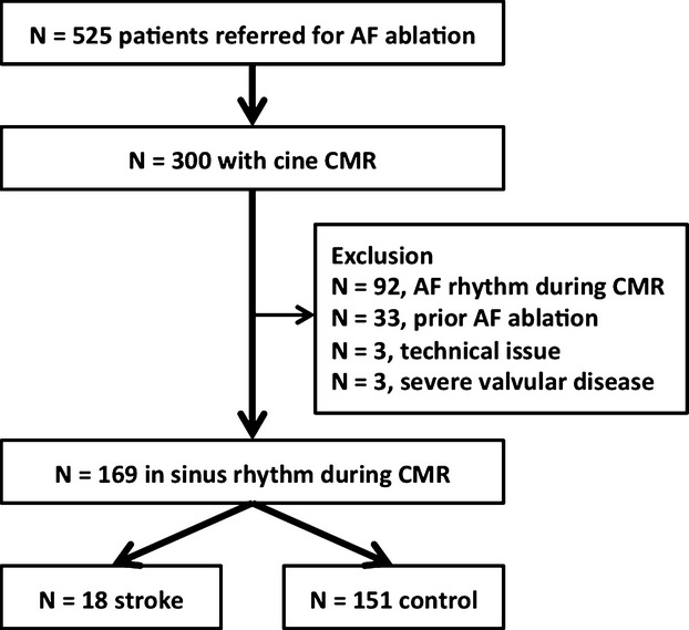
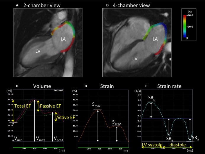
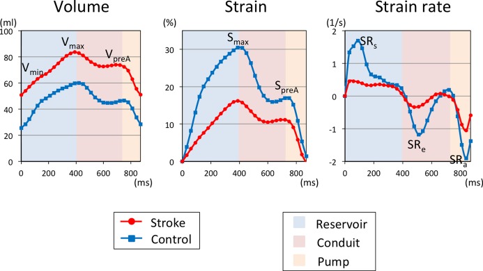
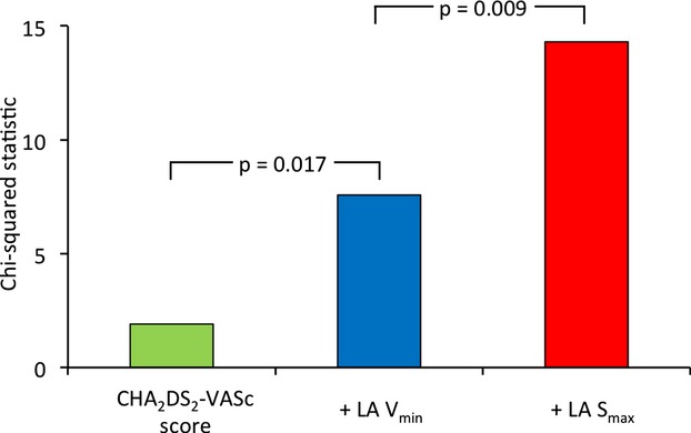
Methods and results: A total of 169 patients (59 ± 10 years, 74% male, 29% persistent AF) with a history of AF in sinus rhythm at the time of pre-ablation cardiac magnetic resonance (CMR) were analyzed. The LA volume, emptying fraction, strain (S), and strain rate (SR) were assessed by tissue-tracking cardiac magnetic resonance. The patients with a history of stroke or transient ischemic attack (n=18) had greater LA volumes (Vmax and Vmin; P=0.02 and P<0.001, respectively), lower LA total emptying fraction (P<0.001), lower LA maximum and pre-atrial contraction strains (Smax and SpreA; P<0.001 and P=0.01, respectively), and lower absolute values of LA SR during left ventricular (LV) systole and early diastole (SRs and SRe; P=0.005 and 0.03, respectively) than those without stroke/transient ischemic attack (n=151). Multivariable analysis demonstrated that the LA reservoir function, including total emptying fraction, Smax, and SRs, was associated with stroke/transient ischemic attack (odds ratio 0.94, 0.91, and 0.17; P=0.03, 0.02, and 0.04, respectively) after adjusting for the CHA2DS2-VASc score and LA Vmin.
Conclusions: Depressed LA reservoir function assessed by tissue-tracking cardiac magnetic resonance is significantly associated with a prior history of stroke/transient ischemic attack in patients with AF. Our findings suggest that assessment of LA reservoir function can improve the risk stratification of cerebrovascular events in AF patients.
Keywords: atrial fibrillation; atrial strain; magnetic resonance imaging; stroke; tracking.
© 2015 The Authors. Published on behalf of the American Heart Association, Inc., by Wiley Blackwell.
Figures
References
-
- Mozaffarian D, Benjamin EJ, Go AS, Arnett DK, Blaha MJ, Cushman M, de Ferranti S, Despres J, Fullerton HJ, Howard VJ, Huffman MD, Judd SE, Kissela BM, Lackland DT, Lichtman JH, Lisabeth LD, Liu S, Mackey RH, Matchar DB, McGuire DK, Mohler ER, III, Moy CS, Muntner P, Mussolino ME, Nasir K, Neumar RW, Nichol G, Palaniappan L, Pandey DK, Reeves MJ, Rodriguez CJ, Sorlie PD, Stein J, Towfighi A, Turan TN, Virani SS, Willey JZ, Woo D, Yeh RW, Turner MB. Heart disease and stroke statistics—2015 update: a report from the American Heart Association. Circulation. 2015; 131:e29-e322. - PubMed
-
- Hart RG. Atrial fibrillation and stroke prevention. N Engl J Med. 2003; 349:1015-1016. - PubMed
-
- Benjamin EJ, D'Agostino RB, Belanger AJ, Wolf PA, Levy D. Left atrial size and the risk of stroke and death. The Framingham Heart Study. Circulation. 1995; 92:835-841. - PubMed
-
- Wong JM, Welles CC, Azarbal F, Whooley MA, Schiller NB, Turakhia MP. Relation of left atrial dysfunction to ischemic stroke in patients with coronary heart disease (from the Heart and Soul Study). Am J Cardiol. 2014; 113:1679-1684. - PubMed
-
- Russo C, Jin Z, Liu R, Iwata S, Tugcu A, Yoshita M, Homma S, Elkind MS, Rundek T, Decarli C, Wright CB, Sacco RL, Di Tullio MR. LA volumes and reservoir function are associated with subclinical cerebrovascular disease: the CABL (Cardiovascular Abnormalities and Brain Lesions) study. JACC Cardiovasc Imaging. 2013; 6:313-323. - PMC - PubMed
MeSH terms
LinkOut - more resources
Full Text Sources
Other Literature Sources
Medical
Research Materials
