. 2015 Apr 28:6:6885.
doi: 10.1038/ncomms7885.

Galanin modulates the neural niche to favour perineural invasion in head and neck cancer

Affiliations

Galanin modulates the neural niche to favour perineural invasion in head and neck cancer

Christina Springstead Scanlon et al. Nat Commun. .

Abstract

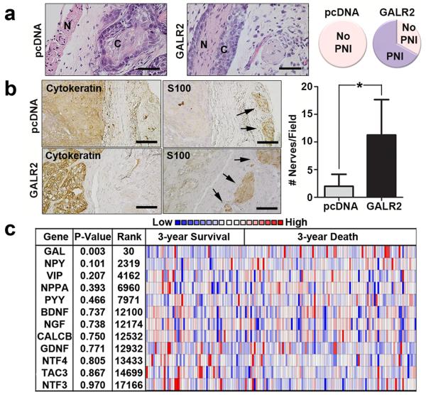
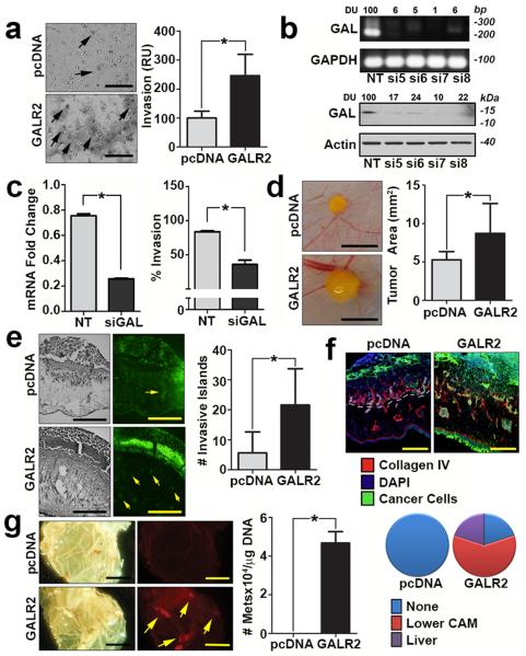
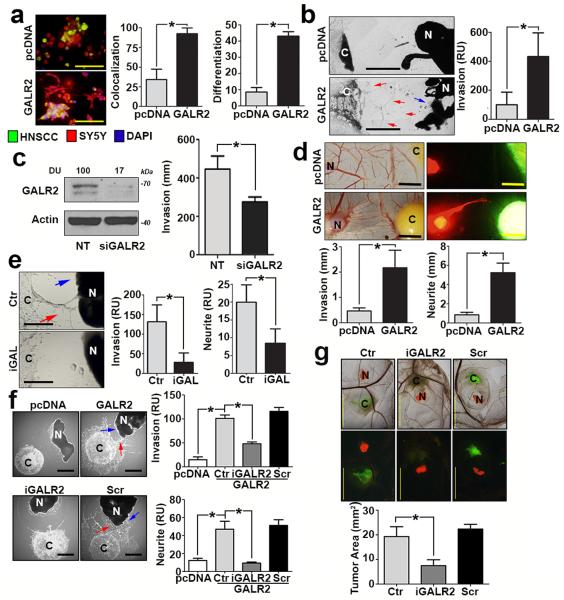
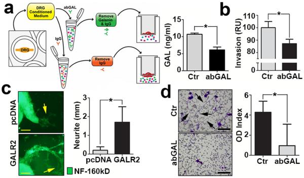
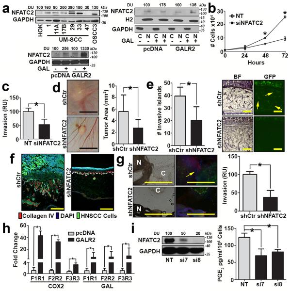
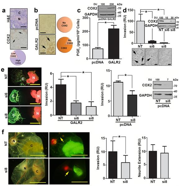
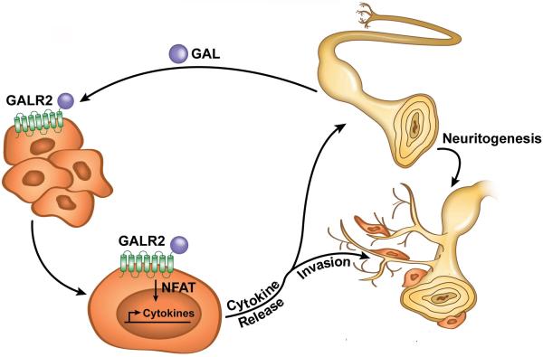
Perineural invasion (PNI) is an indicator of poor survival in multiple cancers. Unfortunately, there is no targeted treatment for PNI since the molecular mechanisms are largely unknown. PNI is an active process, suggesting that cancer cells communicate with nerves. However, nerve-tumour crosstalk is understudied due to the lack of in vivo models to investigate the mechanisms. Here we developed an in vivo model of PNI to characterize this interaction. We show that the neuropeptide galanin (GAL) initiates nerve-tumour crosstalk via activation of its G protein-coupled receptor, GALR2. Our data reveal a novel mechanism by which GAL from nerves stimulates GALR2 on cancer cells to induce NFATC2-mediated transcription of cyclooxygenase-2 and GAL. Prostaglandin E2 promotes cancer invasion, and in a feedback mechanism, GAL released by cancer induces neuritogenesis, facilitating PNI. This study describes a novel in vivo model for PNI and reveals the dynamic interaction between nerve and cancer.


Figures

Figure 1
Figure 1. GAL correlates with poor survival and neuronal involvement
(a) Murine tumours derived from OSCC3-GALR2 cells but not control (OSCC3-pcDNA) cells, exhibit PNI (n = 3 per group, scale bar = 100 μm) (N = nerve; C = cancer). The complete image is in Supplementary Fig. 1b. Comparison of PNI to no PNI in control and GALR2 overexpressing murine xenografts (right panel). (b) GALR2-expressing murine tumours had more adjacent nerves than control tumours (n = 3 per group, arrows identify nerves, scale bar = 50 μm). (*P < 0.05, two sample t-test; data represent mean + SD). Cytokeratin labels tumour and S100 labels nerves. GAL staining distal and proximal to the nerve is shown. (c) The Cancer Genome Atlas (TCGA) study shows that, of several neuronal proteins, only GAL DNA copy number significantly correlates with poor survival (two sample t-test, P = 0.003, 30th most significant).
Figure 2
Figure 2. GALR2 promotes tumour invasion and metastasis
(a) UM-SCC-1-GALR2 cells are more invasive than control UM-SCC-1-pcDNA cells in a Boyden chamber chemoinvasion assay with GAL as the chemoattractant (scale bar = 100 μm). Invasive cells (arrows) from both groups were quantified. Data are representative of three independent experiments with three replicates in each experiment. (*P < 0.05, two sample t-test; data represent mean + SD). (b) UM-SCC-1 cells were transfected with non target (NT) siRNA or siGAL (#5, #6, #7, #8). Downregulation of GAL was verified in RNA (upper panel) and whole cell lysates (lower panel) from these cells. (c) After verification of downregulation of GAL by Q-RT-PCR (left panel), conditioned medium from UM-SCC-1 (parent) cells transfected with NT siRNA or siGAL7 (siGAL) was used as the chemoattractant in a chemoinvasion assay performed with UM-SCC-1-GALR2 (right panel). Cells are significantly more invasive when stimulated with CM from UM-SCC-1-NT than from UM-SCC-1-siGAL7 cells. Data are representative of two experiments each with three replicates. (d-f) UM-SCC-1-GALR2 CAM tumours were (d) larger (scale bar = 5 mm), (e) more invasive (cancer cells are labelled green and highlighted by arrows, scale bar = 200μm), and (f) more disruptive of the basement membrane than control (UM-SCC-1-pcDNA) tumors. Collagen IV and dashed lines label basement membrane (scale bar =100μm). (g) Metastases (yellow, arrows) from the upper CAM to the lower CAM were observed (scale bar = 5 mm). Metastases to the lower CAM and liver were also quantified with quantitative ALU-PCR. (pcDNA group: lower CAM n=4; liver n=5; GALR2 group: lower CAM n=6, liver n=6).
Figure 3
Figure 3. ALR2 promotes PNI
(a) UM-SCC-1 cells (green) overexpressing GALR2 co-mingle with SH-SY5Y neuroblastoma cells (red) and induce more neuritogenesis (differentiation) than controls pcDNA, (2 experiments, 2 replicates in each; scale bar = 100μm). (b) UM-SCC-1-GALR2 cells are more invasive (red arrows) toward DRG than controls (UM-SCC-1-pcDNA) and induce neurite outgrowth (blue arrow) (scale bar = 1mm). (c) Knockdown of endogenous GALR2 in UM-SCC-1 cells, validated by immunoblot (left panel), inhibits invasion (right panel; 2 experiments, 3 replicates in each). (d) UM-SCC-1-GALR2 CAM tumours (green) are more invasive and induce neurite outgrowth (red, scale bar = 5mm, n=6 in each group). (e) UM-SCC-1-GALR2 cell invasion (red arrow) and neurite outgrowth (blue arrow) are attenuated with antagonist anti-GAL antibody (iGAL) (scale bar = 1mm). (f) Invasion of UM-SCC-1-GALR2 (red arrow) and neurite outgrowth (blue arrow) are inhibited by M871 (designated iGALR2), but not scrambled peptide. (pcDNA n=3, GALR2 n=6, GALR2-M871 n=4, GALR2-Scr n=5) (scale bar = 1mm). (g) M871 (iGALR2) inhibits growth of UM-SCC-1-GALR2 CAM tumours (scale bar = 5 mm, Ctr n=7, iGALR2 n=8, Scr n=8). Area was quantified with ImageJ. (b and e) are representative of three independent experiments with 3 replicates in each. (f) is representative of two experiments. (*P < 0.05, two sample t-test; graph represents mean + SD).
Figure 4
Figure 4. GAL from both the DRG and tumour promote PNI
(a) Strategy to remove GAL from CM (left panel). Depletion is verified by ELISA (right panel). (b) CM was depleted of GAL by antibody and used as chemoattractant for UM-SCC-1-GALR2 cells in a modified Boyden chamber invasion assay, and less invasion is observed. (c) DRG treated with CM from UM-SCC-1-GALR2 cells have more neuritogenesis (arrows) than DRG treated with CM from UM-SCC-1-pcDNA cells (scale bar = 1mm). Neurite extensions were quantified (right panel). (d) SH-SY5Y neuroblastoma cells extend more neurites (arrows) when treated with CM from UM-SCC-1-GALR2 cells than when GAL was depleted. (For all studies n = 3 per group, scale bar = 100μm; *P < 0.05, two sample t-test, data represent mean + SD).
Figure 5
Figure 5. GALR2 promotes tumour progression and PNI via NFATC2
(a, left upper panel) Multiple HNSCC cell lines express more NFATC2 than primary human oral keratinocytes (HOK); (a, left lower panel) UM-SCC-1-GALR2 and UM-SCC-1-pcDNA cells were induced with 5nM GAL for 2min and whole cell lysates were immunoblotted with anti-NFATC2. GAPDH was used as a loading control; (a, right panel) GAL (5nM) induces nuclear translocation of NFATC2 in UM-SCC-1-GALR2 and UMSCC-1-pcDNA cells. H2 (histone 2), and GAPDH were used as loading controls for nuclear and cytoplasmic proteins, respectively. (b and c) UM-SCC-1-GALR2 cells transfected with siNFATC2, show reduced proliferation (b) and invasion (c) compared to the same cells transfected with NT siRNA. In CAM, UM-SCC-1-GALR2 cells with constitutive knockdown of NFATC2 (shNFATC2) induce tumours that are (d) smaller, (scale bar = 5mm), (e) less invasive (arrows show invasive islands, scale bar = 200μm; *P<0.02, two sample t-test), and (f) less disruptive to the basement membrane (labelled with Collagen IV, scale bar = 200μm) than the same cells with control scrambled shRNA (shCtr). (g) UM-SCC-1-GALR2-shNFATC2 cells exhibit less PNI than controls (cancer cells labelled “C” fluoresce green and dorsal root ganglia (DRG) are labelled “N”; arrow identifies neurite growth, scale bar = 1mm). Graph shows quantification. (h) NFATC2 binds more to the promoter regions of PTGS2 (COX2) and GAL (GAL) in UM-SCC-1-GALR2 cells compared to control pcDNA. (i) When NFATC2 is downregulated with siRNA in UM-SCC-1-GALR2 cells, PGE2 secretion decreases. For CAM experiments, n = 6 for both; in vitro and DRG explant data are representative of three independent experiments each with three replicates. (*P < 0.05, two sample t-test; data represent mean + SD).
Figure 6
Figure 6. COX2 regulates HNSCC progression but not neural-tumour crosstalk
(a) Most human tumours with PNI express high COX2 adjacent to nerves (arrows, scale bar = 100μm). (N = nerve; C = cancer). (b) Most murine tumours with high GALR2 express COX2 at the invasive front (arrows, scale bar = 100μm), but COX2 is not highly expressed by control tumours. (c) UM-SCC-1-GALR2 cells express more COX2 (immunoblot) and secrete more PGE2 (ELISA) than control cells. (d) siCOX2 in UM-SCC-1-GALR2 cells decreases invasion (arrows label invasive cells, scale bar = 200μm, immunoblot verifies knockdown). (e) Downregulation of COX2 decreases invasion of UM-SCC-1-GALR2 (immunofluorescence and left graph) and UM-SCC-1-pcDNA (right graph) cells toward DRG in co-culture. (scale bar = 1mm; *P<0.02, two sample t-test) (f) siRNA-mediated COX2 downregulation in UM-SCC-1-GALR2 tumours blocks cancer cell invasion (left graph), but does not affect the extension of DRG toward tumours (right graph, scale bar = 5 mm, arrows show neurites). For CAM experiments, n = 6 per group and in vitro and DRG explant data are representative of three independent experiments each with three replicates. (*P < 0.05, two sample t-test; data represent mean + SD). (N = DRG; C = cancer).
Figure 7
Figure 7. A model of neural-tumour crosstalk mediated by the neuropeptide GAL
Neurons release GAL following injury or inflammation and activate tumour-expressed GALR2. GALR2 activation leads to NFATC2-mediated transcription and secretion of COX2/PGE2 and GAL, thereby promoting PNI and neuritogenesis, respectively. Targeting GALR2 or GAL blocks PNI by disrupting neural-tumour crosstalk.

References

    1. Liebig C, Ayala G, Wilks JA, Berger DH, Albo D. Perineural invasion in cancer: a review of the literature. Cancer. 2009;115:3379–3391. - PubMed
    1. Binmadi NO, Basile JR. Perineural invasion in oral squamous cell carcinoma: a discussion of significance and review of the literature. Oral Oncol. 2011;47:1005–1010. - PubMed
    1. Ayala GE, et al. In vitro dorsal root ganglia and human prostate cell line interaction: redefining perineural invasion in prostate cancer. Prostate. 2001;49:213–223. - PubMed
    1. Bapat AA, Hostetter G, Von Hoff DD, Han H. Perineural invasion and associated pain in pancreatic cancer. Nat Rev Cancer. 2011;11:695–707. - PubMed
    1. Johnston M, Yu E, Kim J. Perineural invasion and spread in head and neck cancer. Expert Rev Anticancer Ther. 2012;12:359–371. - PubMed

Publication types