. 2015 Jun 12;290(24):15210-8.
doi: 10.1074/jbc.M115.648097. Epub 2015 Apr 27.

The Crystal Structure of Cancer Osaka Thyroid Kinase Reveals an Unexpected Kinase Domain Fold

Affiliations

The Crystal Structure of Cancer Osaka Thyroid Kinase Reveals an Unexpected Kinase Domain Fold

Sascha Gutmann et al. J Biol Chem. .

Abstract

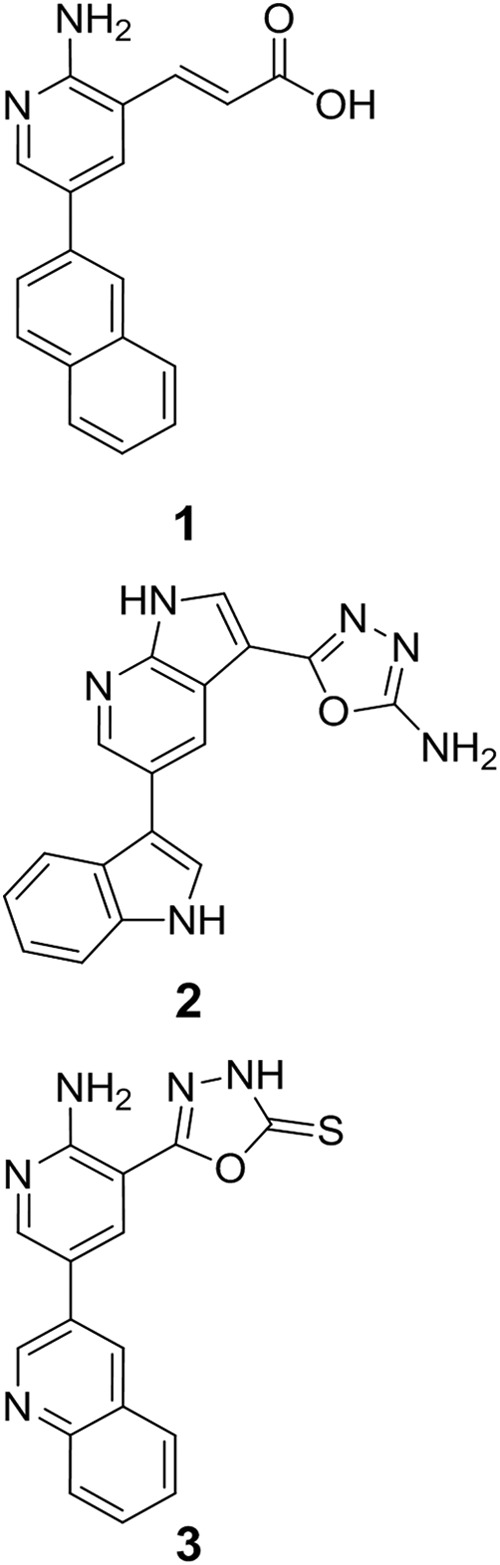
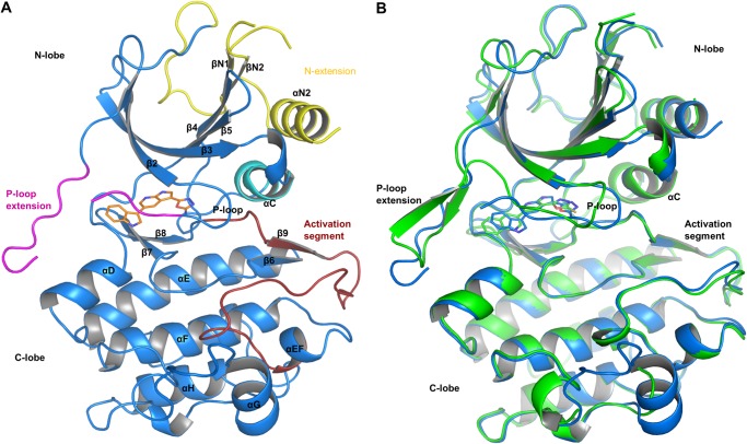
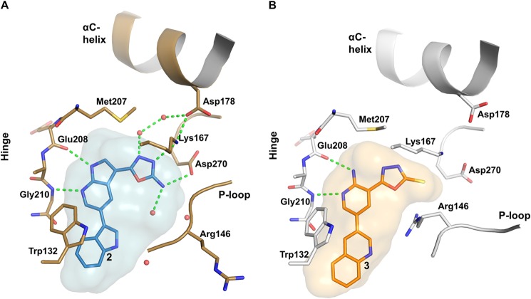
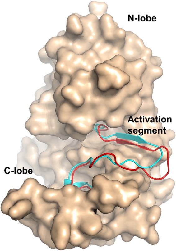
Macrophages are important cellular effectors in innate immune responses and play a major role in autoimmune diseases such as rheumatoid arthritis. Cancer Osaka thyroid (COT) kinase, also known as mitogen-activated protein kinase kinase kinase 8 (MAP3K8) and tumor progression locus 2 (Tpl-2), is a serine-threonine (ST) kinase and is a key regulator in the production of pro-inflammatory cytokines in macrophages. Due to its pivotal role in immune biology, COT kinase has been identified as an attractive target for pharmaceutical research that is directed at the discovery of orally available, selective, and potent inhibitors for the treatment of autoimmune disorders and cancer. The production of monomeric, recombinant COT kinase has proven to be very difficult, and issues with solubility and stability of the enzyme have hampered the discovery and optimization of potent and selective inhibitors. We developed a protocol for the production of recombinant human COT kinase that yields pure and highly active enzyme in sufficient yields for biochemical and structural studies. The quality of the enzyme allowed us to establish a robust in vitro phosphorylation assay for the efficient biochemical characterization of COT kinase inhibitors and to determine the x-ray co-crystal structures of the COT kinase domain in complex with two ATP-binding site inhibitors. The structures presented in this study reveal two distinct ligand binding modes and a unique kinase domain architecture that has not been observed previously. The structurally versatile active site significantly impacts the design of potent, low molecular weight COT kinase inhibitors.

Keywords: cancer; drug discovery; immunology; mitogen-activated protein kinase (MAPK); structural biology.


Figures

FIGURE 1.
FIGURE 1.
Chemical structures of the COT inhibitors (E)-3-(2-amino-5-(naphthalen-2-yl)pyridin-3-yl)acrylic acid (compound 1), 5-(5-(1H-indol-3-yl)-1H-pyrrolo[2,3-b]pyridin-3-yl)-1,3,4-oxadiazol-2-amine (compound 2) 5-(2-amino-5-(quinolin-3-yl)pyridin-3-yl)-1,3,4-oxadiazole-2(3H)-thione (compound 3).
FIGURE 2.
FIGURE 2.
Overview of the COT kinase structures. A, the overall structure of the COT·compound 2 structure is shown as graphic representation (Cα trace of the protein, blue). Compound 2 (orange, stick representation) binds to the active site of the COT kinase domain. N-extension, αC-helix, P-loop insert, and the activation segment are highlighted in yellow, cyan, magenta, and red, respectively. B, structure alignment of the COT·compound 2 (blue) with the COT·compound 3 (green) crystal structure. The Cα atoms of both structures were superimposed to generate this alignment (root mean square deviation = 0.454 Å2) Considerable conformational differences occur in the N-lobe of the COT kinase domain, in particular in the P-loop end P-loop insert segments.
FIGURE 3.
FIGURE 3.
Details of the active site pocket of COT kinase. A, compound 2 (sticks representation; carbon in light blue) binds into the active site of COT (Cα carbon atoms in brown). The blue surface represents the shape of the binding pocket including protein residues within 5 Å around the ligand. B, binding of compound 3 to the active site of COT. (Cα carbon trace in gray). The orange surface represents the shape of the binding pocket including protein residues within 5 Å around the ligand. Nitrogen, oxygen, and sulfur atoms are highlighted in dark blue, red, and yellow, respectively. Polar interactions between ligand and/or protein atoms within 3.2 Å distance are marked with dashed green lines. Water molecules are represented as red spheres.
FIGURE 4.
FIGURE 4.
Details of the P-loop conformational differences. Differences in the P-loop and P-loop insert are depicted as an overlay of Cα atoms of COT·compound 2 and COT·compound 3 in graphic representation. Structure alignment and color coding from Fig. 2B are preserved.
FIGURE 5.
FIGURE 5.
Comparison of the activation segments of COT·compound 2 and cAMP-dependent PKA. Coordinates of the Cα atoms of COT·compound 2 and cAMP-dependent protein kinase in the active state (PDB ID 1ATP) were superimposed. The structure of COT is shown as surface representation with the activation segment (amino acids 270–297) highlighted in red (graphic representation). The superimposed activation segment of PKA is highlighted in cyan (graphic representation).
FIGURE 6.
FIGURE 6.
Sequence comparison of human, mouse, and rat COT kinase domain. An alignment of the sequences of COT kinase from mouse, rat, and human, encompassing amino acids 66–395, is shown. The green box highlights residues that belong to the P-loop and its insert (position 132–152). Numbering is based on the human sequence (UniProt P41279). Positions with fully conserved residues are marked with an asterisk. Conservation of between groups of strongly similar and weakly similar properties are indicated with a colon or a period, respectively.

References

    1. Beinke S., Robinson M. J., Hugunin M., Ley S. C. (2004) Lipopolysaccharide activation of the TPL-2/MEK/extracellular signal-regulated kinase mitogen-activated protein kinase cascade is regulated by IκB kinase-induced proteolysis of NF-κB1 p105. Mol. Cell. Biol. 24, 9658–9667 - PMC - PubMed
    1. Ben-Addi A., Mambole-Dema A., Brender C., Martin S. R., Janzen J., Kjaer S., Smerdon S. J., Ley S. C. (2014) IκB kinase-induced interaction of TPL-2 kinase with 14-3-3 is essential for Toll-like receptor activation of ERK-1 and -2 MAP kinases. Proc. Natl. Acad. Sci. U.S.A. 111, E2394–E2403 - PMC - PubMed
    1. Roget K., Ben-Addi A., Mambole-Dema A., Gantke T., Yang H.-T., Janzen J., Morrice N., Abbott D., Ley S. C. (2012) IκB kinase 2 regulates TPL-2 activation of extracellular signal-regulated kinases 1 and 2 by direct phosphorylation of TPL-2 serine 400. Mol. Cell. Biol. 32, 4684–4690 - PMC - PubMed
    1. Yang H. T., Papoutsopoulou S., Belich M., Brender C., Janzen J., Gantke T., Handley M., Ley S. C. (2012) Coordinate regulation of TPL-2 and NF-κB signaling in macrophages by NF-κB1 p105. Mol. Cell. Biol. 32, 3438–3451 - PMC - PubMed
    1. Patriotis C., Makris A., Bear S. E., Tsichlis P. N. (1993) Tumor progression locus 2 (Tpl-2) encodes a protein kinase involved in the progression of rodent T-cell lymphomas and in T-cell activation. Proc. Natl. Acad. Sci. U.S.A. 90, 2251–2255 - PMC - PubMed

LinkOut - more resources