. 2015 Jun 19;290(25):15621-15634.
doi: 10.1074/jbc.M115.650259. Epub 2015 Apr 27.

An Iron Reservoir to the Catalytic Metal: THE RUBREDOXIN IRON IN AN EXTRADIOL DIOXYGENASE

Affiliations

An Iron Reservoir to the Catalytic Metal: THE RUBREDOXIN IRON IN AN EXTRADIOL DIOXYGENASE

Fange Liu et al. J Biol Chem. .

Abstract

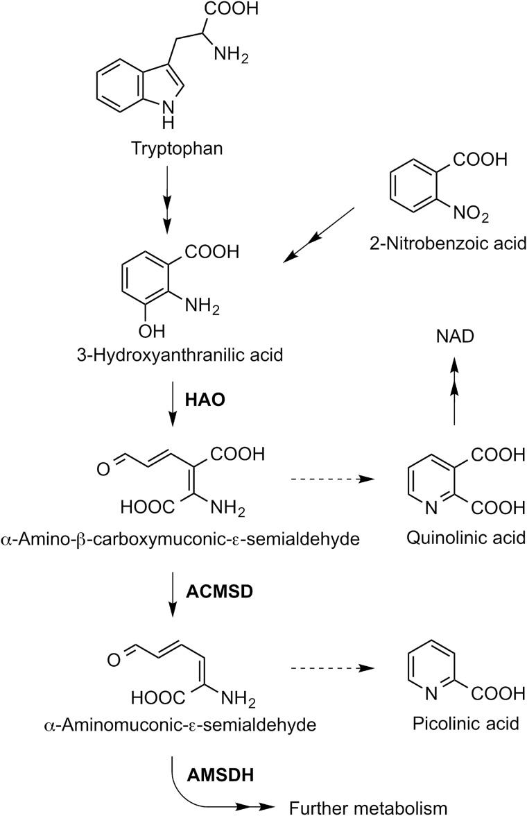
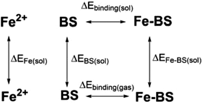
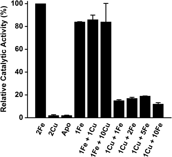
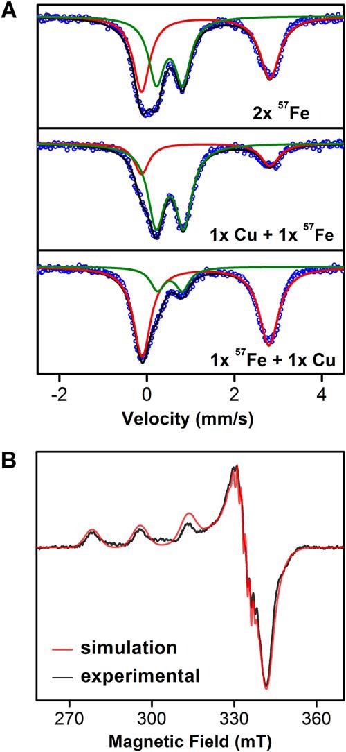
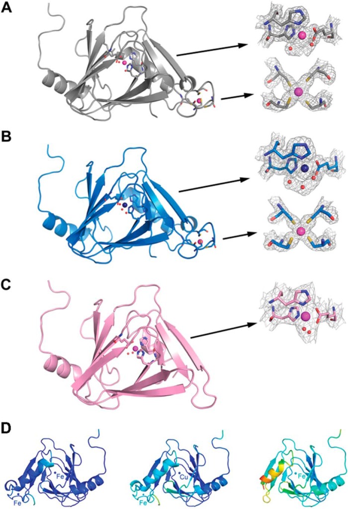
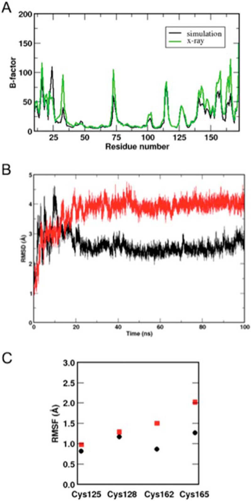
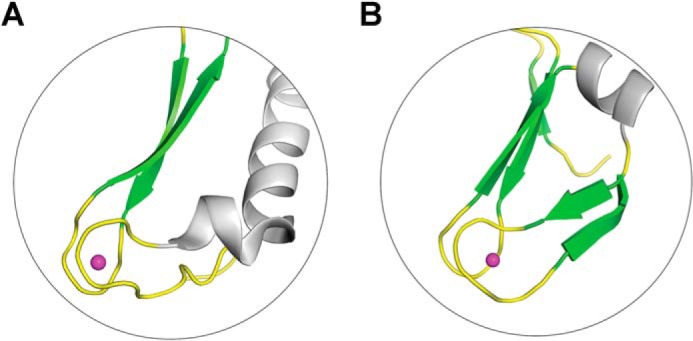
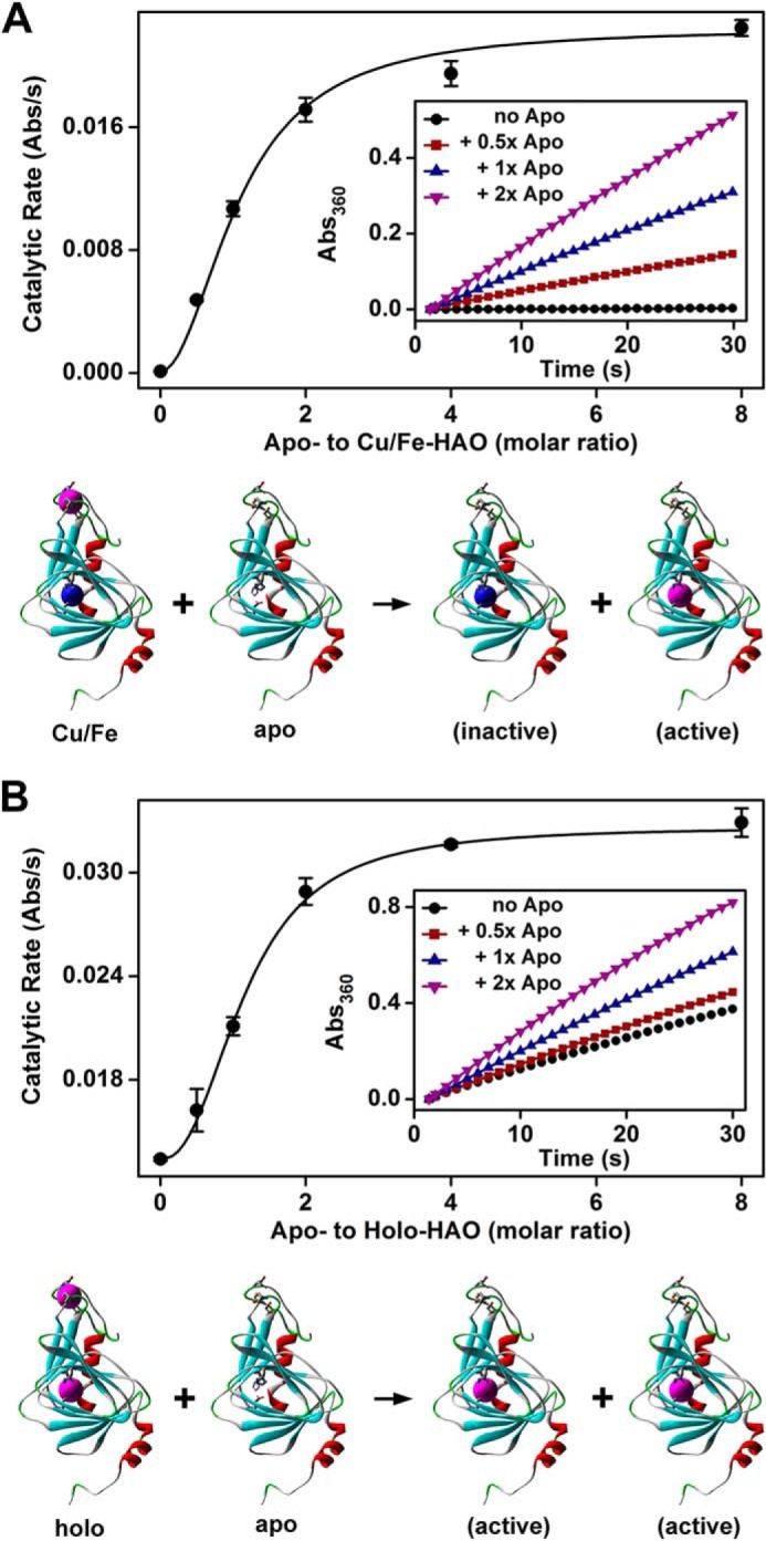
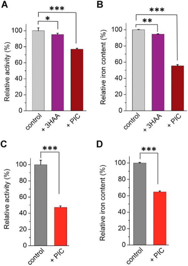
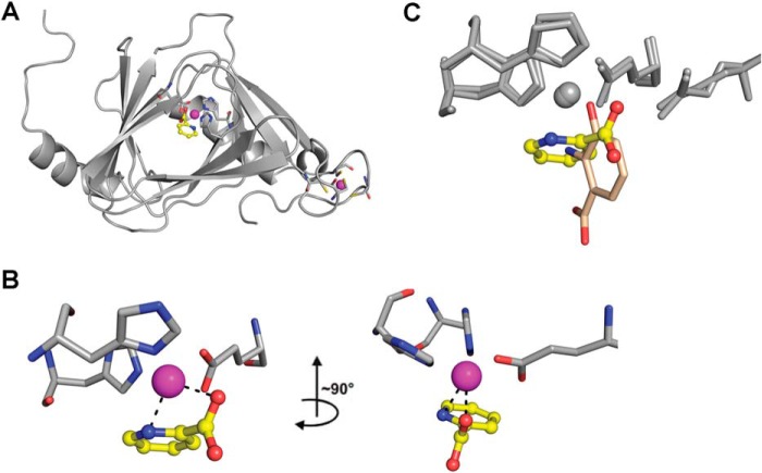
The rubredoxin motif is present in over 74,000 protein sequences and 2,000 structures, but few have known functions. A secondary, non-catalytic, rubredoxin-like iron site is conserved in 3-hydroxyanthranilate 3,4-dioxygenase (HAO), from single cellular sources but not multicellular sources. Through the population of the two metal binding sites with various metals in bacterial HAO, the structural and functional relationship of the rubredoxin-like site was investigated using kinetic, spectroscopic, crystallographic, and computational approaches. It is shown that the first metal presented preferentially binds to the catalytic site rather than the rubredoxin-like site, which selectively binds iron when the catalytic site is occupied. Furthermore, an iron ion bound to the rubredoxin-like site is readily delivered to an empty catalytic site of metal-free HAO via an intermolecular transfer mechanism. Through the use of metal analysis and catalytic activity measurements, we show that a downstream metabolic intermediate can selectively remove the catalytic iron. As the prokaryotic HAO is often crucial for cell survival, there is a need for ensuring its activity. These results suggest that the rubredoxin-like site is a possible auxiliary iron source to the catalytic center when it is lost during catalysis in a pathway with metabolic intermediates of metal-chelating properties. A spare tire concept is proposed based on this biochemical study, and this concept opens up a potentially new functional paradigm for iron-sulfur centers in iron-dependent enzymes as transient iron binding and shuttling sites to ensure full metal loading of the catalytic site.

Keywords: computation; dioxygenase; iron reservoir; iron-sulfur protein; kynurenine; metabolism; tryptophan.


Figures

FIGURE 1.
FIGURE 1.
HAO catalyzes the oxidative phenyl ring cleavage reaction in the kynurenine pathway that is found in both 2-nitrobenzoic acid biodegradation pathway and l-tryptophan catabolism. HAO employs a mononuclear ferrous iron to activate O2. Both oxygen atoms are inserted into the organic substrate.
SCHEME 1
SCHEME 1
FIGURE 2.
FIGURE 2.
HAO sequence similarity network showing the conservatively of the rubredoxin-like domain. A total number of 705 non-redundant HAO sequences were included in the network. Sequences that contain a rubredoxin-like site are diamonds and those without are circles. The connections less than E value of 50 are shown in gray lines. Bacteria are shown in green, fungi in teal, animals in red, and circles marked with an X represent the HAOs that have been previously studied at the protein level.
FIGURE 3.
FIGURE 3.
Specific activity of metal-reconstituted HAO. The first available metal appears to bind to the catalytic center. Apo-HAO was reconstituted with different molar equivalents and orders of metal ions as specified in each column. The catalytic activity of holo-HAO reconstituted with 2 molar eq of Fe2+ per protomer (specific activity 8.6 μmol min−1 mg−1) was assigned at 100% for comparison.
FIGURE 4.
FIGURE 4.
Spectroscopic characterization reveals the metal identity of metal-reconstituted HAO. A, Mössbauer spectra of HAO reconstituted with 2 eq of 57Fe2+ (top panel), 1 eq of Cu2+ + 1 eq of 57Fe2+ (middle panel), and 1 eq of 57Fe2+ + 1 eq of Cu2+ (bottom panel). The black line overlaid on the experimental data (blue circles) is the composite simulation. Each of the spectra contains two sets of quadruple doublets as shown in red and green. The Mössbauer parameters of the two sets of doublets are described in the text. B, X-band EPR spectrum of Cu2+-reconstituted HAO with resolved hyperfine structures obtained at 77 K along with overlaid spectral simulation.
FIGURE 5.
FIGURE 5.
X-ray crystallography describes structural details of metal-reconstituted HAO. Ribbon diagrams of (A) Fe/Fe-HAO (gray, 1.74 Å resolution), (B) Cu/Fe-HAO (blue, 1.75 Å resolution), and (C) Fe/”Cu“-HAO (pink, 2.81 Å resolution) with a zoomed-in view on the catalytic and rubredoxin-like sites. The metal ions are represented by large magenta spheres, whereas water molecules are shown in small red spheres. The rubredoxin-like region (amino acids 154–174) is missing in the Fe/”Cu“-HAO structure. The 2FoFc electron density maps of the two iron-binding sites are also presented and countered to 1.0 σ. D, B-factor value plots show that the structure of the rebredoxin-like site is dynamic in the absence of iron ion. The colors gradually change from dark blue to green and to orange with increasing B-factor values.
FIGURE 6.
FIGURE 6.
Computational studies of rubredoxin-like site of HAO. A, the comparison of the calculated and experimental B-factors of Fe/Fe-HAO. The B-factors were calculated from the simulation trajectory of the Fe/Fe-HAO simulation (black) and compared with the B-factors from the x-ray crystallographic data (green). B, root mean square deviation during molecular dynamics simulations. Root mean square deviation of Fe/Fe-HAO (black) and single load Fe-HAO with the rubredoxin-like site free of metal (red). C, root mean square fluctuation of Cys-125, Cys-128, Cys-162, and Cys-165 in Fe/Fe-HAO and single load Fe-HAO. The residues are more flexible in singe load Fe-HAO (red) than Fe/Fe-HAO (black).
FIGURE 7.
FIGURE 7.
The rubredoxin-like iron-binding site of HAO is similar to that of an iron-storage protein, Dph4. The PDB access codes for HAO (A) and Dph4 (B) are 4L2N and 2L6L, respectively.
FIGURE 8.
FIGURE 8.
The rubredoxin-like site of HAO replenishes iron to the catalytic site. A, addition of apo-HAO (minimally active) to Cu/Fe-HAO (minimally active) produced catalytically active enzyme; B, addition of apo-HAO to holo-HAO doubled the total catalytic activity. Cu/Fe-HAO or holo-HAO was premixed with apo-HAO at different ratios. The catalytic activity of each sample was measured by monitoring the product formation at 360 nm at a saturating substrate concentration of 200 μm with the final concentration of Cu/Fe-HAO or holo-HAO at 10 nm. For each sample, the residual activity from apo-HAO (determined separately from control experiments) was deducted from the observed activity to determine the effective activity for comparison. The data were fitted to the Hill equation with a Hill coefficient of 2.00 and 2.36 for A and B, respectively. The insets show representative kinetic traces. For each trace, the absorbance at the starting point was subtracted to correct for background.
FIGURE 9.
FIGURE 9.
PIC removes the catalytic iron of HAO in the isolated protein and cell lysates. Isolated HAO reconstituted with 1 eq of Fe2+ was incubated with 3-HAA or PIC (2 mm) and analyzed by steady-state kinetic assays (A) and ICP-OES analysis (B). Exogenous 3-HAA or PIC was removed from the samples via a desalting column prior to the kinetic and spectroscopic analyses. Similarly, E. coli cell lysates containing overexpressed HAO were incubated with PIC (2 mm) and analyzed by steady-state kinetic assays (C) and ICP-OES analysis (D). After incubation, HAO was purified with a nickel-affinity column and desalted prior to subsequent analyses. *, p < 0.05; **, p < 0.01; and ***, p < 0.001.
FIGURE 10.
FIGURE 10.
The x-ray crystal structure of HAO in complex with PIC at 1.88-Å resolution shows that PIC bidentately chelates the catalytic iron ion. A, structure of HAO with a PIC bound at the active site. PIC is shown by sticks and the metal center is represented by magenta sphere; B, PIC is bidentately bound to the iron ion in the active site. C, an overlay of the active-site structures of PIC- and 3-HAA-bound HAO.

References

    1. Orme-Johnson W. H. (1973) Iron-sulfur proteins: structure and function. Annu. Rev. Biochem. 42, 159–204 - PubMed
    1. Lovenberg W., Sobel B. E. (1965) Rubredoxin: a new electron transfer protein from Clostridium pasteurianum. Proc. Natl. Acad. Sci. U.S.A. 54, 193–199 - PMC - PubMed
    1. Adam V., Royant A., Nivière V., Molina-Heredia F. P., Bourgeois D. (2004) Structure of superoxide reductase bound to ferrocyanide and active site expansion upon x-ray-induced photo-reduction. Structure 12, 1729–1740 - PubMed
    1. Logan D. T., Mulliez E., Larsson K. M., Bodevin S., Atta M., Garnaud P. E., Sjoberg B. M., Fontecave M. (2003) A metal-binding site in the catalytic subunit of anaerobic ribonucleotide reductase. Proc. Natl. Acad. Sci. U.S.A. 100, 3826–3831 - PMC - PubMed
    1. Larsson K. M., Andersson J., Sjöberg B. M., Nordlund P., Logan D. T. (2001) Structural basis for allosteric substrate specificity regulation in anaerobic ribonucleotide reductases. Structure 9, 739–750 - PubMed

Publication types