. 2015 Jul 3;290(27):16550-9.
doi: 10.1074/jbc.M115.644047. Epub 2015 Apr 27.

RWD Domain as an E2 (Ubc9)-Interaction Module

Affiliations

RWD Domain as an E2 (Ubc9)-Interaction Module

Aileen Y Alontaga et al. J Biol Chem. .

Abstract

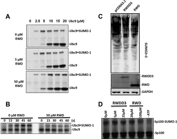
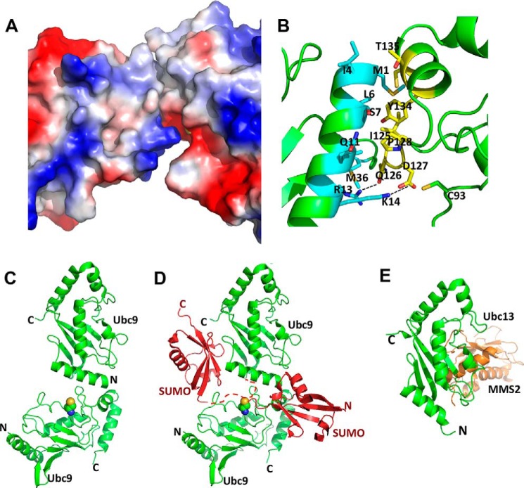
An RWD domain is a well conserved domain found through bioinformatic analysis of the human proteome sequence; however, its function has been unknown. Ubiquitin-like modifications require the catalysis of three enzymes generally known as E1, E2, and E3. We solved the crystal structure of the E2 for the small ubiquitin-like modifiers (SUMO) in complex with an RWD domain and confirmed the structure using solution NMR analysis. The binding surface of RWD on Ubc9 is located near the N terminus of Ubc9 that is known to be involved in noncovalent binding of the proteins in the conjugation machinery, including a domain of E1, SUMO, and an E3 ligase. NMR data indicate that the RWD domain does not bind to SUMO and E1. The interaction between RWD and Ubc9 has a Kd of 32 ± 4 μM. Consistent with the structure and binding affinity and in contrast to a previous report, the RWD domain and RWDD3 have minimal effects on global SUMOylation. The structural and biochemical information presented here forms the basis for further investigation of the functions of RWD-containing proteins.

Keywords: E2; RWD; nuclear magnetic resonance (NMR); small ubiquitin-like modifier (SUMO); sumoylation; ubiquitylation (ubiquitination); x-ray crystallography.


Figures

FIGURE 1.
FIGURE 1.
The RWD domain is a well structured region of RWDD3 and confers the same in vitro effects as RWDD3 on SUMOylation. A, superimposed 1H-15N HSQC spectra of full-length RWDD3 (red) and its RWD domain (blue). B, nonreducing SDS-PAGE comparing the enhancement effect of RWDD3 and its RWD domain on formation of the Ubc9·SUMO thioesters, SAE2·SUMO thioester, and SAE2-SUMO isopeptide bonds. Parallel control assays were performed under the same conditions but without RWDD3 or RWD.
FIGURE 2.
FIGURE 2.
The structure of RWD-Ubc9 complex. A, the overall fold of the crystal structure showing a heterotrimer that contains one RWD and a Ubc9 homodimer (also see Table 1). B and C, plot of CSP upon titration of 15N-labeled Ubc9 with the RWD domain (B) or 15N-labeled RWD domain with Ubc9 (C). The weighted CSP was calculated as Δδweighted = [(ΔδH)2 + (ΔδN/6.7)2]1/2. The horizontal lines (0.05 ppm for Ubc9 and 0.03 ppm for RWD) represent twice the average CSP. The perturbed residues in Ubc9 upon RWD titrations and in RWD upon Ubc9 titrations are indicated in red in ribbon diagrams above their respective CSP versus residue plots.
FIGURE 3.
FIGURE 3.
The binding interface and affinity of the RWD-Ubc9 complex. A, surface representation color-coded by electrostatic potential of the RWD-Ubc9 complex. Red to blue corresponds to negative to positive electrostatic potentials. B, zoomed in view of the RWD-Ubc9 binding interface with key interacting residues shown and labeled. C, estimation of Kd of the complex using CSP of cross-peaks of the indicated residues in 15N-labeled RWD as a function of incremental addition of 0.125 molar equivalent of Ubc9. The Kd of the Ubc9-RWD complex was determined by nonlinear least square fitting of CSP as a function of molar ratios of Ubc9 versus RWD.
FIGURE 4.
FIGURE 4.
The RWD-binding surface on Ubc9 overlaps with the binding surfaces of the UFD of E1, SUMO, and RanBP2 (a SUMO E3 ligase). A, left panel, overlay of Ubc9 in the predicted UFD-Ubc9 complex and the Ubc9-RWD complex. The RWD domain fully overlaps with the UFD domain, indicating their competition for Ubc9. Middle panel, conserved binding site of the ubiquitin-like modifier (SUMO, red) in relation to that of the RWD (cyan) on an E2 (Ubc9, green). Right panel, E3 ligase RanBP2 (yellow) binding site in relation to that of the RWD (cyan) on Ubc9 (green). B, RWD and SUMO-1 do not bind to Ubc9 simultaneously. Left panel, overlay of the 1H-15N HSQC spectra 15N-Ubc9, free (blue) and in complex with 1:1 stoichiometry of unlabeled SUMO-1 (green). Right panel, overlay of the 1H-15N HSQC spectra of the Ubc9-SUMO complex in A (green) and that of the complex with addition of 1:1 stoichiometry of the RWD domain (red). Residues at the protein-protein binding interfaces are indicated. Residues whose resonances disappeared upon complex formation are indicated in italics. C, the RWD domain does not interact with SAE and SUMO. Left panel, overlay of the 1H-15N HSQC spectra of the 15N-labeled Cys domain of SAE, free (black) and in complex with 1.2-fold molar excess of unlabeled RWD domain (red). Middle panel, overlay of the 1H-15N HSQC spectra of the15N-labeled RWD, free (red) and in complex with 1.2 molar excess of unlabeled UFD domain of SAE (cyan). Right panel, overlay of the 1H-15N HSQC spectra of the15N-labeled RWD domain, free (red) and in complex with 1.2 molar excess of unlabeled SUMO-1 (green). No significant chemical shift perturbation was seen upon the formation of all of these complexes.
FIGURE 5.
FIGURE 5.
The effect of RWDD3 and its RWD domain in SUMOylation. A, the effect of various concentrations of the RWD domain on the formation of Ubc9·SUMO thioester conjugate at the indicated Ubc9 concentrations. B, the transfer of SUMO from SAE2 to Ubc9 at the indicated time after the addition of Ubc9 in the presence and absence of 50 μm RWD. C, HEK293T cells were transfected with pCDNA3-RWD, pCDNA3-RWDD3, and pCDNA3.1, respectively. After 48 h of DNA transfection, poly-SUMOylation (SUMO-2 and -3), GAPDH, Flag tag RWDD3, and its RWD domain expressions were detected by Western blot. GAPDH expression was used as the loading control. D, RWDD3 and the RWD domain inhibited SUMO conjugation of the protein Sp100 using 35S-labeled Sp100 obtained from in vitro transcription and translation and detected by autoradiography.
FIGURE 6.
FIGURE 6.
The Ubc9 homodimer interfaces. A, the Ubc9 homodimer interface is shown with the surfaces color-coded according to electrostatic potentials; red to blue corresponds to negative to positive electrostatic potentials. B, zoomed in view of the Ubc9 homodimer interface with key interacting residues shown and labeled. One Ubc9 molecule is shown in cyan, and the other is in yellow. C, ribbon diagram of the Ubc9 homodimer. D, a model of how Ubc9 homodimer could stimulate SUMO chain formation. The structure of noncovalent Ubc9-SUMO complex is superimposed onto the top Ubc9 molecule of the Ubc9 homodimer, resulting in the position of the SUMO molecule on the upper left side. The dashed red line represents the flexible N-terminal segment of SUMO that contains the SUMOylation site but did not have electron density in x-ray diffraction. Ubc9 is shown in green, and SUMO is shown in red. Side chain atoms of the catalytic Cys-93 of the bottom Ubc9 are shown as spheres. A hypothetical SUMO molecule that forms a thioester conjugate with the bottom Ubc9 shown on the lower right side. E, ribbon diagram of Ubc13/MMS2 complex in which Ubc13 is in a similar orientation as the top Ubc9 molecule in C.

Comment in

References

    1. Doerks T., Copley R. R., Schultz J., Ponting C. P., Bork P. (2002) Systematic identification of novel protein domain families associated with nuclear functions. Genome Res. 12, 47–56 - PMC - PubMed
    1. Carbia-Nagashima A., Gerez J., Perez-Castro C., Paez-Pereda M., Silberstein S., Stalla G. K., Holsboer F., Arzt E. (2007) RSUME, a small RWD-containing protein, enhances SUMO conjugation and stabilizes HIF-1alpha during hypoxia. Cell 131, 309–323 - PubMed
    1. Yeh E. T. (2009) SUMOylation and De-SUMOylation: wrestling with life's processes. J. Biol. Chem. 284, 8223–8227 - PMC - PubMed
    1. Ulrich H. D. (2009) The SUMO system: an overview. Methods Mol. Biol. 497, 3–16 - PubMed
    1. Chen Y. (2007) The enzymes in ubiquitin-like post-translational modifications. Biosci. Trends 1, 16–25 - PMC - PubMed

Publication types

MeSH terms

Substances

LinkOut - more resources