Review
. 2015 Apr 27;209(2):199-210.
doi: 10.1083/jcb.201410017.

Review series: The cell biology of renal filtration

Affiliations
Review

Review series: The cell biology of renal filtration

Rizaldy P Scott et al. J Cell Biol. .

Abstract

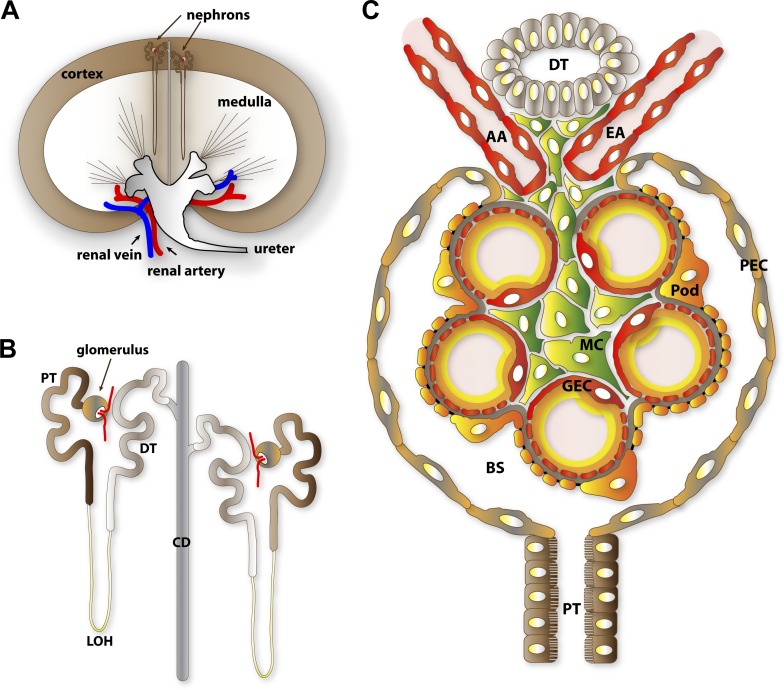
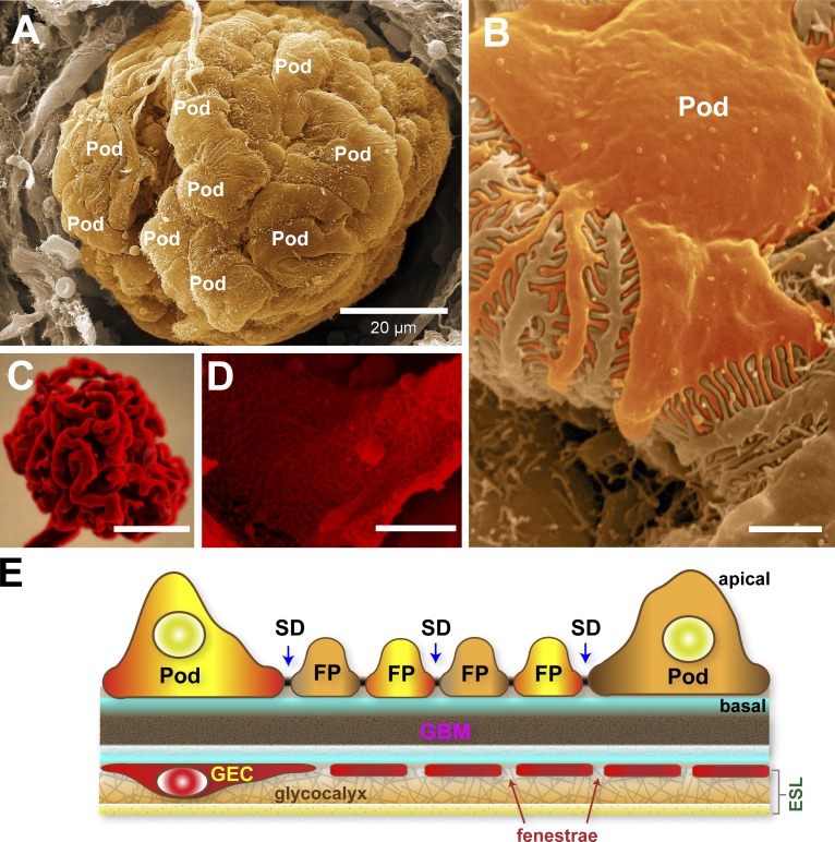
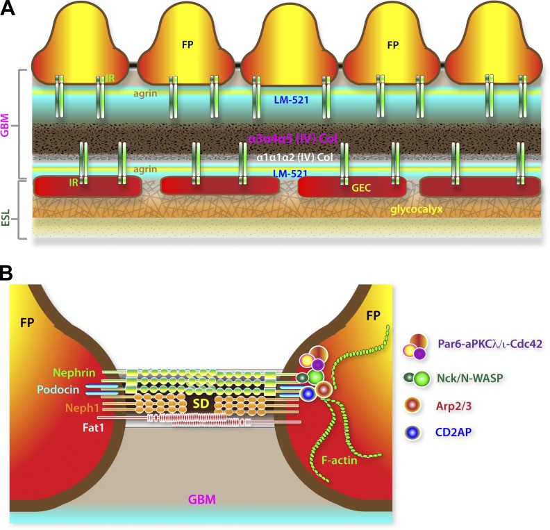
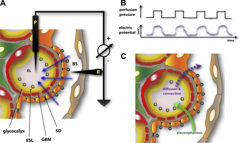
The function of the kidney, filtering blood and concentrating metabolic waste into urine, takes place in an intricate and functionally elegant structure called the renal glomerulus. Normal glomerular function retains circulating cells and valuable macromolecular components of plasma in blood, resulting in urine with just trace amounts of proteins. Endothelial cells of glomerular capillaries, the podocytes wrapped around them, and the fused extracellular matrix these cells form altogether comprise the glomerular filtration barrier, a dynamic and highly selective filter that sieves on the basis of molecular size and electrical charge. Current understanding of the structural organization and the cellular and molecular basis of renal filtration draws from studies of human glomerular diseases and animal models of glomerular dysfunction.


Figures

Figure 1.
Figure 1.
Anatomical overview of renal filtration. (A) Diagrammatic representation of nephron distribution in the kidney. Glomeruli, the filtration compartments of nephrons, are found within the kidney cortex. (B) Segmental structure of nephrons. The vascularized glomerulus is found at the proximal end and is connected through a series of renal tubules where urinary filtrate composition is refined through resorption and secretion. (C) Cellular organization of the glomeruli. GEC, glomerular endothelial cell; AA, afferent arteriole; EA, efferent arteriole; Pod, podocyte; MC, mesangial cell; PEC, parietal epithelial cell; PT, proximal tubule; DT, distal tubule; LOH, loop of Henle; CD, collecting duct; BS, Bowman’s space.
Figure 2.
Figure 2.
An ultrastructural overview of podocytes and the glomerular endothelium. (A) Scanning electron micrograph of an exposed glomerulus. In this image, the Bowman’s capsule is broken, permitting a striking view of podocytes (Pod) completely wrapped around the glomerular capillaries. (B) Higher magnification of a podocyte within the glomerulus revealing the interdigitated FPs. (C) A resin cast of the glomerular capillary tuft with the cells corroded to reveal its highly convoluted shape. Image courtesy of F. Hossler (East Tennessee State University, Johnson City, TN). (D) Scanning electron micrograph of an exposed glomerular capillary and its numerous perforations (fenestrae). (E) Simplified diagram of the GFB. The GEC and its fenestrae are lined by a filamentous glycocalyx enriched in negatively charged proteoglycans. The glycocalyx and adsorbed plasma components form the thicker ESL. The GBM is a stratified ECM in between podocytes and GECs. Podocytes form the final layer of the GFB. The interdigitating FPs of podocytes are linked by porous SDs where primary urinary filtrate passes through. Bars: (A) 20 µm; (B and D) 1 µm; (C) 50 µm.
Figure 3.
Figure 3.
Molecular organization of the GBM and the SD. (A) Highly stratified assembly of GBM components. Laminin LM-521 and agrin are bimodally distributed, whereas collagen IV complexes are concentrated at the core of the GBM. The minor α1α1α2 collagen (Col) is notably biased toward the glomerular endothelium. Both the predominant α3α4α5 and the less abundant α1α1α2 type IV collagens are normally too distant from β1–integrin receptor (IR) complexes on the podocyte side. This suggests that LM-521 and agrin but not type IV collagens are the normal physiological ligands of IR complexes expressed by podocytes. (B) Simplified representation of major adhesion receptors (nephrin, Neph1, and Fat1) found in the SD. Lipid-raft localization of the SD is dependent on the cholesterol-binding podocin. The SD is coupled to both F-actin regulatory (Nck–N-WASP–Arp2/3 and CD2AP–Arp2/3) and cell polarity (Par6–aPKCλ/ι–Cdc42) complexes.
Figure 4.
Figure 4.
Electrokinetic model of renal filtration. (A) Experimental setup used to demonstrate the existence of a flow-dependent electrical potential (streaming potential) across the GFB in salamander (N. maculosus) glomeruli (Hausmann et al., 2010). P, potential electrode; R, reference electrode. Small ions, due to differential interaction with the negatively charged GFB, create a net gradient of charges measurable as a streaming potential (blue arrows), making the endothelial lumen (EL) more positive than the Bowman’s space (BS). (B) Filtration pressure dependence of glomerular streaming potential. (C) Retrograde electrophoretic field created by streaming potentials. Due to streaming potentials, macromolecules encounter a dynamic electrophoretic field (green arrow) that is opposite to that of diffusive and convective fluxes (purple arrow). Albumin, a negatively charged macromolecule, would not only encounter size-dependent exclusion by the GFB but would effectively be electrophoresed away from the GFB during the course of active filtration.

References

    1. Abrahamson D.R.2012. Role of the podocyte (and glomerular endothelium) in building the GBM. Semin. Nephrol. 32:342–349 10.1016/j.semnephrol.2012.06.005 - DOI - PMC - PubMed
    1. Abrahamson D.R., Hudson B.G., Stroganova L., Borza D.B., and St. John P.L.. 2009. Cellular origins of type IV collagen networks in developing glomeruli. J. Am. Soc. Nephrol. 20:1471–1479 10.1681/ASN.2008101086 - DOI - PMC - PubMed
    1. Akilesh S., Huber T.B., Wu H., Wang G., Hartleben B., Kopp J.B., Miner J.H., Roopenian D.C., Unanue E.R., and Shaw A.S.. 2008. Podocytes use FcRn to clear IgG from the glomerular basement membrane. Proc. Natl. Acad. Sci. USA. 105:967–972 10.1073/pnas.0711515105 - DOI - PMC - PubMed
    1. Akilesh S., Suleiman H., Yu H., Stander M.C., Lavin P., Gbadegesin R., Antignac C., Pollak M., Kopp J.B., Winn M.P., and Shaw A.S.. 2011. Arhgap24 inactivates Rac1 in mouse podocytes, and a mutant form is associated with familial focal segmental glomerulosclerosis. J. Clin. Invest. 121:4127–4137 10.1172/JCI46458 - DOI - PMC - PubMed
    1. Ashraf S., Gee H.Y., Woerner S., Xie L.X., Vega-Warner V., Lovric S., Fang H., Song X., Cattran D.C., Avila-Casado C., et al. . 2013. ADCK4 mutations promote steroid-resistant nephrotic syndrome through CoQ10 biosynthesis disruption. J. Clin. Invest. 123:5179–5189 10.1172/JCI69000 - DOI - PMC - PubMed

Publication types

LinkOut - more resources