. 2015 May;145(5):419-30.
doi: 10.1085/jgp.201511377.

Ruling out pyridine dinucleotides as true TRPM2 channel activators reveals novel direct agonist ADP-ribose-2'-phosphate

Affiliations

Ruling out pyridine dinucleotides as true TRPM2 channel activators reveals novel direct agonist ADP-ribose-2'-phosphate

Balázs Tóth et al. J Gen Physiol. 2015 May.

Abstract

Transient receptor potential melastatin 2 (TRPM2), a Ca(2+)-permeable cation channel implicated in postischemic neuronal cell death, leukocyte activation, and insulin secretion, is activated by intracellular ADP ribose (ADPR). In addition, the pyridine dinucleotides nicotinamide-adenine-dinucleotide (NAD), nicotinic acid-adenine-dinucleotide (NAAD), and NAAD-2'-phosphate (NAADP) have been shown to activate TRPM2, or to enhance its activation by ADPR, when dialyzed into cells. The precise subset of nucleotides that act directly on the TRPM2 protein, however, is unknown. Here, we use a heterologously expressed, affinity-purified-specific ADPR hydrolase to purify commercial preparations of pyridine dinucleotides from substantial contaminations by ADPR or ADPR-2'-phosphate (ADPRP). Direct application of purified NAD, NAAD, or NAADP to the cytosolic face of TRPM2 channels in inside-out patches demonstrated that none of them stimulates gating, or affects channel activation by ADPR, indicating that none of these dinucleotides directly binds to TRPM2. Instead, our experiments identify for the first time ADPRP as a true direct TRPM2 agonist of potential biological interest.


Figures

Figure 1.
Figure 1.
Nonpurified pyridine dinucleotides act as low affinity TRPM2 activators. (A–C) Macroscopic T5L-TRPM2 currents elicited in inside-out patches by cytosolic application of saturating (125 µM) Ca2+ (black bars) and either 32 µM ADPR (dark blue bars) or increasing concentrations of nonpurified NAD (A, green bars), NAAD (B, blue bars), or NAADP (C, purple bars). (D) Dose–response curves for fractional current activation by ADPR (dark blue; replotted from Tóth and Csanády, 2012), and nonpurified NAD (green), NAAD (blue), and NAADP (purple). Currents were normalized to those measured in 32 µM ADPR. Solid lines are fits to the Hill equation yielding K1/2 = 1.4 ± 0.1 µM, n = 1.8 ± 0.2, and Irel;∞ = 0.96 ± 0.03 for ADPR; K1/2 = 73 ± 3 µM, n = 1.4 ± 0.1, and Irel;∞ = 0.96 ± 0.01 for NAD; K1/2 = 109 ± 17 µM, n = 1.3 ± 0.2, and Irel;∞ = 0.96 ± 0.06 for NAAD; and K1/2 = 226 ± 19 µM, n = 1.8 ± 0.2, and Irel;∞ = 0.78 ± 0.04 for NAADP. Error bars represent mean ± SEM.
Figure 2.
Figure 2.
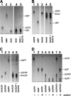
Enzymatic treatment efficiently removes ADPR and ADPRP contamination of pyridine dinucleotides. (A–C) TLC images showing the purification of (A) NAD, (B) NAAD, and (C) NAADP. Loaded samples (1 µl) were as follows: (A) 10 mM ADPR (lane 1), 10 mM AMP (lane 2), 10 mM NAD (lane 3), and 100 mM NAD before (lane 4) and after (lane 5) treatment with NUDT9. (B) 10 mM ADPR (lane 1), 10 mM AMP (lane 2), 10 mM NAAD (lane 3), and 100 mM NAAD before (lane 4) and after (lane 5) treatment with NUDT9. (C) 10 mM NAADP (lane 1), 10 mM ADPRP (lane 2), 10 mM AMPP (lane 3), and 100 mM NAADP before (lane 4) and after (lane 5) treatment with NUDT9. (D) Hydrolytic activity of NUDT9 on ADPR and ADPRP, showing 10 mM ADPR (lane 1), 10 mM AMP (lane 2), 10 mM ADPR treated with NUDT9 (lane 3), 10 mM ADPRP (lane 4), 10 mM AMPP (lane 5), 10 mM ADPRP treated with NUDT9 (lane 6), mixture of 10 mM ADPR and 10 mM ADPRP before (lane 7), and after (lane 8) treatment with NUDT9. Developing solution was DS2 in A and C, and DS1 in B and D.
Figure 3.
Figure 3.
Purified pyridine dinucleotides do not activate TRPM2 channels even at very high concentrations. (A–C) Macroscopic T5L-TRPM2 currents evoked by cytosolic application of saturating Ca2+ (black bars) and either 32 µM ADPR (dark blue bars) or high concentrations of untreated or NUDT9-treated (“purified”) NAD (A, green bars), NAAD (B, blue bars), or NAADP (C, purple bars). (D) Fractional current activation by indicated concentrations (mM) of NUDT9-treated (“+”) or nonpurified (“−”) NAD (green), NAAD (blue), and NAADP (purple). Currents were normalized to those measured in 32 µM ADPR. Error bars represent mean ± SEM.
Figure 4.
Figure 4.
Purified pyridine dinucleotides do not compete activation of TRPM2 channels by ADPR. (A–C) Macroscopic T5L-TRPM2 currents elicited by cytosolic application of saturating Ca2+ (black bars) and either 32 or 1 µM ADPR (dark blue bars) alone, or a mixture of 1 µM ADPR with a high concentration of NUDT9-treated, filtered (“purified”) NAD (A, green bar), NAAD (B, blue bar), or NAADP (C, purple bar). (D) Fractional current activation by 1 µM ADPR alone (dark blue) or in combination with indicated concentrations of NUDT9-treated, filtered NAD (green), NAAD (blue), and NAADP (purple). Currents were normalized to those measured in 32 µM ADPR. Error bars represent mean ± SEM.
Figure 5.
Figure 5.
ADPRP is a novel true TRPM2 partial agonist with distinctive kinetic features. (A) Macroscopic T5L-TRPM2 currents in response to cytosolic application of saturating Ca2+ (black bars) and either 32 µM ADPR (dark blue bars) or increasing concentrations of ADPRP (red bars). (B) Dose–response curves for fractional current activation by ADPR (dark blue; replotted from Fig. 1 D) and ADPRP (red). Currents were normalized to those measured in 32 µM ADPR. Solid lines are fits to the Hill equation with parameters plotted in the panel. (C) Macroscopic T5L-TRPM2 current repeatedly activated by brief exposures to 32 µM ADPR (dark blue bars) or 100 µM ADPRP (red bars), in the continued presence of saturating Ca2+ (black bars). Current decay time courses upon nucleotide removal were fitted by single-exponential functions (colored curves) with time constants indicated (in seconds). (D) Average T5L-TRPM2 current relaxation time constants (in seconds) upon sudden removal of saturating ADPR (dark blue) or ADPRP (red) in the maintained presence of Ca2+. Error bars represent mean ± SEM.
Figure 6.
Figure 6.
Kinetic fingerprint betrays active contaminant of nonpurified pyridine dinucleotides. (A–C) Macroscopic T5L-TRPM2 current repeatedly activated by brief exposures to 32 µM ADPR (dark blue bars) or saturating concentrations of nonpurified NAD (1 mM; A, green bars), NAAD (1 mM; B, blue bars), or NAADP (2 mM; C, purple bars) in the continued presence of saturating Ca2+ (black bars). Current decay time courses upon nucleotide removal were fitted by single-exponential functions (colored curves), with time constants indicated (in seconds). (D) Average T5L-TRPM2 current relaxation time constants (in seconds) upon sudden removal of saturating NAD (green), NAAD (blue), or NAADP (purple) in the maintained presence of Ca2+. Analogous parameters for ADPR (dark blue) and ADPRP (red) are replotted from Fig. 5 D for comparison. Error bars represent mean ± SEM.
Figure 7.
Figure 7.
Summary of nucleotide structures and metabolic pathways of interconversion. Color coding: dark blue, ADPR core structure; green, nicotinamide; blue, nicotinic acid; red, 2′-phosphate. In living cells, ADPR and ADPRP are generated from pyridine dinucleotide precursors by the multifunctional enzyme CD38, and degraded into AMP(P) and ribose-5-phosphate by NUDT9. NUDT9 substrates are also activators of TRPM2.

Comment in

  • Activators of TRPM2: Getting it right.
    Rosenbaum T. Rosenbaum T. J Gen Physiol. 2015 Jun;145(6):485-7. doi: 10.1085/jgp.201511405. Epub 2015 May 11. J Gen Physiol. 2015. PMID: 25964430 Free PMC article. No abstract available.

Similar articles

Cited by

References

    1. Aarhus R., Graeff R.M., Dickey D.M., Walseth T.F., and Lee H.C.. 1995. ADP-ribosyl cyclase and CD38 catalyze the synthesis of a calcium-mobilizing metabolite from NADP. J. Biol. Chem. 270:30327–30333 10.1074/jbc.270.51.30327 - DOI - PubMed
    1. Beck A., Kolisek M., Bagley L.A., Fleig A., and Penner R.. 2006. Nicotinic acid adenine dinucleotide phosphate and cyclic ADP-ribose regulate TRPM2 channels in T lymphocytes. FASEB J. 20:962–964 10.1096/fj.05-5538fje - DOI - PubMed
    1. Brailoiu E., Churamani D., Cai X., Schrlau M.G., Brailoiu G.C., Gao X., Hooper R., Boulware M.J., Dun N.J., Marchant J.S., and Patel S.. 2009. Essential requirement for two-pore channel 1 in NAADP-mediated calcium signaling. J. Cell Biol. 186:201–209 10.1083/jcb.200904073 - DOI - PMC - PubMed
    1. Calcraft P.J., Ruas M., Pan Z., Cheng X., Arredouani A., Hao X., Tang J., Rietdorf K., Teboul L., Chuang K.T., et al. . 2009. NAADP mobilizes calcium from acidic organelles through two-pore channels. Nature. 459:596–600 10.1038/nature08030 - DOI - PMC - PubMed
    1. Churamani D., Carrey E.A., Dickinson G.D., and Patel S.. 2004. Determination of cellular nicotinic acid-adenine dinucleotide phosphate (NAADP) levels. Biochem. J. 380:449–454 10.1042/BJ20031754 - DOI - PMC - PubMed

Publication types