. 2015 May 12;112(19):6194-9.
doi: 10.1073/pnas.1421785112. Epub 2015 Apr 27.

Adhesion G protein-coupled receptors are activated by exposure of a cryptic tethered agonist

Affiliations

Adhesion G protein-coupled receptors are activated by exposure of a cryptic tethered agonist

Hannah M Stoveken et al. Proc Natl Acad Sci U S A. .

Erratum in

Abstract

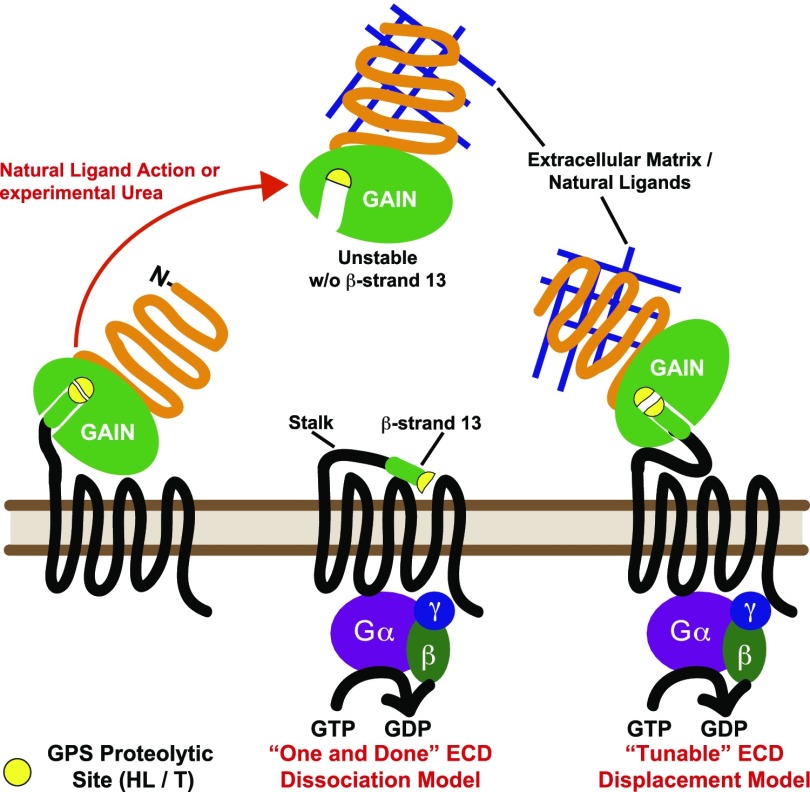
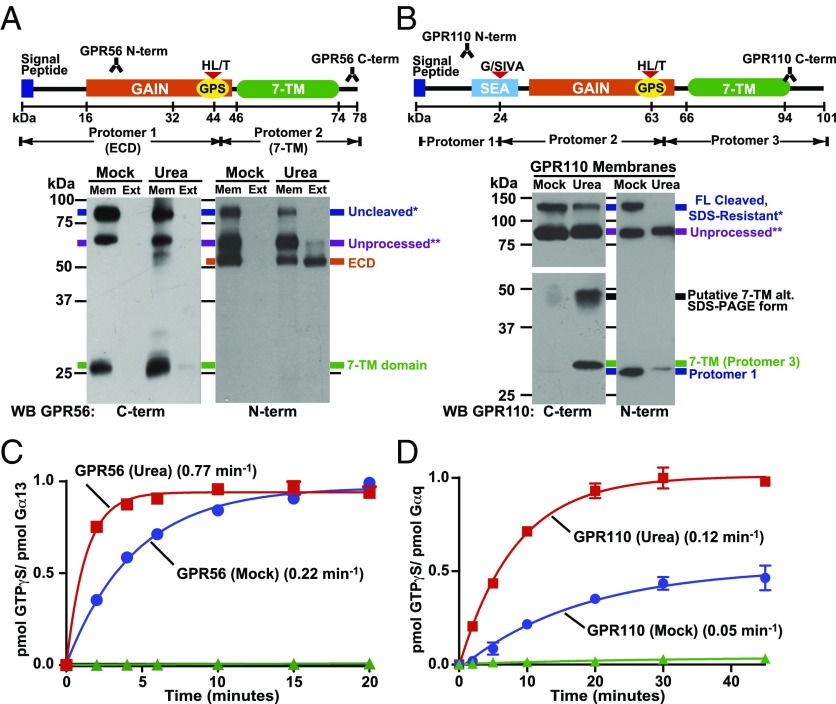
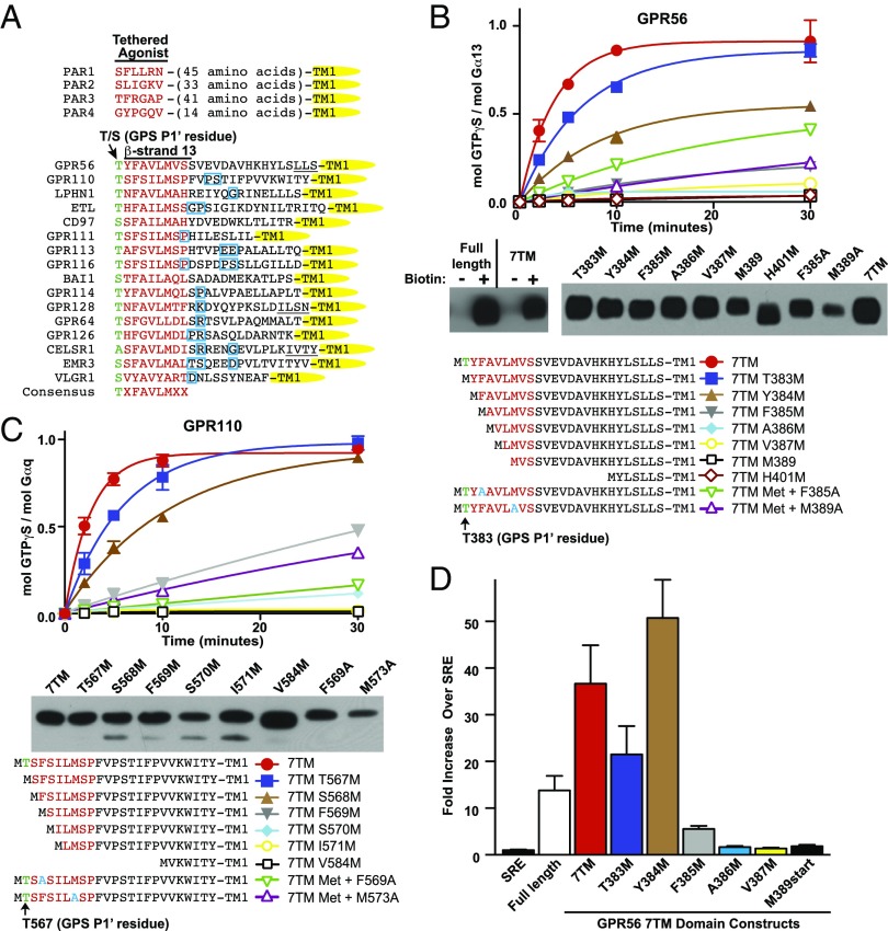
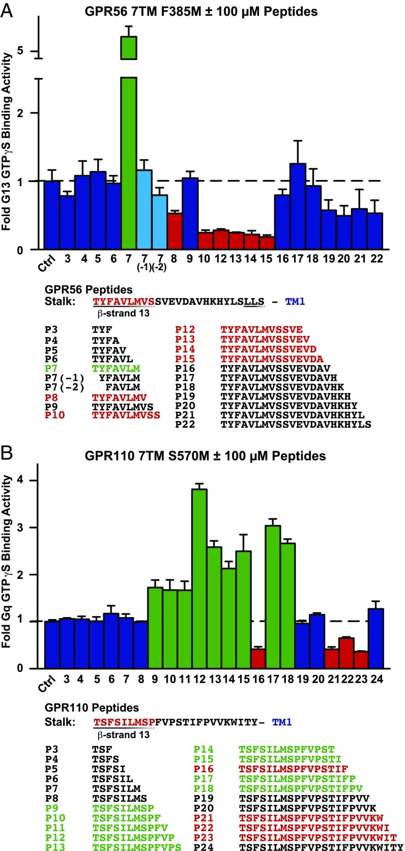
The large class of adhesion G protein-coupled receptors (aGPCRs) bind extracellular matrix or neighboring cell-surface ligands to regulate organ and tissue development through an unknown activation mechanism. We examined aGPCR activation using two prototypical aGPCRs, GPR56 and GPR110. Active dissociation of the noncovalently bound GPR56 or GPR110 extracellular domains (ECDs) from the respective seven-transmembrane (7TM) domains relieved an inhibitory influence and permitted both receptors to activate defined G protein subtypes. After ECD displacement, the newly revealed short N-terminal stalk regions of the 7TM domains were found to be essential for G protein activation. Synthetic peptides comprising these stalks potently activated GPR56 or GPR110 in vitro or in cells, demonstrating that the stalks comprise a tethered agonist that was encrypted within the ECD. Establishment of an aGPCR activation mechanism provides a rational platform for the development of aGPCR synthetic modulators that could find clinical utility toward aGPCR-directed disease.

Keywords: G proteins; GPR110; GPR56; adhesion GPCRs; tethered agonist.


Conflict of interest statement

The authors declare no conflict of interest.

Figures

Fig. 1.
Fig. 1.
Models of adhesion GPCR activation. Adhesion GPCRs are constitutively self-cleaved at the GPCR proteolytic site (GPS) within the GAIN domain. Natural ligand- or experimental urea-mediated displacement of aGPCR ECDs relieves an inhibition of the 7TM domain. The highly conserved GAIN domain β-strand-13 and stalk region could act as a tethered agonist to the 7TM domain after ECD dissociation (One and Done model) or upon displacement (Tunable model).
Fig. 2.
Fig. 2.
GPR56 and GPR110 ECD dissociation activates 7TM-mediated G protein activation. (A and B) Prepared insect cell membranes with expressed, full-length GPR56 or GPR110 were mock treated or extracted with urea. The presence of the GPR56 ECD and 7TM domain in the membrane (Mem) and extract (Ext) fractions was determined by Western blotting with the GPR56 N- and C-terminal antibodies. The relative levels of GPR110 ECD protomer 1 and the 7TM domain in mock and urea-extracted membranes were determined by Western blotting with the GPR110 N- and C-terminal antibodies. The GPR110 C-terminal low-molecular weight (MW) panel is a longer exposure than the high-MW panel. (C) GPR56 or (D) GPR110 urea-treated (■, red) and untreated (●, blue) membranes, or nonreceptor membranes (▲, green) were reconstituted with purified Gα13 or Gαq and Gβ1γ2, and receptor-stimulated [35S]-GTPγS binding kinetics were measured. Error bars, SEM.
Fig. 3.
Fig. 3.
Adhesion GPCR β-strand-13/stalk regions are essential for G protein activation. (A) Sequence comparison of representative aGPCR and PAR stalk regions. The β-strand-13 TXFAVLMXX consensus sequences are denoted in red, and predicted turn elements are boxed in blue (CFSSP server) (28). Underlined sequences are alternative TM1 assignments (TMPred server). (B) GPR56 or (C) GPR110 7TM domain N-terminal single amino acid truncation series stimulation of G protein [35S]-GTPγS binding kinetics. Adhesion GPCR C-terminal antibody Western blots show relative levels of the indicated receptors isolated from insect cell surfaces by biotinylation pull-down assay. (D) HEK293 SRE luciferase activity in response to expressed GPR56 full-length and 7TM domain receptor N-terminal single amino acid truncation series. Error bars, SEM.
Fig. 4.
Fig. 4.
GPR56 and GPR110 synthetic stalk peptide screens for modulation of 7TM domain-mediated G protein activation. Synthetic peptides (100 µM) comprising the indicated portions of the (A) GPR56 or (B) GPR110 β-strand-13/stalk regions were tested for the ability to modulate low activity GPR56 7TM F385M domain-stimulated G13 or GPR110 7TM S570M domain-stimulated Gq [35S]-GTPγS binding. Values are expressed as fold vehicle treatment (Ctrl). Error bars, SEM.
Fig. 5.
Fig. 5.
Specific aGPCR synthetic peptides act as agonists. (A) GPR56 7TM F385M membranes (●, with 100 µM P7 peptide; ○, no peptide), 7TM H401M membranes (■, with 100 µM peptide; □, no peptide) and nonreceptor membranes (△) with 100 µM P7 peptide were reconstituted with G13 before measurement of receptor-stimulated [35S]-GTPγS binding kinetics (30-min point only, △). (B) GPR110 7TM S570M membranes (●, with 100 µM P12 peptide; ○, no peptide) and GPR110 7TM V584M membranes (■, with 100 µM P12 peptide; □, no peptide) were reconstituted with Gq before measurement of receptor-stimulated [35S]-GTPγS binding kinetics. (C) GPR56 7TM F385M membranes were incubated with the indicated concentrations of GPR56 peptides, TYFAVLM (P7) or YFAVLM (P7-1), and (D) GPR110 7TM S570M membranes were incubated with the indicated concentration of GPR110 P12 peptide before reconstitution with G13 or Gq and measurement of initial [35S]-GTPγS binding rates. Rates were plotted vs. peptide concentrations. (E) Urea- (squares) and mock-extracted (circles) full-length GPR56 membranes were incubated with 100 µM P7 peptide (closed symbols) or without peptide (open symbols) before G13 reconstitution and measurement of receptor-stimulated [35S]-GTPγS binding kinetics. (F) HEK293 SRE luciferase activity in response to empty vector (SRE) or expressed GPR56 7TM receptors and the indicated concentrations of P7 peptide. Error bars, SEM.

References

    1. Bjarnadóttir TK, et al. The human and mouse repertoire of the adhesion family of G-protein-coupled receptors. Genomics. 2004;84(1):23–33. - PubMed
    1. Langenhan T, Aust G, Hamann J. Sticky signaling—adhesion class G protein-coupled receptors take the stage. Sci Signal. 2013;6(276):re3. - PubMed
    1. Paavola KJ, Hall RA. Adhesion G protein-coupled receptors: Signaling, pharmacology, and mechanisms of activation. Mol Pharmacol. 2012;82(5):777–783. - PMC - PubMed
    1. Aust G. Adhesion-GPCRs in tumorigenesis. Adv Exp Med Biol. 2010;706:109–120. - PubMed
    1. Lum AM, et al. Orphan receptor GPR110, an oncogene overexpressed in lung and prostate cancer. BMC Cancer. 2010;10(1):40. - PMC - PubMed

Publication types