. 2015 May 12;112(19):6044-9.
doi: 10.1073/pnas.1411718112. Epub 2015 Apr 27.

Crystal structure of the human mitochondrial chaperonin symmetrical football complex

Affiliations

Crystal structure of the human mitochondrial chaperonin symmetrical football complex

Shahar Nisemblat et al. Proc Natl Acad Sci U S A. .

Abstract

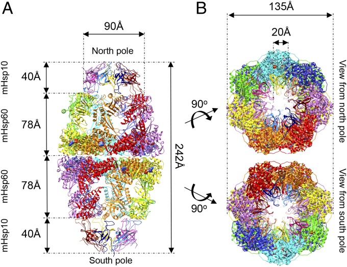
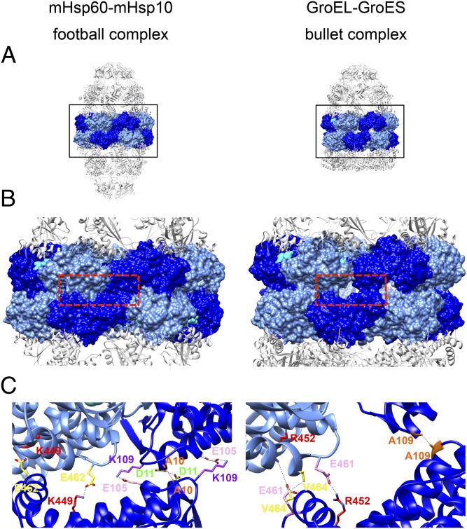
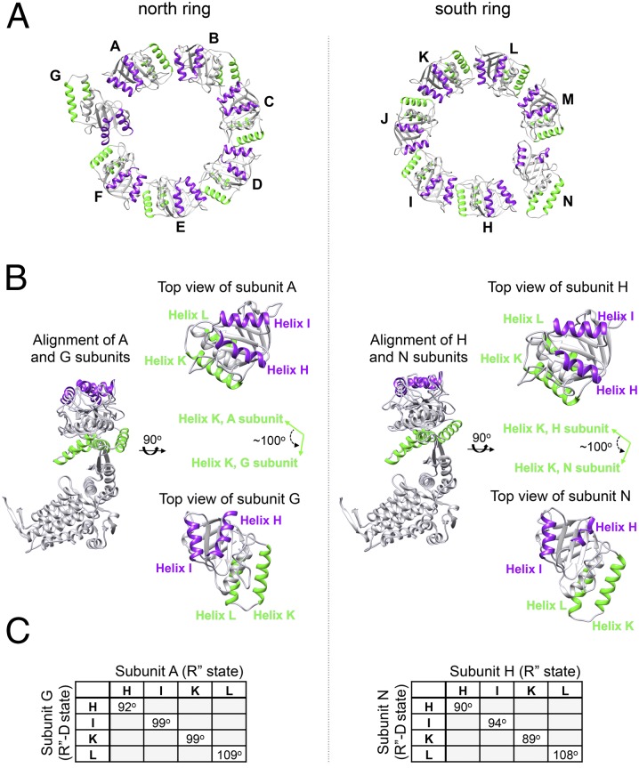
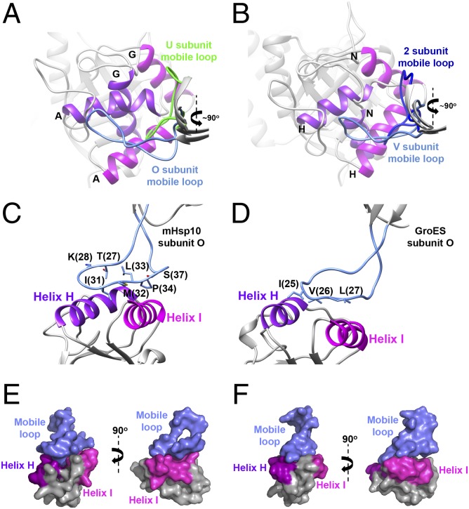
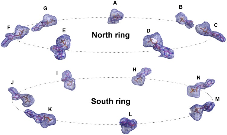
Human mitochondria harbor a single type I chaperonin system that is generally thought to function via a unique single-ring intermediate. To date, no crystal structure has been published for any mammalian type I chaperonin complex. In this study, we describe the crystal structure of a football-shaped, double-ring human mitochondrial chaperonin complex at 3.15 Å, which is a novel intermediate, likely representing the complex in an early stage of dissociation. Interestingly, the mitochondrial chaperonin was captured in a state that exhibits subunit asymmetry within the rings and nucleotide symmetry between the rings. Moreover, the chaperonin tetradecamers show a different interring subunit arrangement when compared to GroEL. Our findings suggest that the mitochondrial chaperonins use a mechanism that is distinct from the mechanism of the well-studied Escherichia coli system.

Keywords: Hsp10; Hsp60; chaperone; mitochondrial chaperonin; symmetrical complex.


Conflict of interest statement

The authors declare no conflict of interest.

Figures

Fig. 1.
Fig. 1.
Overview of the mHsp60–mHsp10 football complex. (A) View perpendicular to the long axis of the complex. The seven subunits in each mHsp60 ring are colored red, orange, yellow, green, cyan, blue, and purple. The mHsp10 subunits in each ring are colored blue, light blue, purple, dark purple, yellow, pink, and dark red. Structure dimensions are marked by arrows (distances between Cα). Atoms of the 14 ADP molecules are rendered as spheres. (B) Views from the north and south poles of the mHsp6014–(mHsp107)2 complex. The coloring scheme is as in A.
Fig. 2.
Fig. 2.
Hsp60 interring contacts in the symmetric human football complex (Left) and in the asymmetric bacterial bullet complex (Right). (A) Full side views of the mitochondrial complex and the bacterial (PDB ID code 1AON) complex. The subunit equatorial domains are rendered as a molecular surface in alternating blue and light blue. The other domains in the Hsp60 subunits and Hsp10 subunits are presented as gray cartoons. (B) Zoom-in view of the boxed area in A, showing the Hsp60 interring contact area. ADP atoms are presented as cyan spheres. (C) Three subunits from each complex were taken from the red boxed area in B and presented as cartoons with stripped surfaces, colored as in A and B. Residues forming the main interring contacts are presented as sticks: in the mitochondrial complex (Left) [A10 (orange), D11 (green), E105 (pink), K109 (purple), K449 (red), and E462 (yellow)] and in the bacterial complex (Right) [A109, (orange), R452 (red), E461 (pink), and V464, (yellow)]. The bonds between the interacting residues are shown as black dashed lines. Subunits of mHsp60 assemble in a zipper-like conformation in which the contact surface is larger and contacts are tighter than in GroEL (B and C), allowing new contacts to be formed.
Fig. 3.
Fig. 3.
Asymmetry of subunits within mHsp60 rings. (A) Top view of the apical domains in a ring (residues 192–375) is presented: north pole (Left) and south pole (Right). Helices H and I are colored purple, and helices K and L are colored green. Subunits of mHsp60 are identified by letters. In each ring, there is one subunit that breaks the symmetry of the ring (subunit G in the north pole and subunit N in the south pole). (B) Alignment of subunits A and G (north ring) and subunits H and N (south ring). Coloring is as in A. (Right) In each column, separate top views of the aligned subunits are shown. The apical domains of subunits G and N have rotated counterclockwise nearly 100° from the original position of the other six subunits in each ring, as demonstrated here by comparing helices K of subunits A and G or of subunits H and N (green arrows). (C) Tables showing the crossing angles of helices H, I, K, and L between the R′′ state (relaxed state after ATP hydrolysis) and the newly formed R′′-D state (R′′ state that started the dissociation process) in subunits G and N. Angles were calculated using the UCSF Chimera package (64).
Fig. 4.
Fig. 4.
Conformations of the mobile loop in the mHsp6014–(mHsp107)2 complex. (A) View from the north pole on the alignment of mHsp60 subunits A and G, showing the binding sites with mHsp10 mobile loops of subunits O and U, respectively. Mobile loops are colored light blue (subunit O) and green (subunit U). Helix H is colored purple, and helix I is colored magenta. Subunit identities of helices H and I are shown. (B) Same as A, but for the view from the south pole on the alignment of subunits H and N while bound to mobile loops of subunits V and 2, respectively. Mobile loops are colored light blue (subunit V) and blue (subunit 2). Views show the mHsp10 mobile loop/mHsp60-binding site (C) compared with the GroES mobile loop/GroEL-binding site (D). In both images, the mobile loop is colored blue and helices H and I are colored as in A. The residues in the mobile loop that mediate the binding to mHsp60/GroEL apical domains are displayed as sticks. (E and F) Surface presentation of C and D, respectively (Left), and 90° image rotation of the same views (Right). The color scheme is as in C and D.
Fig. 5.
Fig. 5.
Both mHsp60 rings are in the post ATP-hydrolysis state. A side view of the 14 ADP molecules as they are positioned in the structure is shown. Nucleotides are shown as sticks and are labeled according to their subunits. In addition, a Fo-Fc omit electron density map of the 14 nucleotides is shown. The map was calculated by omitting the nucleotides and magnesium atoms from the final model and performing five cycles of refinement using REFMAC [version 5.7.32 (58)]. The map was contoured at a 3σ cutoff.

References

    1. Cheng MY, et al. Mitochondrial heat-shock protein hsp60 is essential for assembly of proteins imported into yeast mitochondria. Nature. 1989;337(6208):620–625. - PubMed
    1. Ostermann J, Horwich AL, Neupert W, Hartl FU. Protein folding in mitochondria requires complex formation with hsp60 and ATP hydrolysis. Nature. 1989;341(6238):125–130. - PubMed
    1. Goloubinoff P, Gatenby AA, Lorimer GH. GroE heat-shock proteins promote assembly of foreign prokaryotic ribulose bisphosphate carboxylase oligomers in Escherichia coli. Nature. 1989;337(6202):44–47. - PubMed
    1. Goloubinoff P, Christeller JT, Gatenby AA, Lorimer GH. Reconstitution of active dimeric ribulose bisphosphate carboxylase from an unfolded state depends on two chaperonin proteins and Mg-ATP. Nature. 1989;342(6252):884–889. - PubMed
    1. Saibil HR, Ranson NA. The chaperonin folding machine. Trends Biochem Sci. 2002;27(12):627–632. - PubMed

Publication types

Associated data

LinkOut - more resources