. 2015 Apr 13:9:115.
doi: 10.3389/fncel.2015.00115. eCollection 2015.

Acetylcholine release in mouse hippocampal CA1 preferentially activates inhibitory-selective interneurons via α4β2* nicotinic receptor activation

Affiliations

Acetylcholine release in mouse hippocampal CA1 preferentially activates inhibitory-selective interneurons via α4β2* nicotinic receptor activation

L Andrew Bell et al. Front Cell Neurosci. .

Abstract

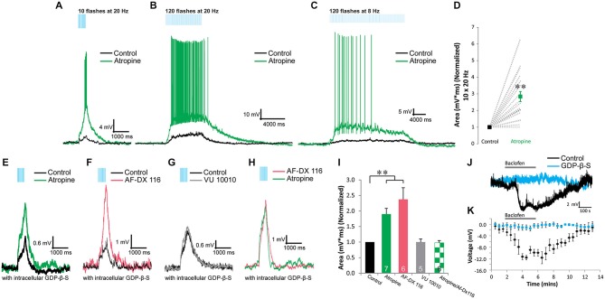
Acetylcholine (ACh) release onto nicotinic receptors directly activates subsets of inhibitory interneurons in hippocampal CA1. However, the specific interneurons activated and their effect on the hippocampal network is not completely understood. Therefore, we investigated subsets of hippocampal CA1 interneurons that respond to ACh release through the activation of nicotinic receptors and the potential downstream effects this may have on hippocampal CA1 network function. ACh was optogenetically released in mouse hippocampal slices by expressing the excitatory optogenetic protein oChIEF-tdTomato in medial septum/diagonal band of Broca cholinergic neurons using Cre recombinase-dependent adeno-associated viral mediated transfection. The actions of optogenetically released ACh were assessed on both pyramidal neurons and different interneuron subtypes via whole cell patch clamp methods. Vasoactive intestinal peptide (VIP)-expressing interneurons that selectively innervate other interneurons (VIP/IS) were excited by ACh through the activation of nicotinic receptors containing α4 and β2 subunits (α4β2*). ACh release onto VIP/IS was presynaptically inhibited by M2 muscarinic autoreceptors. ACh release produced spontaneous inhibitory postsynaptic current (sIPSC) barrages blocked by dihydro-β-erythroidine in interneurons but not pyramidal neurons. Optogenetic suppression of VIP interneurons did not inhibit these sIPSC barrages suggesting other interneuron-selective interneurons were also excited by α4β2* nicotinic receptor activation. In contrast, interneurons that innervate pyramidal neuron perisomatic regions were not activated by ACh release onto nicotinic receptors. Therefore, we propose ACh release in CA1 facilitates disinhibition through activation of α4β2* nicotinic receptors on interneuron-selective interneurons whereas interneurons that innervate pyramidal neurons are less affected by nicotinic receptor activation.

Keywords: CA1; disinhibition; hippocampus; interneuron-selective interneuron; nicotinic receptor; optogenetics.


Figures

Figure 1
Figure 1
Acetylcholine (ACh) release activates α4β2*-nicotinic receptors on vasoactive-intestinal peptide (VIP) interneuron-selective interneurons. (A) VIP interneuron-selective (IS) interneuron (morphology, A1) responded to optogenetically released ACh with fast depolarizations (black traces, 10 × 20 Hz) that were inhibited by 1 µM DHβE (orange traces) but not 25 nM MLA (gray trace). (B) All VIP interneurons not displaying basket cell morphology were unaffected by bath application of 25 nM MLA (gray bar, n = 12) but were blocked by 1 µM DHβE (orange bar, one-way ANOVA, p < 0.001, Bonferroni post hoc p < 0.001, n = 20). (C) Histogram illustrating the distribution of response types across VIP expressing interneurons: VIP/IS cells, and VIP/noID. Most VIP/IS cells (19 of 21) and all non-visually identified VIP cells (19 of 19) exhibited nicotinic-dependent depolarizations (purple). (D–F) Example of VIP interneurons with nicotinic responses that displayed irregular action potential firing patterns that decreased in amplitude in response to depolarizing current injection. Negative current pulses produced hyperpolarizing membrane responses with little or no voltage sag. (G) A VIP interneuron that produced accommodating regular action potential firing patterns to depolarizing current injection and a hyperpolarizing sag in response to negative current injection.
Figure 2
Figure 2
Presynaptic M2 autoreceptors inhibit the release of ACh onto α4β2* nicotinic receptors in VIP/IS interneurons. (A–C) Optogenetically released ACh produced depolarizing responses to short/fast (A, 10 × 20 Hz), prolonged/fast (B, 120 × 20 Hz), and prolonged/slow (C, 120 × 8 Hz) blue light flashes (black traces). The depolarizations were potentiated by 10 µM atropine (green traces). (D) Atropine (green) significantly potentiated the area of the nicotinic response (normalized to control, black, 10 × 20 Hz) (t-test, p < 0.001, n = 0). (E). Inclusion of GDP-β-S (black trace) in the intracellular recording solution did not inhibit atropine (green trace) potentiation of the nicotinic responses. (F) M2 antagonist AF-DX 116 (500 nM, red trace) potentiated nicotinic responses when GDP-β-S was included in the intracellular solution. (G) M4 positive allosteric modulator VU 10010 did not affect nicotinic responses (one-way ANOVA, ns, n = 5). (H) M2 antagonist AF-DX 116 (500 nM, red trace) occluded atropine (green trace) potentiation of nicotinic responses. (I) Histogram showing that atropine (GDP-β-S) (one-way ANOVA, p < 0.001, Bonferroni post hoc test p < 0.001, n = 7) and AF-DX 116 (GDP-β-S) (Bonferroni post hoc test p < 0.001, n = 6) significantly potentiated nicotinic responses. Atropine (green checkers) did not significantly increase nicotinic responses previously potentiated by AF-DX 116 (GDP-β-S) (t-test, ns, n = 6). (J) Application of 10 µM baclofen hyperpolarized VIP interneurons (black trace) but not when GDP-β-S (blue trace) was included in the intracellular solution. (K) All VIP interneurons tested produced significant hyperpolarizations when exposed to baclofen (black dots, one-way ANOVA, p < 0.001, Bonferroni post hoc p < 0.01 for time points 4–7 min, n = 4). Recordings that included GDP-β-S in the patch pipette did not respond to baclofen application (blue dots, one-way ANOVA, ns, n = 4).
Figure 3
Figure 3
ACh release drives nicotinic receptor-mediated feedforward inhibition onto CA1 interneurons but not CA1 pyramidal neurons. (A,E,I,M) Schematic drawings of four recording paradigms. sIPSCs were recorded in response to ACh release in non-VIP CA1 interneurons located at the border of SR and SLM (A–H), in CA1 pyramidal neurons (I–L), or in layer 2/3 neocortical pyramidal neurons (M–P). All recordings were performed in the presence of 10 µM atropine, 30 µM DNQX, and 50 µM APV. (B) Voltage clamp recordings (Vh = −70 mV) demonstrating that optogenetic release of ACh (blue bars, 10 × 20 Hz) increased the number of sIPSCs observed in CA1 non-VIP interneurons. (C) A peristimulus time histogram (PSTH) illustrating the increase in time-dependent sIPSC frequency in nonVIP interneurons following ACh release. (D) An increase in the averaged time-dependent sIPSC frequency across all non-VIP interneurons (black line, gray shading = S.E.M, one-way ANOVA compared to baseline, p < 0.01, n = 23) was completely blocked by 1 µM DHβE (orange line, orange shading = SEM, one-way ANOVA, ns compared to baseline, n = 8). (F) Voltage clamp recordings showing that suppression of VIP interneurons by yellow light activation of Arch (yellow bar) did not inhibit the increase in sIPSC frequency following ACh release (blue bars). (G) PSTH showing time-dependent sIPSC frequency following ACh release persisted in the presence of yellow light (yellow bar). (H) The increased averaged time-dependent sIPSC frequency measured across all non-VIP interneurons (black line, gray shading = S.E.M, one-way ANOVA, p < 0.001, n = 21) was not suppressed by activation of Arch in VIP interneurons. (J) Voltage clamp recordings from a CA1 pyramidal neuron showed no change in sIPSC frequency following ACh release (blue bars). (K) PSTH demonstrated that the time-dependent sIPSC frequency was unchanged following ACh release in an individual CA1 pyramidal neuron. (L) Averaged time-dependent sIPSC frequency across all CA1 pyramidal cell recordings demonstrated no change sIPSC frequency following ACh release (black line, gray shading = S.E.M, one-way ANOVA, ns, n = 42). (N) Voltage clamp recordings from a neocortical pyramidal neuron demonstrated that ACh release produced an increase in sIPSC frequency. (O) PSTH demonstrated that the time-dependent frequency of sIPSCs in the neocortical pyramidal neuron increased following ACh release. (P) The averaged time-dependent sIPSC frequency in all measured neocortical pyramidal neurons increased following ACh release (black line, gray shading = S.E.M., one-way ANOVA, p < 0.001, n = 5) and was blocked by 1 µM DHβE (orange line, orange shading = S.E.M., one-way ANOVA, ns, n = 5).

References

    1. Acsády L., Arabadzisz D., Freund T. F. (1996a). Correlated morphological and neurochemical features identify different subsets of vasoactive intestinal polypeptide-immunoreactive interneurons in rat hippocampus. Neuroscience 73, 299–315. 10.1016/0306-4522(95)00610-9 - DOI - PubMed
    1. Acsády L., Görcs T. J., Freund T. F. (1996b). Different populations of vasoactive intestinal polypeptide-immunoreactive interneurons are specialized to control pyramidal cells or interneurons in the hippocampus. Neuroscience 73, 317–334. 10.1016/0306-4522(95)00609-5 - DOI - PubMed
    1. Alkondon M., Pereira E. F., Albuquerque E. X. (1998). α-bungarotoxin- and methyllycaconitine-sensitive nicotinic receptors mediate fast synaptic transmission in interneurons of rat hippocampal slices. Brain Res. 810, 257–263. 10.1016/s0006-8993(98)00880-4 - DOI - PubMed
    1. Alkondon M., Pereira E. F., Barbosa C. T., Albuquerque E. X. (1997). Neuronal nicotinic acetylcholine receptor activation modulates gamma-aminobutyric acid release from CA1 neurons of rat hippocampal slices. J. Pharmacol. Exp. Ther. 283, 1396–1411. - PubMed
    1. Arroyo S., Bennett C., Aziz D., Brown S. P., Hestrin S. (2012). Prolonged disynaptic inhibition in the cortex mediated by slow, non-alpha7 nicotinic excitation of a specific subset of cortical interneurons. J. Neurosci. 32, 3859–3864. 10.1523/JNEUROSCI.0115-12.2012 - DOI - PMC - PubMed

LinkOut - more resources