

Ultraviolet B Inhibits Skin Wound Healing by Affecting Focal Adhesion Dynamics
- PMID: 25918970
- PMCID: PMC4513668
- DOI: 10.1111/php.12462
Ultraviolet B Inhibits Skin Wound Healing by Affecting Focal Adhesion Dynamics
Abstract
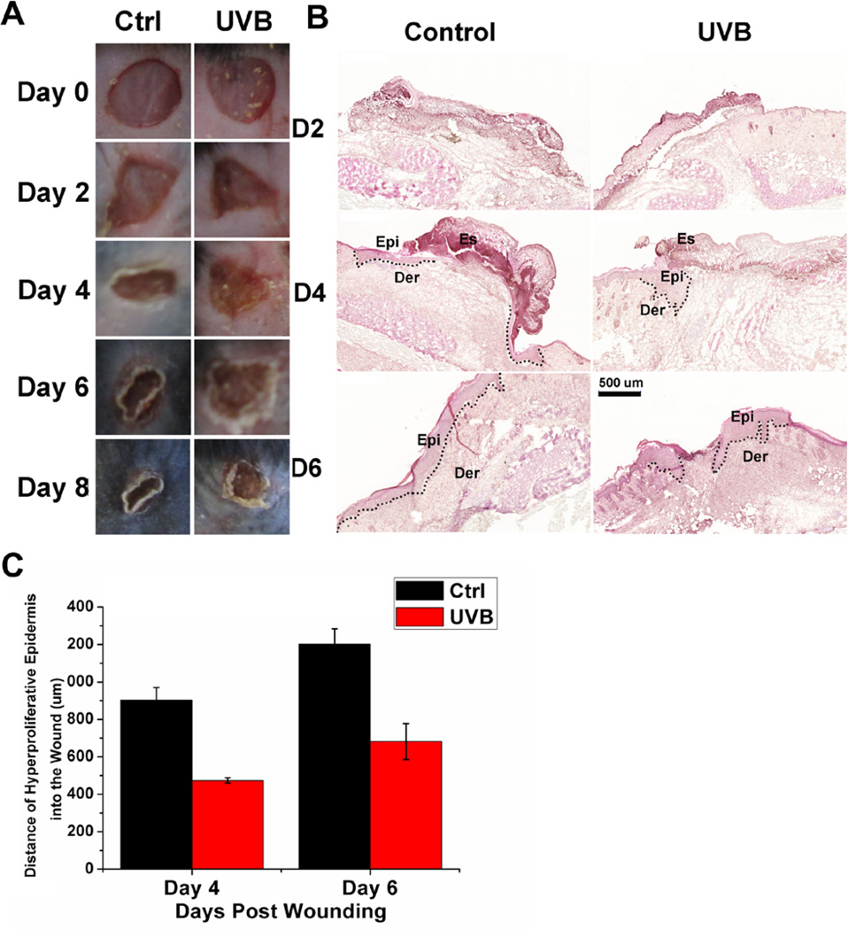
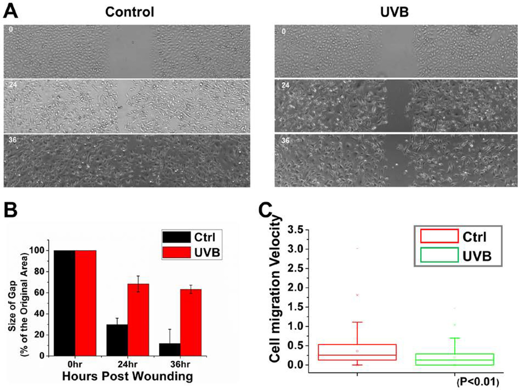
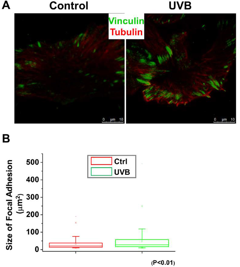
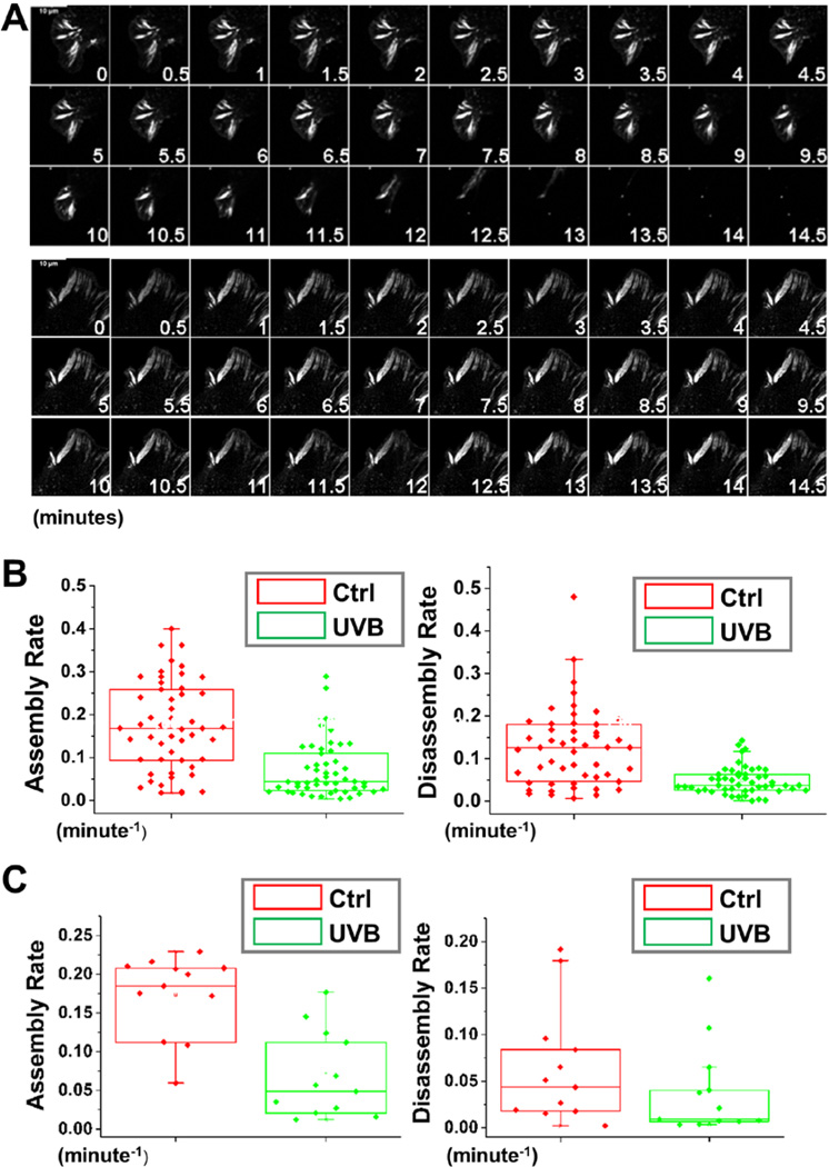
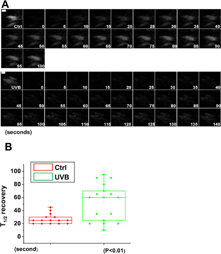
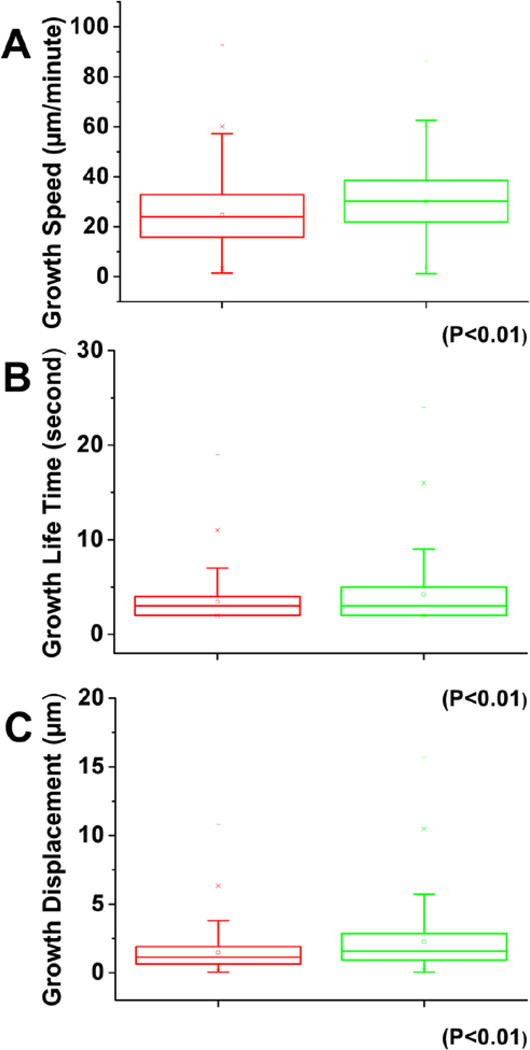
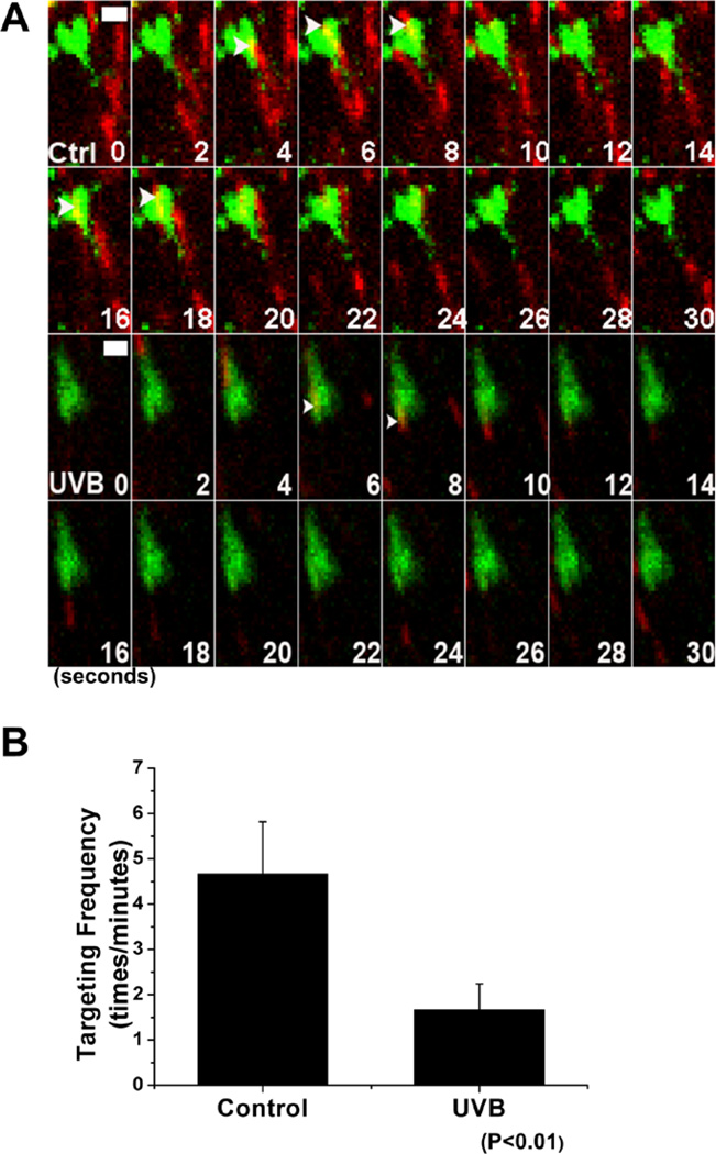
As the most important interface between human body and external environment, skin acts as an essential barrier preventing various environmental damages, among which DNA-damaging UV radiation from the sun remains the major environmental risk factor causing various skin diseases. It has been well documented that wavelengths in the ultraviolet B (UVB) radiation range (290-320 nm) of the solar spectrum can be absorbed by skin and lead to cutaneous injury and various other deleterious effects. During process such as wound healing, the orchestrated movement of cells in a particular direction is essential and highly regulated, integrating signals controlling adhesion, polarity and the cytoskeleton. Cell adhesion and migration are modulated through both of actin and microtubule cytoskeletons. However, little was known about how UVB affects skin wound healing and migration of epidermal keratinocytes. Here, we demonstrate that UVB can delay the wound healing progress in vivo with a murine model of full-thickness skin wound. In addition, UVB significantly inhibited keratinocyte motility by altering focal adhesion turnover and cytoskeletal dynamics. Our results provide new insights into the etiology of UVB exposure-induced skin damages.
© 2015 The American Society of Photobiology.
Figures
Similar articles
-
[Enhancement of endogenous antioxidant defenses: a promising strategy for prevention of skin cancers].Bull Acad Natl Med. 2001;185(8):1507-25; discussion 1526-7. Bull Acad Natl Med. 2001. PMID: 11974970 French.
-
Solar ultraviolet radiation induces biological alterations in human skin in vitro: relevance of a well-balanced UVA/UVB protection.Indian J Dermatol Venereol Leprol. 2012 Jun;78 Suppl 1:S15-23. doi: 10.4103/0378-6323.97351. Indian J Dermatol Venereol Leprol. 2012. PMID: 22710109
-
Ghrelin induced by ultraviolet B exposure promotes the restoration of diabetic cutaneous wound healing.Skin Res Technol. 2024 Aug;30(8):e13919. doi: 10.1111/srt.13919. Skin Res Technol. 2024. PMID: 39113612 Free PMC article.
-
Cutaneous solar ultraviolet exposure and clinical aspects of photodamage.Indian J Dermatol Venereol Leprol. 2012 Jun;78 Suppl 1:S9-S14. doi: 10.4103/0378-6323.97350. Indian J Dermatol Venereol Leprol. 2012. PMID: 22710112 Review.
-
An organotypic model of skin to study photodamage and photoprotection in vitro.J Am Acad Dermatol. 2008 May;58(5 Suppl 2):S155-9. doi: 10.1016/j.jaad.2007.08.050. J Am Acad Dermatol. 2008. PMID: 18410802 Review.
Cited by
-
Biopolymeric Insulin Membranes for Antimicrobial, Antioxidant, and Wound Healing Applications.Pharmaceutics. 2024 Jul 30;16(8):1012. doi: 10.3390/pharmaceutics16081012. Pharmaceutics. 2024. PMID: 39204356 Free PMC article.
-
Potential Applications of Magnesium-Based Polymeric Nanocomposites Obtained by Electrospinning Technique.Nanomaterials (Basel). 2020 Aug 4;10(8):1524. doi: 10.3390/nano10081524. Nanomaterials (Basel). 2020. PMID: 32759696 Free PMC article. Review.
-
Application of adipose-derived stem cells in photoaging: basic science and literature review.Stem Cell Res Ther. 2020 Nov 23;11(1):491. doi: 10.1186/s13287-020-01994-z. Stem Cell Res Ther. 2020. PMID: 33225962 Free PMC article. Review.
-
Sun Protection Behavior Following Skin Cancer Resection and Reconstruction.J Cancer Educ. 2022 Oct;37(5):1401-1406. doi: 10.1007/s13187-021-01971-x. Epub 2021 Feb 21. J Cancer Educ. 2022. PMID: 33611743
-
The Cutaneous Wound Innate Immunological Microenvironment.Int J Mol Sci. 2020 Nov 19;21(22):8748. doi: 10.3390/ijms21228748. Int J Mol Sci. 2020. PMID: 33228152 Free PMC article. Review.
References
-
- Paus R, Theoharides TC, Arck PC. Neuroimmunoendocrine circuitry of the 'brain-skin connection'. Trends in immunology. 2006;27:32–39. - PubMed
-
- Clydesdale GJ, Dandie GW, Muller HK. Ultraviolet light induced injury: immunological and inflammatory effects. Immunology and cell biology. 2001;79:547–568. - PubMed
-
- Melnikova VO, Ananthaswamy HN. Cellular and molecular events leading to the development of skin cancer. Mutation research. 2005;571:91–106. - PubMed
-
- Ullrich SE. Mechanisms underlying UV-induced immune suppression. Mutation research. 2005;571:185–205. - PubMed
-
- Lauffenburger DA, Horwitz AF. Cell migration: a physically integrated molecular process. Cell. 1996;84:359–369. - PubMed
Publication types
MeSH terms
Grants and funding
LinkOut - more resources
Full Text Sources
Other Literature Sources
Medical
Research Materials
