. 2015 Jun 8;54(24):7022-7.
doi: 10.1002/anie.201500799. Epub 2015 Apr 27.

Multiformat T-cell-engaging bispecific antibodies targeting human breast cancers

Affiliations

Multiformat T-cell-engaging bispecific antibodies targeting human breast cancers

Yu Cao et al. Angew Chem Int Ed Engl. .

Abstract

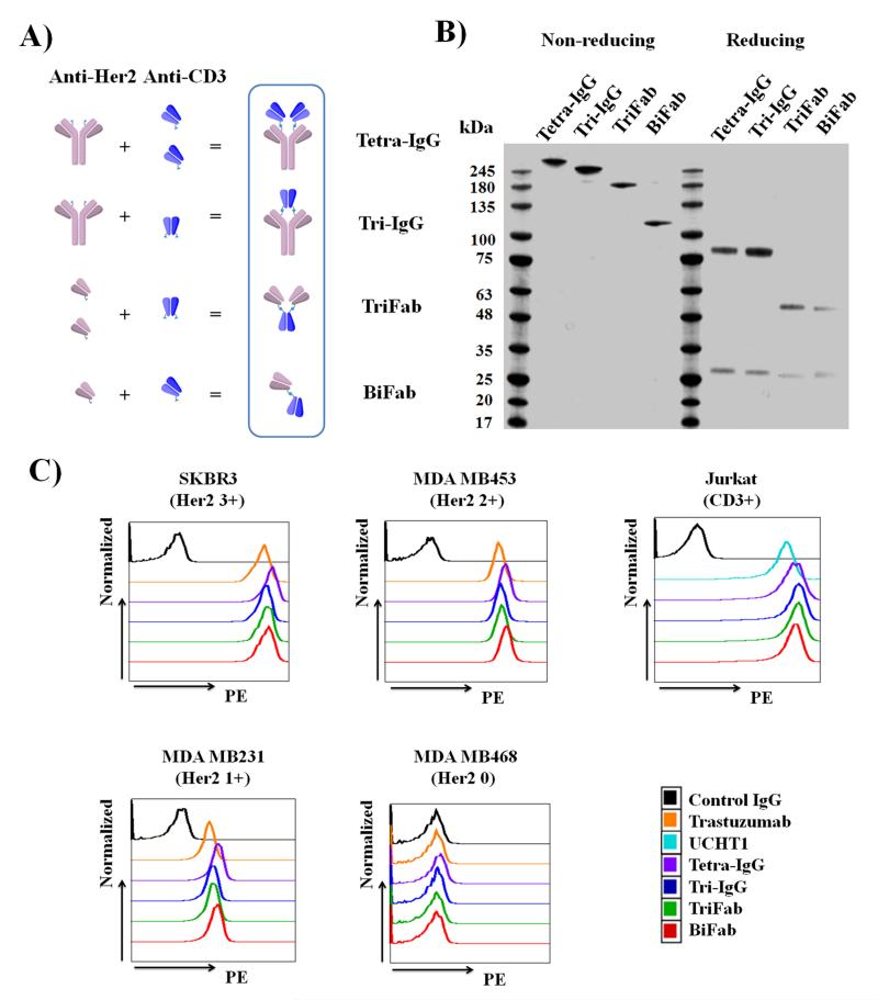
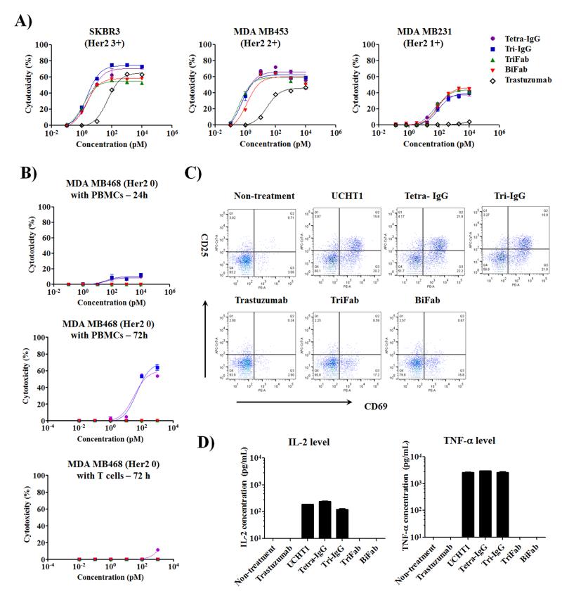
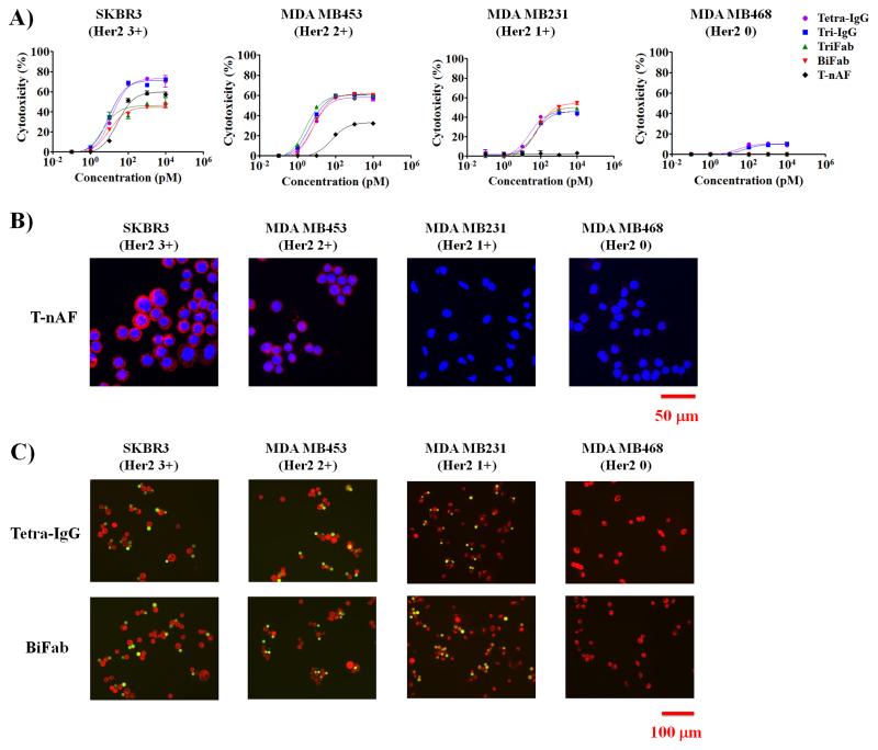
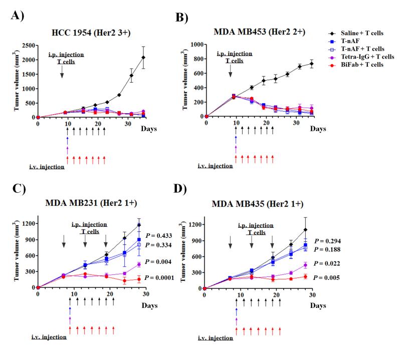
Four different formats of bispecific antibodies (bsAbs) were generated that consist of anti-Her2 IgG or Fab site-specifically conjugated to anti-CD3 Fab using the genetically encoded noncanonical amino acid. These bsAbs varied in valency or in the presence or absence of an Fc domain. Different valencies did not significantly affect antitumor efficacy, whereas the presence of an Fc domain enhanced cytotoxic activity, but triggered antigen-independent T-cell activation. We show that the bsAbs can efficiently redirect T cells to kill all Her2 expressing cancer cells, including Her2 1+ cancers, both in vitro and in rodent xenograft models. This work increases our understanding of the structural features that affect bsAb activity, and underscores the potential of bsAbs as a promising therapeutic option for breast cancer patients with low or heterogeneous Her2 expression.

Keywords: T-cell activation; antibody drug conjugates; bispecific antibodies; breast cancers; noncanonical amino acids.


Figures

Figure 1
Figure 1
Characterization of anti-Her2/anti-CD3 bsAbs. (A) Schematic diagram of bsAb constructs. (B) SDS-PAGE analysis of purified bsAbs under non-reducing and reducing conditions. (C) Flow cytometry analysis of bsAb constructs and parental antibodies (trastuzumab and UCHT1) binding to different Her2 expressing breast cancer cells and CD3+ Jurkat cells. Cells were consecutively labeled with bsAbs or parental antibodies (25 nM) and secondary PE-conjugated anti-human kappa antibody (eBioscience).
Figure 2
Figure 2
In vitro activity of distinct bsAb formats with different Her2 expressing cancer cells. Effector cells were incubated with target cells at 10:1 ratio for 24 or 72 h. (A) 24 h cytotoxic activity of PBMCs against different Her2 expressing cancer cells in the presence of indicated concentrations of bsAbs or trastuzumab. Cytolytic activity was determined by measuring the amount of lactate dehydrogenase (LDH) released into cultured media. (B) Comparison of human PBMCs or purified T cell cytotoxicity induced by IgG- and Fab-based bsAbs against MDA MB468 cells (Her2 0). (C) Flow cytometry analysis of T cell activation markers (CD25 and CD69) in 24 h cultures consisting of MDA MB468, PBMCs, and 100 pM of bsAbs or parental antibodies. (D) Quantification of cytokine (IL-2 and TNF-α) levels in the cultures described in (C) by ELISA. Error bars represent standard deviation of duplicate samples.
Figure 3
Figure 3
In vitro characterization of T-nAF and bsAbs with different Her2 expressing breast cancer cells. (A) 24h cytotoxicity assays were performed with human PBMCs and indicated target cells at 10: 1 ratio in the presence of different concentrations of T-nAF or bsAbs. Error bars represent standard deviation of duplicate samples. (B) Internalization analysis on breast cancer cells after 4 h treatment with 20 nM T-nAF. After the removal of surface-bound T-nAF by an acid wash, internalized T-nAF was detected with Alexa fluor 555-labeled anti-human IgG (red), and imaged by confocal microscopy (Zeiss 710). Hoechst 33342 (blue) was used for nuclear counterstaining. (C) Analysis of the crosslinking of cancer cells and T cells. Fluorescently labeled target cells (red) and T cells (green) were mixed at 1:1 ratio, and incubated for 4 h in the presence of 20 nM Tetra-IgG or BiFab. Non-conjugated cells were gently removed by PBS wash for three times, prior to imaging on a fluoresce microscope (Nikon Eclipse).
Figure 4
Figure 4
In vivo efficacy comparison of T-nAF and bsAbs in human breast cancer xenograft models. For HCC1954 (A) and MDA MB453 (B) xenograft studies, eight days after subcutaneous implantation of 5×10⁶ cancer cells in 50% Matrigel, female NSG mice received one intraperitoneal (IP) injection of 30×106 activated T cells. For MDA MB231 (C) and MDA MB435 (D) xenograft studies, seven days after subcutaneous implantation of 20×106 or 2×10⁶ cancer cells in 50% Matrigel, respectively, mice were injected three times with 20×106 activated T cells every 6 days via IP. For all studies, two days after the initial T cell infusion, mice were treated intravenously with one dose of T-nAF (5mg/kg) or Tetra-IgG (10mg/kg), or 7 doses of BiFab (1mg/kg) or saline every other day. Tumors were measured twice a week with calipers, and tumor volume was calculated by W× L× H. Each data point represent mean tumor volume of 5 mice in each group ± SD. Arrows indicate time of activated PBMC injections or of treatment with specified therapeutics. P values < 0.05 compared to the control groups (saline + T cells) were considered significant.

References

    1. Vogel CL, Cobleigh MA, Tripathy D, Gutheil JC, Harris LN, Fehrenbacher L, Slamon DJ, Murphy M, Novotny WF, Burchmore M, Shak S, Stewart SJ, Press M. J.Clin.Oncol. 2002;20:719–726. - PubMed
    1. Krop I, Winer EP. Clin.Cancer Res. 2014;20:15–20. - PubMed
    1. Keating GM. Drugs. 2012;72:353–360. - PubMed
    1. Owens MA, Horten BC, Da Silva MM. Clin.Breast Cancer. 2004;5:63–69. - PubMed
    1. Lewis Phillips GD, Li G, Dugger DL, Crocker LM, Parsons KL, Mai E, Blattler WA, Lambert JM, Chari RV, Lutz RJ, Wong WL, Jacobson FS, Koeppen H, Schwall RH, Kenkare-Mitra SR, Spencer SD, Sliwkowski MX. Cancer Res. 2008;68:9280–9290. - PubMed

Publication types

MeSH terms