. 2015 Apr 28;10(4):e0124408.
doi: 10.1371/journal.pone.0124408. eCollection 2015.

Nkx2.2 and Nkx2.9 are the key regulators to determine cell fate of branchial and visceral motor neurons in caudal hindbrain

Affiliations

Nkx2.2 and Nkx2.9 are the key regulators to determine cell fate of branchial and visceral motor neurons in caudal hindbrain

Wassan Jarrar et al. PLoS One. .

Abstract

Cranial motor nerves in vertebrates are comprised of the three principal subtypes of branchial, visceral, and somatic motor neurons, which develop in typical patterns along the anteroposterior and dorsoventral axes of hindbrain. Here we demonstrate that the formation of branchial and visceral motor neurons critically depends on the transcription factors Nkx2.2 and Nkx2.9, which together determine the cell fate of neuronal progenitor cells. Disruption of both genes in mouse embryos results in complete loss of the vagal and spinal accessory motor nerves, and partial loss of the facial and glossopharyngeal motor nerves, while the purely somatic hypoglossal and abducens motor nerves are not diminished. Cell lineage analysis in a genetically marked mouse line reveals that alterations of cranial nerves in Nkx2.2; Nkx2.9 double-deficient mouse embryos result from changes of cell fate in neuronal progenitor cells. As a consequence progenitors of branchiovisceral motor neurons in the ventral p3 domain of hindbrain are transformed to somatic motor neurons, which use ventral exit points to send axon trajectories to their targets. Cell fate transformation is limited to the caudal hindbrain, as the trigeminal nerve is not affected in double-mutant embryos suggesting that Nkx2.2 and Nkx2.9 proteins play no role in the development of branchiovisceral motor neurons in hindbrain rostral to rhombomere 4.


Conflict of interest statement

Competing Interests: The authors have declared that no competing interests exist.

Figures

Fig 1
Fig 1. Immunostaining of cranial motor nerves in whole-mount mouse embryos: Effects on the spinal accessory and vagal nerve in mutant mouse embryos lacking Nkx2.2 and/or Nkx2.9 genes.
Lateral view of brainstem including the anterior part of the developing spinal cord of wild type (A), Nkx2.2-/- (B), Nkx2.9-/- (C), Nkx2.2+/-; Nkx2.9-/- (D), and Nkx2.2-/-; Nkx2.9-/- (E) embryos at E10.5. Immunostaining of motor neurons was performed with NCAM-L1-specific antibody. The arrow indicates the accessory nerve (XI) with the arrowhead marking its maximal caudal extension. The rootlets of the vagal nerve (X) are indicated by the bracket. In wild type and Nkx2.2-/- single knock-out embryos, the spinal accessory nerve extends from rhombomere 7 into the cervical spinal cord (see arrowheads) and the vagal nerve appears also normal. In the absence of Nkx2.9, however, the N. accessorius is shorter and thinner than usual (arrowhead in C) and the vagal nerve exhibits moderate defects. Nkx2.2 and Nkx2.9 compound and double-null mutant embryos show almost total loss of the accessory nerve with only few unorganized axon fascicles remaining and severe defects of the vagus nerve illustrated by the severe reduction of rootlets (D and E). Cranial nerves are referred to as trigeminal nerve (V), N. facialis/N. vestibulocochlearis (VII/VIII), N. glossopharyngeus (IX), N. vagus (X), N. accessorius (XI), and N. hypoglossus (XII). C1: first spinal nerve of the cervical spinal cord. Scale bar: 400 μm.
Fig 2
Fig 2. Reduction of branchiovisceral motor neurons in rhombomere 7 of the Nkx2.2; Nkx2.9 double-null mutant mouse embryo.
Transversal sections through rhombomere 7 of wild type (A and F, n = 4), Nkx2.2-/- (B and G, n = 3), Nkx2.9-/- (C and H, n = 4), Nkx2.2+/-; Nkx2.9-/- (D and I, n = 4), and Nkx2.2-/-; Nkx2.9-/- (E and J, n = 5) embryos at E10.5. Branchiovisceral motor neurons were stained by immunohistochemistry for Phox2b+ in blue and Islet-1+ in red. Somatic motor neurons were stained for HB9+ in green and Islet-1+ in red. The decrease of Phox2b+ cell nuclei in mutant tissue is also illustrated for better recognition in black and white (F-J). Note that the decrease of bvMNs in mutant tissue appears to be counterbalanced by the inverse expansion of somatic motor neurons. The significance of this observation was determined by the one-way ANOVA statistical test (K). The total number of postmitotic Islet-1+ motor neurons was similar in wild type and the various Nkx2 mutant mice with small, statistically insignificant differences (L). ns: not significant, *: p < 0.05, ***: p < 0.001. Scale bar in E represents 50 μm and applies to all figure panels.
Fig 3
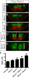
Fig 3. The p3 domain in hindbrain of Nkx2.2; Nkx2.9 double-deficient mutant embryos harbors progenitor cells for sMNs and lacks the normal population of bvMN progenitors.
Immunofluorescence staining of Nkx2.2- (red) and Olig2- (green) expressing cells on transversal sections through rhombomere 7 in E10.5 mouse embryos (A-E). The ventricular and pial surfaces are indicated by dotted lines. The p3 domain characterized by bvMN progenitor cells (red) in wild type (A; n = 6), single Nkx2.2 (B; n = 4), and Nkx2.9 (C; n = 3) mutant embryos is moderately reduced in Nkx2.2+/-; Nkx2.9-/- compound mutants (D; n = 6). These bvMNs progenitors are totally absent in Nkx2.2-/-; Nkx2.9-/- double-null mutant embryos (E; n = 5) and have been replaced by Olig2+ SMN progenitor cells (indicated by white vertical bars in A and E). Olig2+ progenitors were counted on the sections through neural tubes and the columns represent the numbers in various mutants and control animals (F). Statistical significance was evaluated by one-way ANOVA analysis (ns: not significant, **: p < 0.01, ***: p < 0.001). Scale bar (white horizontal bar in A): 50 μm.
Fig 4
Fig 4. Changes in motor nuclei of cranial nerves in Nkx2.2-/-; Nkx2.9-/- double-deficient mouse mutants.
In situ hybridizations were performed with digoxigenin-labeled peripherin riboprobe on serial cross-sections of hindbrain from E15.5 wild type (A-D), single Nkx.2.2 (E-H) and Nkx2.9 (I-L), and double mutant (M-T) mouse embryos. Note the strong reduction of motor nuclei for the facial nerve (nVII) and the almost complete loss of nuclei for the vagal nerve (dmnX, filled arrowhead) as well as the nucleus ambiguus (dotted circle) in hindbrains of Nkx2.2-/-; Nkx2.9-/- double-deficient embryos. In contrast, the branchiovisceral motor nucleus of the trigeminal nerve (nV) was basically unaltered and the somatic motor nuclei of the hypoglossal (nXII) and abducens (nVI) nerves appeared augmented in double mutant mice with additional ectopic peripherin-expressing cells next to the motor nucleus of the abducens nerve (open arrowhead in P and T). Scale bars correspond to 100 μm.
Fig 5
Fig 5. Additional and ectopic populations of somatic motor neurons develop in caudal but not in rostral hindbrain of Nkx2.2; Nkx2.9 deficient embryos.
Triple immunofluorescence on transversal sections through hindbrains from wild type (A-D), Nkx2.2-/- (E-H), Nkx2.9-/- (I-L), Nkx2.2+/-; Nkx2.9-/- (M-P), and Nkx2.2-/-; Nkx2.9-/- (Q-T) E10.5 embryos reveals expression of HB9 (green)/Islet-1 (red) in somatic motor neurons, and Phox2b (blue)/Islet-1 (red) in branchial and visceral motor neurons. In wild type hindbrain somatic Hb9+/Islet-1+ double-positive motor neurons are only present in rhombomeres 5 (B) and 7 (see Fig 2), whereas hindbrains of Nkx2.2; Nkx2.9 double-knockout embryos contain considerable numbers of somatic motor neurons ectopically in rhombomeres 4 and 6 (Q-S). Similar albeit much weaker phenotypes have also been observed in single Nkx2.2 and Nkx2.9 knock-out and compound mutants (E-P). The surplus of sMNs is accounted for by significant loss of bvMNs (Q-S). The anterior rhombomeres 2 and 3 do not contain SMNs and failed to acquire ectopic sMNs even in the absence of both Nkx2.2 and Nkx2.9 transcription factors. Scale bar in A: 50 μm.
Fig 6
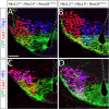
Fig 6. Transformation of motor neuron cell fate in hindbrain of Nkx2.2-/-; Nkx2.9-/- double mutant embryos.
Cells of the Nkx2.2-expressing bvMN lineage in the p3 domain were genetically labeled by membrane-targeted GFP (green) that is expressed persistently after Nkx2.2-Cre-mediated recombination in the Rosa-mT/mG reporter mouse. Confocal images of immunohistochemically stained sections from control (A, C) and Nkx2.2; Nkx2.9 double-deficient (B, D) mouse strains are shown. Note that p3 cells and their descendants in the presence of both Nkx2.2 and Nkx2.9 transcription factors (A, C) coexpress the cell lineage marker GFP together with the pan-motor neuron marker Islet1 (red) but fail to express the sMN markers Olig2 and HB9 (both blue). In contrast Nkx2.2-/-; Nkx2.9-/- double mutant embryos exhibit lineage-marked GFP-positive neuronal progenitor cells that falsely also express the sMN progenitor-specific Olig2 (blue) and the postmitotic sMN marker HB9 in the p3 domain (B, D). Dotted lines delineate the ventricular and pial surfaces in the hindbrain sections. The scale bar in A represents 25 μm.
Fig 7
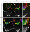
Fig 7. Ectopically generated somatic motor nerves in Nkx2.2; Nkx2.9 double-deficient mouse embryos project axons correctly via ventral exit points into the periphery.
Hindbrain sections at levels of rhombomeres 7 (r7; A-F) and 4 (r4, G-L) from control (A, D, G, J) and Nkx2.2; Nkx2.9 double-deficient (B, C, E, F, H, I, K, L) mouse embryos (E10.5) were immunostained for GFP (green), neurofilament (NF, red) and Islet-1 (Isl, blue). Note that axons of the Nkx2.2-derived bvMN cell lineage leave the hindbrain exclusively through dorsolateral exit points, both in r4 and r7. These numerous axons in controls are labeled by membrane-targeted GFP (cell lineage marker: yellow in A, D, G, and J), while only few residual axons at best use this route in Nkx2.2; Nkx2.9 double-null mutants (arrows in B, E, H, and K). Axons of somatic motor nerves are identified by their expression of neurofilament (red label in A). Their projections leave the CNS exclusively via ventral exit points in r7, but not in r4 (white arrowheads in A and G). Axons transformed to the sMN subtype in Nkx2.2; Nkx2.9 double-null mutants mix with the original sMNs at preexisting ventral exit points of r7 (orange arrowheads in B, C, E, and F) and form additional ventral exit points in r4 (H, I, K and L). Scale bars: (A): 100 μm, (C) and (I): 20 μm.
Fig 8
Fig 8. Typical somatic motor nerves are derived from progenitors of branchiovisceral motor neurons in Nkx2.2; Nkx2.9-deficient mutant embryos.
Cre-mediated activation of membrane-targeted GFP is shown by immunostaining on whole- mounts of hindbrain and upper cervical spinal cord from control (A) and Nkx2.2; Nkx2.9 double-knockout (B) embryos. Axonal bundles derived from Nkx2.2-expressing progenitor cells (brown label) mark the branchial and visceral motor nerves in the E11.5 control mouse embryos (A). In the Nkx2.2; Nkx2.9-deficient mutant embryo the accessory (XI) and vagal (X) motor nerves are missing but axons of the hypoglossal nerve (XII) and ventral roots in cervical spinal cord are now labeled (B). The facial (VII) and glossopharyngeal (*) motor nerves are still present but markedly reduced (B). Note that the motor branch of the trigeminal nerve (V) in the mutant appears indistinguishable from control. Scale bar: 500 μm.

References

    1. Keynes R, Krumlauf R. Hox genes and regionalization of the nervous system. Annu Rev Neurosci. 1994;17:109–32. 10.1146/annurev.ne.17.030194.000545 - DOI - PubMed
    1. Glover JC, Renaud JS, Rijli FM. Retinoic acid and hindbrain patterning. J Neurobiol. 2006;66(7):705–25. 10.1002/neu.20272 - DOI - PubMed
    1. Rhinn M, Picker A, Brand M. Global and local mechanisms of forebrain and midbrain patterning. Curr Opin Neurobiol. 2006;16:5–12. 10.1016/j.conb.2006.01.005 - DOI - PubMed
    1. Jessell TM. Neuronal specification in the spinal cord: inductive signals and transcriptional codes. Nat Rev Genet. 2000;1(1):20–9. 10.1038/35049541 - DOI - PubMed
    1. Kandel ER, Schwartz JH, Jessell TM. Principles of neural science. 4th ed ed. New York: McGraw-Hill; 2000.

Publication types

LinkOut - more resources