. 2015 Apr 28;10(4):e0125090.
doi: 10.1371/journal.pone.0125090. eCollection 2015.

Impact of acute malaria on pre-existing antibodies to viral and vaccine antigens in mice and humans

Affiliations

Impact of acute malaria on pre-existing antibodies to viral and vaccine antigens in mice and humans

Simran Banga et al. PLoS One. .

Abstract

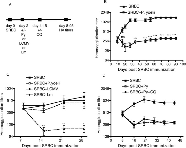
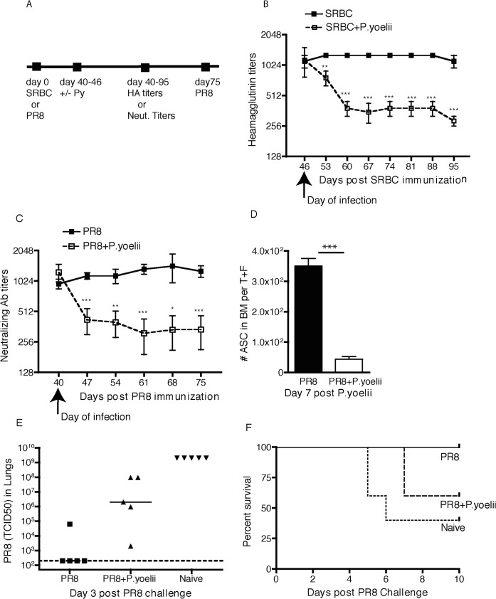
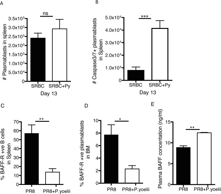
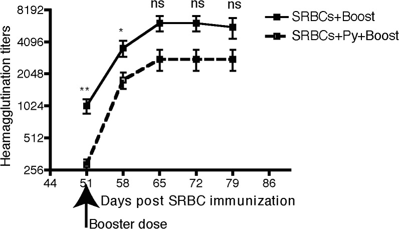
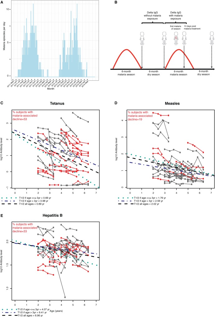
Vaccine-induced immunity depends on long-lived plasma cells (LLPCs) that maintain antibody levels. A recent mouse study showed that Plasmodium chaubaudi infection reduced pre-existing influenza-specific antibodies--raising concerns that malaria may compromise pre-existing vaccine responses. We extended these findings to P. yoelii infection, observing decreases in antibodies to model antigens in inbred mice and to influenza in outbred mice, associated with LLPC depletion and increased susceptibility to influenza rechallenge. We investigated the implications of these findings in Malian children by measuring vaccine-specific IgG (tetanus, measles, hepatitis B) before and after the malaria-free 6-month dry season, 10 days after the first malaria episode of the malaria season, and after the subsequent dry season. On average, vaccine-specific IgG did not decrease following acute malaria. However, in some children malaria was associated with an accelerated decline in vaccine-specific IgG, underscoring the need to further investigate the impact of malaria on pre-existing vaccine-specific antibodies.


Conflict of interest statement

Competing Interests: The authors have declared that no competing interests exist.

Figures

Fig 1
Fig 1. Decrease of SRBC-specific antibody responses after P. yoelii infection.
A: Experimental design. Female BALB/c mice were immunized with 10% SRBC suspension intraperitoneally. Two days later, half of these mice were infected with 105 P. yoelii infected RBCs intravenously, while the other half of mice were injected with saline. Serum samples were collected on specific days and analyzed for Heamagglutination titers. B: Heamagglutination titers over a period of 90 days in mice immunized with SRBC and infected or not infected with P.yoelii. C: SRBC immunized mice were injected with saline or infected after two days with P. yoelii, L. monocytogenes (5 X 10⁶ cfu) or LCMV (2 X 10⁵ pfu) and Heamagglutination titers were analyzed over a period of 30 days. D: SRBC immunized mice were injected with saline or infected after two days with P. yoelii. Some P. yoelii infected mice received chloroquine (50 mg per kg) on days 4, 7, 10, 12 and 15 post infection to limit blood stage parasitemia and Heamagglutination titers were analyzed over a period of 30 days. Parasitemia did not exceed 4% in the chloroquine treated mice. The data are representative of two or more independent experiments with10 mice per group/exp. *p<0.05, **p<0.01, ***p<0.001.
Fig 2
Fig 2. Decrease of preexisting antibodies after P. yoelii infection.
A: Experimental design. BALB/c mice and outbred Swiss Webster mice were immunized with SRBC or received sublethal intranasal infection with PR8 IAV. At the indicated days post infection, some mice were infected with P. yoelii. Serum samples collected on specific days were analyzed for Heamagglutination titers or virus neutralizing antibody titers. B: Heamagglutination titers over a period of 95 days post SRBC immunization in P. yoelii infected and uninfected mice. C: Virus neutralizing antibody titers over a period of 75 days post PR8 infection in mice and infected or not infected with P.yoelii. D: PR8 specific Antibody secreting cells in the bone marrow of immune mice seven days after saline or P. yoelii infection. The numbers are derived from combination of one tibia and one femur from each mousse. E: Naïve or PR8 immune mice receiving saline or P. yoelii were challenged with PR8 virus 40 days post P.yoelii infection. On day 3 post challenge, lungs were harvested to determine virus load., each symbol represents an individual mouse. F: Survival curve of mice as in E. Data are representative of three independent experiments with 8 to 12 mice per group/experiment. * p<0.05, ** p<0.01, *** p<0.001.
Fig 3
Fig 3. P. yoelii infection increases plasma cell apoptosis, decreases plasma cell BAFF-R and increases circulating BAFF concentrations.
BALB/c mice were immunized with 10% SRBC suspension. Two days later, half of these mice were infected with 106 P. yoelii parasitized RBC while the other half of mice were injected with saline. (A) Total plasma cells in the spleen at day 13 post SRBC immunization. (B) Total caspase 3/7 positive plasma cells in the spleen at day 13 post SRBC immunization. Data in A,B are mean + S.D. and represent one of two independent experiments with 5 mice per group. Swiss Webster mice were infected with a sublethal dose of PR8. Forty-two days later, half of these mice were infected with 106 P. yoelii parasitized RBC. The percent of BAFF-R positive plasma cells in the (C) spleen and (D) bone marrow were determined on day forty-eight after immunization (day 6 after Py infection). Serum was obtained from mice in C,D and analyzed for circulating BAFF concentrations (E). Data in C-E reflect mean + S.D. and represent one of two independent experiments with 5 mice per group. *** p<0.001, ** p<0.01, * p<0.05, ns p non-significant
Fig 4
Fig 4. P. yoelli infection does not disrupt the anamnestic response to SRBC immunization.
BALB/c mice were immunized with 10% SRBC suspension. Two days later, half of these mice were infected with 106 P. yoelii while the other half of mice were injected with saline. A booster dose of 10% SRBC suspension was given after 40+ days to both groups of mice. Serum samples were collected on the indicated days and analyzed for Heamagglutinin antibody titers. Data are representative of two experiments, with 10 mice per group/exp. *** p<0.001, ** p<0.01, * p<0.05, ns p non-significant.
Fig 5
Fig 5. Kinetics of vaccine-specific IgG levels in children during the dry season and after acute malaria.
(A) The study was designed to take advantage of the sharply demarcated and intense 6-month malaria season (July—December) and 6-month dry season (January—June; negligible malaria transmission) in Mali. Shown is the number of febrile malaria episodes per day over two years at the study site in a cohort of 695 children and adults. (B) IgG levels specific for routine vaccines administered under one year of age (tetanus, measles and Hepatitis B) were measured in plasma collected from 54 children at four time points (vertical arrows): before and after the 6-month dry season, 10 days after the first acute malaria episode of the ensuing malaria season, and after the second dry season. Shown for each subject are IgG titers specific for (C) tetanus, (D) measles and (E) hepatitis B vaccines at the time points indicated in (B). The x-axis indicates the age at which the respective time points occurred for each subject. In red are subjects who experienced an accelerated decline in vaccine-specific IgG titers following acute malaria (between 2nd and 3rd time points) relative to each child’s own rate of change during the preceding dry season (between 1st and 2nd time points). The percentage of subjects for whom malaria was associated with an accelerated decline in IgG is shown in red text for each vaccine. A linear mixed effects model that included three time points over 18 months (before and after the first dry season, and after the second dry season) was used to estimate average IgG half-lives for all subjects (black dashed line) and separately for children aged ≤3 years (green dotted line) and >3 years of age (blue dash-dot line).

References

    1. Keja K, Chan C, Hayden G, Henderson RH. Expanded programme on immunization. World health statistics quarterly Rapport trimestriel de statistiques sanitaires mondiales. 1988;41(2):59–63. PubMed . - PubMed
    1. Okwo-Bele JM, Cherian T. The expanded programme on immunization: a lasting legacy of smallpox eradication. Vaccine. 2011;29 Suppl 4:D74–9. 10.1016/j.vaccine.2012.01.080 PubMed . - DOI - PubMed
    1. Black RE, Cousens S, Johnson HL, Lawn JE, Rudan I, Bassani DG, et al. Global, regional, and national causes of child mortality in 2008: a systematic analysis. Lancet. 2010;375(9730):1969–87. 10.1016/S0140-6736(10)60549-1 PubMed . - DOI - PubMed
    1. Centers for Disease C, Prevention. Global routine vaccination coverage, 2011. MMWR Morbidity and mortality weekly report. 2012;61(43):883–5. PubMed . - PubMed
    1. Greenwood BM, Bradley-Moore AM, Bryceson AD, Palit A. Immunosuppression in children with malaria. Lancet. 1972;1(7743):169–72. PubMed . - PubMed

Publication types

MeSH terms

Supplementary concepts