. 2015 Oct;63(10):1753-71.
doi: 10.1002/glia.22842. Epub 2015 Apr 27.

The control of reactive oxygen species production by SHP-1 in oligodendrocytes

Affiliations

The control of reactive oxygen species production by SHP-1 in oligodendrocytes

Ross C Gruber et al. Glia. 2015 Oct.

Abstract

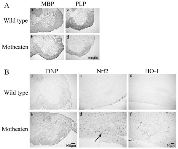
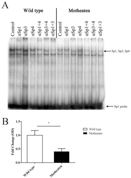
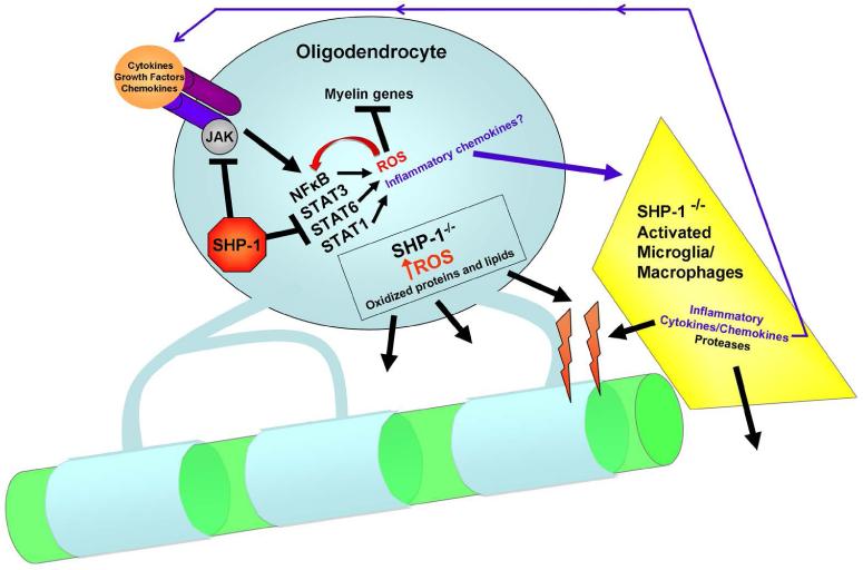
We have previously described reduced myelination and corresponding myelin basic protein (MBP) expression in the central nervous system of Src homology 2 domain-containing protein tyrosine phosphatase 1 (SHP-1) deficient motheaten (me/me) mice compared with normal littermate controls. Deficiency in myelin and MBP expression in both brains and spinal cords of motheaten mice correlated with reduced MBP mRNA expression levels in vivo and in purified oligodendrocytes in vitro. Therefore, SHP-1 activity seems to be a critical regulator of oligodendrocyte gene expression and function. Consistent with this role, this study demonstrates that oligodendrocytes of motheaten mice and SHP-1-depleted N20.1 cells produce higher levels of reactive oxygen species (ROS) and exhibit corresponding markers of increased oxidative stress. In agreement with these findings, we demonstrate that increased production of ROS coincides with ROS-induced signaling pathways known to affect myelin gene expression in oligodendrocytes. Antioxidant treatment of SHP-1-deficient oligodendrocytes reversed the pathological changes in these cells, with increased myelin protein gene expression and decreased expression of nuclear factor (erythroid-2)-related factor 2 (Nrf2) responsive gene, heme oxygenase-1 (HO-1). Furthermore, we demonstrate that SHP-1 is expressed in human white matter oligodendrocytes, and there is a subset of multiple sclerosis subjects that demonstrate a deficiency of SHP-1 in normal-appearing white matter. These studies reveal critical pathways controlled by SHP-1 in oligodendrocytes that relate to susceptibility of SHP-1-deficient mice to both developmental defects in myelination and to inflammatory demyelinating diseases.

Keywords: inflammation; multiple sclerosis; myelin; normal-appearing white matter; oxidative stress.


Figures

Figure 1
Figure 1
Immunohistochemical analysis of oxidative stress and reduced myelination in SHP-1 deficient spinal cords. A. Wild type mouse spinal cord cross-sections demonstrate strong staining of myelin proteins (a,c). In contrast, motheaten spinal cords demonstrate reduced myelin protein staining (b,d). B. Higher DNP staining in motheaten spinal cord cross sections (b) indicates higher constitutive oxidative stress in naïve motheaten mice relative to control (a). Nrf2 and HO-1 staining indicate increased expression in longitudinal sections of motheaten spinal cords (d,f) relative to WT (c,e).
Figure 2
Figure 2
Increased markers of ROS in motheaten brains and glia. A.Thiobarbituric Acid Reactive Substances (TBARS) were used a measure of lipid peroxidation. Lipid peroxidation is significantly higher (p<0.01) in motheaten (n=5) brain lysates than in WT (n=7) brain lysates. B. Lipid peroxidation is significantly (p<0.05) higher in motheaten (n=4) whole glial brain-derived lysates than in WT (n=4) whole glial lysates. C. Hydrogen peroxide production in glial cultures. WT (n=4) and motheaten (n=4) brain-derived glial cultures were incubated for 4 hours in fresh phenol free DMEM. Hydrogen peroxide levels in supernatants were measured at 1, 2, and 4 hours using a hydrogen peroxide assay kit (Amplex Red, Invitrogen). Over the 4 hour time course, motheaten cultures produced significantly more hydrogen peroxide than WT cultures p=0.02 by two-way ANOVA: at 4 hour time point, p=0.048 by Student’s t-test).
Figure 3
Figure 3
NF-κB activation and cytokine upregulation in wild type and motheaten brain-derived glia. A. Nuclear lysates from whole WT (n=6) and motheaten glia (n=6) in either media alone or with one hour TNF-α treatment (10ng/mL) were isolated and allowed to bind to an NF-κB consensus binding sequence. Bound NF-κB was then detected by a p65 specific antibody and quantified based on a calibration curve generated by using a purified p65 recombinant protein. The NF-κB binding activity is reported as nanograms of bound p65 protein per 10μg of glial nuclear protein. A constitutive increase in NF-κB activation is seen in motheaten glia relative to WT. After TNF-α treatment, NF-κB binding increases in both WT and motheaten glia, however motheaten glia continue to demonstrate significantly higher NF-κB binding. B. NF-κB inducible proteins, IL-1β, IL-6, and MCP-1 were measured in glial supernatants after 18h treatments with TNF-α using ELISA. After TNF-α treatment SHP-1 deficient glia (n=6) demonstrate a significant increase in secreted NF-κB inducible proteins relative to WT (n=6), p value is shown, Student’s t-test.
Figure 4
Figure 4
Increased oxidative stress in motheaten oligodendrocytes. A. Stable oxidative modifications to proteins in SHP-1 deficient mice. Immunoreactivity is markedly increased constitutively in motheaten (b) brain-derived oligodendrocytes relative to control (a), indicating increased protein carbonylation. A 2 hour treatment with TNF-α (100ng/μL) appeared to increase protein carbonylation in WT brain-derived oligodendrocytes (c). Motheaten oligodendrocytes (d) appeared to have similar levels of DNP staining. B. Increased reactive oxygen species in motheaten oligodendrocytes. Untreated brain-derived oligodendrocytes from motheaten mice (b) demonstrate a higher level of constitutive ROS than WT oligodendrocytes (arrows) (a). After a 2 hour treatment with TNF-α (100ng/mL), oligodendrocytes from either WT mice (c), or motheaten mice (d) express a high level of reactive oxidative species. Cells were pre-loaded with Carboxy-H2DCFDA (Invitrogen) for 15 minutes, rinsed, then treated with culture media or TNF-α, and then imaged live. C. Cell specific constitutive ROS production was quantified by flow cytometry in O4+ oligodendrocytes directly isolated from spinal cords motheaten mice and littermate controls. WT (n=7) and motheaten (n=4) cells were incubated with 10μM CM-H2DCFDA and then labeled with O4 antibody. 10,000-40,000 events per sample were collected and analyzed using FlowJo (Tree Star), representative mean fluorescence curves for motheaten vs WT mice are shown. D. Average MFI for motheaten and controls are shown, data is standardized to WT MFI (Student’s t-test, *p=0.042).
Figure 5
Figure 5
Oxidation can directly affect binding of Sp1 to the myelin basic protein promoter region. A. Analysis of Sp1, Sp3, and Sp4 binding activities in purified brain-derived oligodendrocyte nuclei of WT and motheaten mice. Oligodendrocyte nuclear extracts were incubated with a 32P-labled Sp1 probe. In some reactions antibodies to either Sp1, Sp3, or Sp4 either singly or in pairs were added to supershift/delineate individual components of the overlapping Sp factors in the upper binding activity as indicated. B. Quantification of the upper binding activity containing Sp1, Sp3, and Sp4 in WT and motheaten oligodendrocytes. Pixel density of the upper band (Sp1) was determined using ImageJ and a histogram was created representing the fold difference in radiolabeled probe binding between nuclear extracts harvested from purified oligodendrocytes of WT and motheaten cultures. Statistical significance was determined by Student’s t-test (*p<0.05).
Figure 6
Figure 6
Increased ROS generation in SHP-1 deficient N20.1 cells: A. Cells were transfected with scrambled siRNA control or SHP-1 siRNA and analyzed for SHP-1 expression by Western immunoblot, positive and negative control protein homogenates are isolated from motheaten and WT spleen. B. Cells were then double stained for Olig2 (green) or SHP-1 (red). C. Cells were loaded with 25μM carboxy-H2DCFDA for 30 minutes at 37°C. Increased fluorescence is observed in media-treated SHP-1 deficient N20.1 cell lines (control). Following treatments with H2O2 or TNF-α (100ng/mL), SHP-1 deficient cells demonstrate a large increase in fluorescence whereas only a nominal increase is seen in control cells. D. Nrf2 staining in cells transfected with control or SHP-1 siRNA. Nuclear localization can be seen in untreated SHP-1 deficient cells (arrowhead), and in both control and SHP-1 cells after a 2 hour treatment with TNF-α. E. Decreased glutathione content in SHP-1 deficient N20.1 cell line. The GSH-Glo glutathione assay was performed using control (n=5) or SHP-1 (n=5) deficient N20.1 cell lines. Untreated cell lines exhibited a significant constitutive reduction in glutathione content (p<0.01) when SHP-1 was reduced using siRNA. A 2-hour treatment with TNF-α (100ng/mL) appeared to increase glutathione levels in control cells and to have the opposite effect on SHP-1 deficient cells, however neither of these were significant, p>0.05. Statistical differences were determined by Two-Way ANOVA with Bonferroni’s multiple comparison test, *p≤0.05 **p≤0.01 ***p≤0.001.
Figure 7
Figure 7
Treatment with the antioxidant butylated hydroxyanisole (BHA) restores mature myelin gene expression and reduces mRNA expression of a marker of oxidative stress, HO-1, in motheaten mice to WT levels. Oligodendrocytes from WT (n=7) and motheaten (n=5) brain and spinal cord were isolated by magnetic bead separation, analyzed by real time qRT-PCR. mRNA expression is represented as fold change from motheaten. Treatment with the antioxidant BHA for 5 hr in vitro at 37°C, 10% CO2, increases mRNA expression of MBP, MAG and CNPase, and reduces HO-1 to levels in WT mouse oligodendrocytes. Motheaten HO-1 expression decreases 22.8% from 1.0±0.17 to 0.77±0.08 after BHA treatment. Treatment of isolated motheaten spinal cord and brain oligodendrocytes with BHA increases MBP expression by 23% from 1.0±0.04 to 1.23±0.07. After BHA treatment, MAG expression increases 47% from 1.0±0.08 to 1.47±0.28, and CNPase expression increased 36% from 1.0±0.08 to 1.36±0.19. The mRNA level of two pan-oligodendrocyte genes, SOX10 and Olig2, are unchanged in both genotypes and following both treatments. Statistical differences were determined by Two-Way ANOVA with Bonferroni’s multiple comparison test, *p≤0.05 **p≤0.01 ***p≤0.001.
Figure 8
Figure 8
SHP-1 is expressed in human oligodendrocytes and is deficient in a population of MS patients. A. Western immunoblot for SHP-1, ~68kDa (top) and GAPDH (bottom). SHP-1 is expressed in control NAWM but is visibly decreased in five MS patients. The first lane of the SHP-1 Immunoblot is the 64kDa band of the molecular weight marker (MWM). B. Quantified SHP-1/GAPDH expressed as fold difference from control subjects. Control subject SHP-1 expression clusters in a tight grouping when SHP-1 is normalized to GAPDH (1±0.080). However, MS NAWM SHP-1 expression segregates into two groups. When these groups are combined they appear similar in SHP-1 expression (1.167±0.317) (p=0.61, Student’s t-test). However when separated, three MS NAWM samples have a 2.23±0.102 fold increase in SHP-1 (p<0.0001, Student’s t-test) and a group of five MS NAWM samples have a deficiency 0.522±0.077 (p=0.0021, Student’s t-test). C. SHP-1 is expressed in human oligodendrocytes. Human control white matter was stained for SHP-1 (a) or IgG control (b). Arrows in figure 8C(a) indicate interfascicular oligodendrocyte chains. Oligodendrocyte staining was confirmed by staining for SHP-1 blue (c,d) and SOX10 red (c) or Olig2 red (d).
Figure 9
Figure 9
Schematic representation of dysmyelination by SHP-1 deficiency: SHP-1 acts as a constitutive inhibitor of cytokine, chemokine and growth factor signaling. In the absence of SHP-1, oligodendrocytes experience functional impairment partially due to increased constitutive ROS production. Increased ROS can activate NFkB and vice versa resulting in a self-perpetuating cycle of oxidative stress. Additionally, oxidized myelin and increased chemokine production by glia may attract SHP-1-deficient microglia, (or during active demyelination, infiltrating monocytes/macrophages) which in turn produce increased inflammation, resulting in a feed-forward loop of enhanced myelin degradation and inflammation.

References

    1. Anrather J, Racchumi G, Iadecola C. NF-kappaB regulates phagocytic NADPH oxidase by inducing the expression of gp91phox. J Biol Chem. 2006;281:5657–67. - PubMed
    1. Back SA, Gan X, Li Y, Rosenberg PA, Volpe JJ. Maturation-dependent vulnerability of oligodendrocytes to oxidative stress-induced death caused by glutathione depletion. J Neurosci. 1998;18:6241–53. - PMC - PubMed
    1. Berlett BS, Stadtman ER. Protein oxidation in aging, disease, and oxidative stress. J Biol Chem. 1997;272:20313–6. - PubMed
    1. Cammer W. Protection of cultured oligodendrocytes against tumor necrosis factor-alpha by the antioxidants coenzyme Q(10) and N-acetyl cysteine. Brain Res Bull. 2002;58:587–92. - PubMed
    1. Cammer W, Zhang H. Maturation of oligodendrocytes is more sensitive to TNF alpha than is survival of precursors and immature oligodendrocytes. J Neuroimmunol. 1999;97:37–42. - PubMed

Publication types

MeSH terms