. 2016 Feb;24(2):237-42.
doi: 10.1038/ejhg.2015.86. Epub 2015 Apr 29.

LRP5 variants may contribute to ADPKD

Affiliations

LRP5 variants may contribute to ADPKD

Wybrich R Cnossen et al. Eur J Hum Genet. 2016 Feb.

Abstract

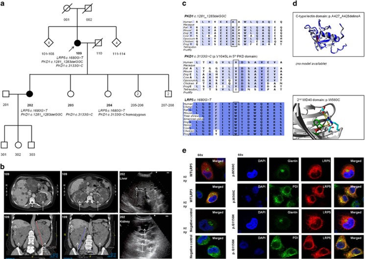
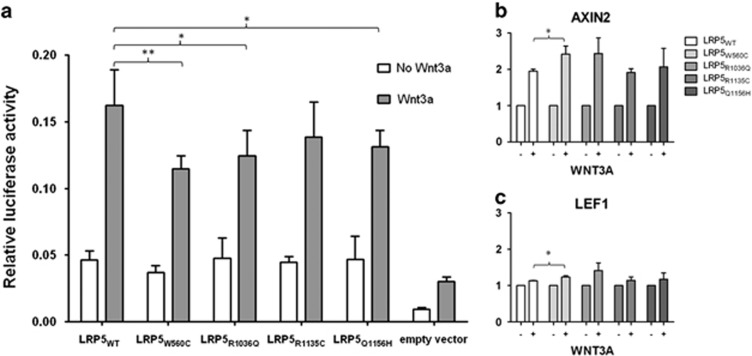
Mutations in Polycystic Kidney Disease proteins (PKD1 or PKD2) are causative for autosomal dominant polycystic kidney disease (ADPKD). However, a small subset of ADPKD probands do not harbor a mutation in any of the known genes. Low density lipoprotein Receptor-related Protein 5 (LRP5) was recently associated with hepatic cystogenesis in isolated polycystic liver disease (PCLD). Here, we demonstrate that this gene may also have a role in unlinked and sporadic ADPKD patients. In a cohort of 79 unrelated patients with adult-onset ADPKD, we identified a total of four different LRP5 variants that were predicted to be pathogenic by in silico tools. One ADPKD patient has a positive family history for ADPKD and variant LRP5 c.1680G>T; p.(Trp560Cys) segregated with the disease. Although also two PKD1 variants probably affecting protein function were identified, luciferase activity assays presented for three LRP5 variants significant decreased signal activation of canonical Wnt signaling. This study contributes to the genetic spectrum of ADPKD. Introduction of the canonical Wnt signaling pathway provides new avenues for the study of the pathophysiology.


Figures

Figure 1
Figure 1
Clinical and genetic data of ADPKD family A. (a) Pedigree of family A present two affected individuals, proband 109 and daughter 202. Affected individuals indicate those with confirmed ADPKD on CT scanning or abdominal ultrasound according to the Ravine criteria. (b) CT scanning in proband 109 presented a severe polycystic liver in segments 2, 3 and 8 and the kidney sizes were craniocaudal 19 cm (left) and 21 cm (right) in diameter. Abdominal ultrasonography in patient 202 assessed polycystic kidneys (white arrows) and multiple small hepatic cysts (dotted white arrows). (c) PKD1 c.1281_1283delGGC and PKD1 c.3133G>C were detected in the proband. Both PKD1 variants are located in low evolutionary conserved regions in contrast to LRP5 c.1680C>T. (d) The in-frame PKD1 deletion is located on an extracellular C-type lectin domain at the end of a loop close to a double helix structure. The alanine deletion shortens the strand, but there is no destabilization of the domain which is closely located to a rigid double helix structure with many hydrogen bounds. Missense variant PKD1 p.(Val1045Leu) is located on the fifth tandem PKD domain without a difference in charge between WT and mutant protein. (e) Confocal imaging studies in transiently transfected HeLa cells identified co-localization of LRP5 in the endoplasmic reticulum. There were no differences between LRP5 localization compared with the WT LRP5 construct for all 4 LRP5 variants.
Figure 2
Figure 2
LRP5 variants present reduced activation of canonical Wnt signaling. (a) Canonical Wnt signaling activity was analyzed by firefly luciferase activity and normalized to renilla luciferase activity without (white bar) and with (grey bar) addition of 250 ng/ml Wnt3a. LRP5 constructs showed a significant increase in Wnt signaling activity compared with the empty vector. LRP5 variants p.(Trp560Cys), p.(Arg1036Gln) and p.(Gln1156His) presented a decreased Wnt3a-induced signal activity (*P<0.05; **P<0.01). (b) All LRP5 variants indicated only concordantly increased AXIN2 expression levels by Wnt3a activation. (c) LEF1 and others showed slightly increased gene expression. Significant increased AXIN1 and LEF1 expression by Wnt3a activation was present in LRP5 variant p.(Trp560Cys) compared with LRP5 WT (*P<0.05). The y-axis presents the relative gene expression level.

References

    1. 1Torres VE, Harris PC, Pirson Y: Autosomal dominant polycystic kidney disease. Lancet 2007; 369: 1287–1301. - PubMed
    1. 2Bae KT, Zhu F, Chapman AB et al: Magnetic resonance imaging evaluation of hepatic cysts in early autosomal-dominant polycystic kidney disease: the Consortium for Radiologic Imaging Studies of Polycystic Kidney Disease cohort. Clin J Am Soc Nephrol 2006; 1: 64–69. - PubMed
    1. 3Luciano RL, Dahl NK: Extra-renal manifestations of autosomal dominant polycystic kidney disease (ADPKD): considerations for routine screening and management. Nephrol Dial Transplant 2014; 29: 247–254. - PubMed
    1. 4Torra R, Darnell A, Estivill X et al: Interfamilial and intrafamilial variability of clinical expression in ADPKD. Contrib Nephrol 1995; 115: 97–101. - PubMed
    1. 5Liu M, Shi S, Senthilnathan S, Yu et al: Genetic variation of DKK3 may modify renal disease severity in ADPKD. J Am Soc Nephrol 2010; 21: 1510–1520. - PMC - PubMed

Publication types

Substances