

Down-regulating HIF-1α by lentivirus-mediated shRNA for therapy of triple negative breast cancer
- PMID: 25920936
- PMCID: PMC4622682
- DOI: 10.1080/15384047.2015.1040958
Down-regulating HIF-1α by lentivirus-mediated shRNA for therapy of triple negative breast cancer
Abstract
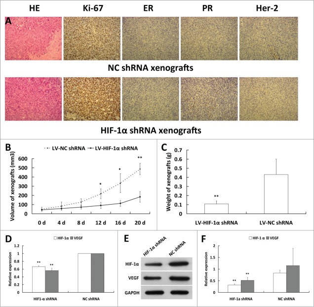
Hypoxia is associated with poor response to treatment in various cancers. Hypoxia inducible factor 1 (HIF-1) is a major transcription factor that mediates adaptation of cancer cells to a hypoxic environment and regulates many genes that are involved in key cellular functions, including cell immortalization, stem cell maintenance, autocrine growth/survival, angiogenesis, invasion/metastasis, and resistance to chemotherapy. HIF-1α has been considered as an attractive therapeutic target for cancer treatment, but there is limited success in this research field. In the present study, we designed a recombinant lentivirus containing HIF-1α siRNA, developed stably transfected cell lines, and tested the anticancer effects of the siRNA on cancer cells in vitro and in vivo. Our results indicated that the stable downregulation of HIF-1α reversed chemoresistance, inhibited proliferation, migration and invasion of cancer cells, and slowed down the tumor growth in breast cancer xenograft models. In conclusion, the recombinant lentivirus containing HIF-1α siRNA provides a new avenue for developing novel therapy for triple negative breast cancer.
Keywords: BCSCs, breast cancer stem cells; EGFR, epidermal growth factor receptor; HIF-1α, hypoxia inducible factor-1α; MDR, multidrug resistance; PARP, poly ADP ribose polymerase; PI3K, phosphatidylinositol 3-kinase; TNBC, triple negative breast cancer; VEGF, va; HIF-1α; apoptosis; gene therapy; recombinant lentivirus; siRNA therapy; stably transfected cell lines; triple negative breast cancer.
Figures


Similar articles
-
Suppressing the malignant phenotypes of glioma cells by lentiviral delivery of small hairpin RNA targeting hypoxia-inducible factor-1α.Int J Clin Exp Pathol. 2013 Oct 15;6(11):2323-32. eCollection 2013. Int J Clin Exp Pathol. 2013. PMID: 24228093 Free PMC article.
-
Targeting HIF-1α and VEGF by lentivirus-mediated RNA interference reduces liver tumor cells migration and invasion under hypoxic conditions.Neoplasma. 2016;63(6):934-940. doi: 10.4149/neo_2016_612. Neoplasma. 2016. PMID: 27565331
-
Effect of RNAi-Mediated Survivin and Hypoxia-Inducible Factor 1α Gene Silencing on Proliferation, Invasion, Migration and Apoptosis of Gastric Cancer BGC-823 Cells.Mol Biotechnol. 2024 Aug;66(8):1872-1882. doi: 10.1007/s12033-023-00786-z. Epub 2023 Jul 13. Mol Biotechnol. 2024. PMID: 37440157
-
Targeting hypoxia-inducible factor-1alpha: A new strategy for triple-negative breast cancer therapy.Biomed Pharmacother. 2022 Dec;156:113861. doi: 10.1016/j.biopha.2022.113861. Epub 2022 Oct 10. Biomed Pharmacother. 2022. PMID: 36228375 Review.
-
Hypoxia-mediated activation of hypoxia-inducible factor-1α in triple-negative breast cancer: A review.Medicine (Baltimore). 2023 Oct 27;102(43):e35493. doi: 10.1097/MD.0000000000035493. Medicine (Baltimore). 2023. PMID: 37904441 Free PMC article. Review.
Cited by
-
Knockdown of lncRNA HIF1A-AS2 increases drug sensitivity of SCLC cells in association with autophagy.Med Oncol. 2021 Aug 11;38(9):113. doi: 10.1007/s12032-021-01562-2. Med Oncol. 2021. PMID: 34378101
-
Insights into the Emerging Therapeutic Targets of Triple-negative Breast Cancer.Curr Cancer Drug Targets. 2025;25(1):3-25. doi: 10.2174/0115680096280750240123054936. Curr Cancer Drug Targets. 2025. PMID: 38385495 Review.
-
Modulating the tumor microenvironment in a mouse model of colon cancer using a combination of HIF-1α inhibitors and Toll-Like Receptor 7 agonists.Naunyn Schmiedebergs Arch Pharmacol. 2025 May;398(5):5867-5880. doi: 10.1007/s00210-024-03658-8. Epub 2024 Nov 30. Naunyn Schmiedebergs Arch Pharmacol. 2025. PMID: 39614894 Free PMC article.
-
Knockdown of HIF-1α by siRNA-expressing plasmid delivered by attenuated Salmonella enhances the antitumor effects of cisplatin on prostate cancer.Sci Rep. 2017 Aug 8;7(1):7546. doi: 10.1038/s41598-017-07973-4. Sci Rep. 2017. PMID: 28790395 Free PMC article.
-
Activation of HERV-K Env protein is essential for tumorigenesis and metastasis of breast cancer cells.Oncotarget. 2016 Dec 20;7(51):84093-84117. doi: 10.18632/oncotarget.11455. Oncotarget. 2016. PMID: 27557521 Free PMC article.
References
-
- Ban KA, Godellas CV. Epidemiology of Breast Cancer. Surg Oncol Clin N Am 2014; 23:409–22; PMID:24882341; http://dx.doi.org/10.1016/j.soc.2014.03.011 - DOI - PubMed
-
- Siegel R, Ma J, Zou Z, Jemal A. Cancer statistics, 2014. CA Cancer J Clin 2014; 64:9–29; PMID:24399786; http://dx.doi.org/10.3322/caac.21208 - DOI - PubMed
-
- DeSantis C, Ma J, Bryan L, Jemal A. Breast cancer statistics, 2013. CA Cancer J Clin 2014; 64:52–62; PMID:24114568; http://dx.doi.org/10.3322/caac.21203 - DOI - PubMed
-
- de Boer M, van Dijck JA, Bult P, Borm GF, Tjan-Heijnen VC. Breast cancer prognosis and occult lymph node metastases, isolated tumor cells, and micrometastases. J Natl Cancer Inst 2010; 102:410–25; PMID:20190185; http://dx.doi.org/10.1093/jnci/djq008 - DOI - PubMed
-
- Bernard-Marty C, Cardoso F, Piccart MJ. Facts and controversies in systemic treatment of metastatic breast cancer. Oncologist 2004; 9:617–32; PMID:15561806; http://dx.doi.org/10.1634/theoncologist.9-6-617 - DOI - PubMed
Publication types
MeSH terms
Substances
LinkOut - more resources
Full Text Sources
Other Literature Sources
Research Materials
Miscellaneous